Abstract
SHAFIEE-KERMANI, FARIDEH. Chronic Gonadotropin-Releasing Hormone (GnRH) Inhibits Activin Induction of the Ovine Follicle Stimulating Hormone Beta-Subunit (oFSHβ): Involvement of cAMP Response Element Binding Protein (CREB) and Nitric Oxide Synthase Type I (NOSI). (Under the direction of Dr. William L. Miller.)
Follicle stimulating hormone (FSH) is necessary for folliculogenesis and is important
for spermatogenesis. It is induced by activin and modulated by GnRH through its β -subunit (FSHβ). Activin induces FSHβ, but GnRH that is released from the
hypothalamus in a pulsatile manner induces and inhibits FSHβ based on its pulse amplitude and frequency. This study focuses on GnRH-mediated inhibition of
activin-induced expression of FSHβ. Activin-treated primary murine pituitary cultures robustly express mut-oFSHβLuc-∆AP1, a luciferase transgene driven by 4.7 kb of the ovine FSHβ
promoter. This promoter lacks two GnRH inducible AP-1 sites making it easier to
observe GnRH mediated inhibition. Luciferase activity of this transgene was decreased
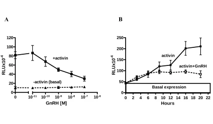
94% by 100 nM GnRH with an IC50 of 10-10 M and t½ of 4 h. The expression of
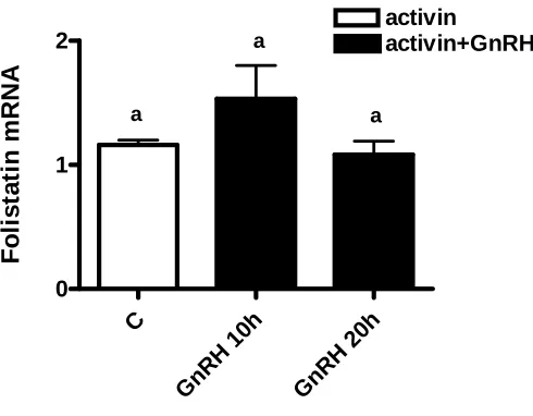
follistatin that can inhibit activin was not increased by GnRH. Activators of cAMP and
PKC such as forskolin and phorbol 12-myristate 13-acetate (PMA), respectively,
mimicked the GnRH inhibitory effect. Kinetic studies of wild type oFSHβLuc in transformed gonadotropes, LβT2, showed a continuous induction by activin (5.5 fold) over 20 h. The induction by activin, up to 6 h, was not affected by GnRH, but was
completely blocked thereafter. Cyclic AMP response element binding protein (CREB)
was implicated in this inhibition because overexpression of its constitutively active
mutant mimicked the inhibitory effect of GnRH and its inhibitor (ICERII) reversed the
forskolin or PMA increased the expression of a CREB responsive promoter 6xCRE-Luc.
Interstingly, inhibition of nitric oxide type I (NOSI) by 7-nitroindazole also reversed
GnRH-mediated inhibition of wt-oFSHβLuc by 60%. It is known that GnRH and CREB
induce expression of NOSI in gonadotropes and brain cortical cells, respectively. These
CHRONIC GONADOTROPIN-RELEASING HORMONE (GnRH) INHIBITS ACTIVIN INDUCTION OF THE OVINE FOLLICLE STIMULATING
HORMONE BETA-SUBUNIT (oFSHβ): INVOLVEMENT OF cAMP RESPONSE
ELEMENT BINDING PROTEIN (CREB) AND NITRIC OXIDE SYNTHASE TYPE I (NOSI)
By
FARIDEH SHAFIEE-KERMANI
A dissertation submitted to the Graduate Faculty of North Carolina State University
in partial fulfillment of the
requirements for the Degree of Doctor of Philosophy
MOLECULAR AND STRUCTURAL BIOCHEMISTRY
Raleigh, North Carolina
2007
APPROVED BY:
Robert R. Anholt James A. Knopp
DEDICATION
To my parents: Arsalan Shafiee-Kermani and Mehri Iravani
To my husband: Robert D. Morrel
BIOGRAPHY
Farideh Shafiee-Kermani was born and raised in Tehran, Iran. She received her primary
and secondary education in Tehran and attended Pars College where she received her
bachelor degree in educational psychology. A few years after the Iranian revolution,
1986, she moved to Sweden. There, she worked in a mental hospital where she found
herself interested in learning about bio-molecular changes leading to mental disorders. In
1994, she moved to the USA to pursue her interest in molecular genetics. She first
entered Wake Technical Community College and then transferred to North Carolina State
University in order to complete the under graduate science courses required for attending
graduate school in science. On August 1999 she was accepted into the Department of
Molecular and Structural Biochemistry at North Carolina State University to pursue a
Doctor of Philosophy degree. There, she joined Dr. William L. Miller’s laboratory to
begin her graduate research work in the field of gene transcription and focused on follicle
ACKNOWLDGEMENTS
I would like to express my gratitude to my research advisor, Dr. William L. Miller, for
his guidance, inspiration and encouragement during the course of my graduate studies.
Thanks are also extended to my graduate committee members, Dr. Dennis T. Brown, Dr.
Carla Mattos, Dr. Robert A. Anholt and Dr. James A. Knopp, for their advice and help
throughout this work. I would like also to thank my colleagues, Joyce Wu, Pei Su, Nedal
Safwat, Jesse Gore and Sang-oh Han, for their helpful discussions.
I would like to thank my parents, to whom I am most indebted, for their endless love and
spirit. Finally, I would like to thank my family members and friends for their support
during the course of my graduate studies.
TABLE OF CONTENTS
LIST OF TABLES ... vii
LIST OF FIGURES ... viii
LITERATURE REVIEW ...1
REFERENCES ...18
CHAPTER I ...31
Chronic GnRH Inhibits Activin Induction of the ovine FSH Beta Subunit: Involvement of cAMP Response Element Binding Protein (CREB) and Nitric Oxide Synthase type I (NOSI) Abstract ...33
Introduction ...34
Materials and Methods ...38
Results ...43
Discussion ...49
References ...64
CHAPTER II ...74
Expression and Regulation of the β-Subunit of Ovine Follicle Stimulating Hormone Relies Heavily on an AML1-Like Enhancer Abstract ...76
Introduction ...77
Materials and Methods ...79
Results ...85
Discussion ...89
References ...104
SUMMARY OF THESIS WORK ...107
APPENDIX I ...109
The nature of FSH induction by GnRH Trends in Endocrinology & Metabolism 13: 257-62, 2002 Pages are not numbered Abstract ...110
Introduction ...110
Discussion ...114
References ...115
APPENDIX II ...117
Meeting Abstract ...118
LIST OF TABLES
CHAPTER II
Table 1 Expression of the mut-oFSHβLuc-ΔAML1 transgene in tissues ...95
LIST OF FIGURES
LITERATURE REVIEW
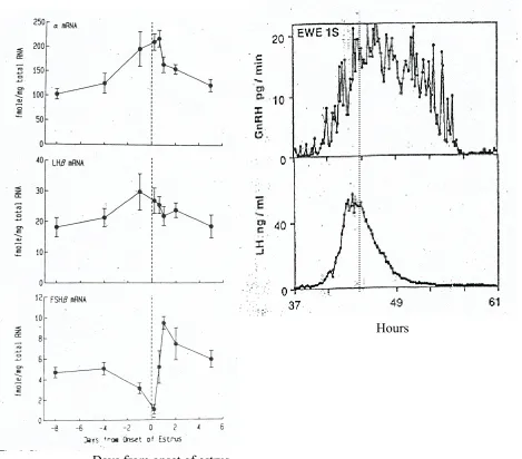
Figure 1 Changes in pituitary gonadotropin subunits (α, LHβ & FSHβ) mRNA during the estrous cycle of sheep and the preovulatory GnRH and LH surge in sheep ...13
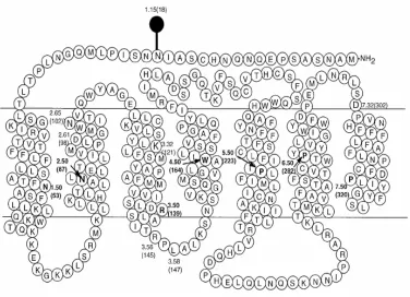
Figure 2 The structure of GnRH receptor ...14
Figure 3 Schematic presentation of cAMP and PKC pathways ...15
Figure 4 Schematic presentations of CREB, CREM and ICER genes organization ...16
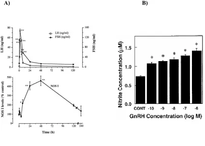
Figure 5 The effect of GnRH on NOSI production and gonadotropin release in vivo and the effect of GnRH on NO production in vitro ...17
CHAPTER I
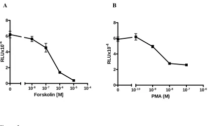
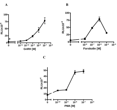
Figure 1 GnRH inhibited activin-induced wt-oFSHβLuc, mut-oFSHβLuc-ΔAP1 and endogenous mouse FSHβ ...54 Figure 2 Forskolin or PMA inhibited activin-induced expression of oFSHβLuc-ΔAP1 ...56 Figure 3 Inhibition by (Sp-8-Br-cAMP) and other reagents that increase intracellular cAMP (IBMX, CTX) or cAMP and PKC (PACAP) ...57
Figure 4 GnRH did not increase follistatin in mouse pituitary cultures ...58
Figure 5 GnRH inhibits activin-induced expression of wt-oFSHβLuc in LβT2 cells but not the basal expression ...59
Figure 6 GnRH, forskolin and PMA induced transcription of 6xCRE-37PRL-Luc in LβT2 cells ...60 Figure 7 Over expression of pCF Y/F CREB inhibited activin-induced expression of wt- oFSHβLuc, and overexpression of ICERII reversed inhibition caused by GnRH, forskolin, or PMA in LβT2 cells ...61 Figure 8 7-Nitroindazole reversed the inhibition caused by GnRH ...63
CHAPTER II
Figure 1 Follistatin inhibited FSH production in pituitary cultures ...96
Figure 2 Basal and activin-induced expression of oFSHβLuc constructs in LβT2
Cells ...98
Figure 3 Pituitary expression of mut-oFSHβLuc-∆AML1 (mutant 3 in figure 2) in 9
transgenic mouse lines ...100
Figure 4 Expression of mut-oFSHβLuc-ΔAML1 did not increase at estrus like wild type Transgenes ...101
Figure 5 Expression of mut-oFSHβluc-ΔAML1 was neither inhibited by follistatin nor induced by activin or GnRH alone, but was induced by 6 h of treatment with activin plus GnRH together ...102
Literature Review
Follicle stimulating hormone and its regulators
Follicle stimulating hormone, FSH, is absolutely necessary for egg maturation and
female fertility. Female mice that do not make FSH are infertile due to a complete arrest
at early stages of egg maturation (1). Similarly, an inactivating point mutation in the
FSH receptor gene causes female infertility in humans, a condition called
hypergonadotropic ovarian dysgenesis (2). However, FSH-deficient male mice are fertile
in spite of their lower sperm count and reduced testes size. In humans, males
homozygous for the inactivating point mutation of the FSH receptor show variable
suppression of spermatogenesis and fertility. The difference between the female and
male responses could be explained by the fact that FSH and testosterone have an apparent
synergistic effect on spermatogenesis and the absence of either hormone can be partially
compensated by the presence of the other hormone (1, 3).
Pituitary FSH and luteinizing hormone, LH, belong to the family of glycoprotein
hormones. These hormones are closely related in structure and each consists of two
subunits (α and β). Each subunit is glycosylated at specific amino acids and is internally
cross-linked by disulfide bonds. Both, LH and FSH, share a common α-subunit that is
noncovalently associated with its unique β-subunit that confers hormonal specificity.
Synthesis of FSHβ is rate limiting to overall production of biologically active FSH.
Therefore, regulation of FSHβ has been the focus of this study (4-6).
In mammals, the reproductive axis is controlled by hormones from the
produce gonadotropin releasing hormone (GnRH), which is released in a pulsatile fashion
into the anterior pituitary to stimulate gonadotropes. In response to GnRH, gonadotropes
synthesize and release FSH and LH. In male gonads, LH stimulates interstitial cells to
produce testosterone that in combination with FSH stimulates spermatogenesis (sperm
production) and sertoli cells that mature the sperm. The increased testosterone can then
change the expression levels of gonadotropin subunit genes (α, LHβ, and FSHβ) by
feedback effects on the hypothalamus or pituitary. In female gonads, FSH causes egg
maturation (folliculogenesis) and LH causes the follicle to burst that leads to egg release.
Growing follicles produce estrogen and ruptured follicles that are converted to corpus
luteum produce estrogen and progesterone. These two hormones can also affect the
hypothalamus and pituitary gland to alter gonadotropin gene expression (7, 8).
Studies have shown that, unlike LH, FSH production and secretion are not solely
dependent on GnRH, but it is also regulated by paracrine/autocrine factors within the
pituitary gland. This was evidenced by in vivo studies that showed anti-GnRH antibodies
or GnRH antagonists did not dramatically change serum FSH levels while LH levels
were reduced 50%. Also, abrupt withdrawal of activin from ovariectomized rats
decreased FSH serum levels by 50-60% within 4-5 h while withdrawal of GnRH from
castrated male rats decreased serum FSH by 50% within 12 h (9, 10, 11). In addition,
studies have shown that cultured pituitary cells can produce and secrete FSH without
stimulation by exogenous activators and addition of follistatin that incapacitates activin
can inhibit FSH synthesis (12, 13). These autocrine/paracrine effectors of FSHβ gene
morphogenetic proteins, BMPs, are activators; inhibin and follistatin are inhibitors of
FSHβ expression (14, 15, 16, 17).
Primary and transformed gonadotropes
Gonadotropes are located in the anterior pituitary and make up 3-10% of total
pituitary cells (18). The majority of pituitary cells are comprised of somatotropes,
lactotropes, adrenocorticotropes, folliculostellets and other cell types yet to be
characterized. This heterogeneity of pituitary cell cultures has limited the study of
different effectors of gonadotropes and their signaling pathways. Thus, Pamela Mellon
(19, 20) purposefully developed a gonadotrope cell line by targeted expression of a
transforming gene into mouse gonadotropes. This murine cell line was named LβT2.
These cells are derived from pituitary tumors of transgenic mice carrying the LHβ
promoter linked to the oncogenic SV40 large T-antigen. These cells provide a
homogeneous gonadotrope cell line (19, 20) that can overcome the limitations caused by
heterogeneity of primary pituitary cultures. These cells express FSHβ in the presence of
activin, and since it is possible to transiently transfect them, they are especially useful for
studying signaling pathways involved in regulation of FSHβ (21, 22). In addition, since
they are homogeneous gonadotropes, they provide strong evidence that the effect of
hormones on the FSHβ promoter are not caused by activation of the factors from other
cell types that are surrounding gonadotropes in pimary pituitary cell cultures (paracrine
Transgenic technique
Another useful tool for studying regulation of FSHβ is mice carrying FSHβ
promoterreporter transgenes. Cloned promoterreporter plasmids consist of the ovine FSHβ promoter from
-4741 bp to +759 bp linked to a luciferase gene (wt-oFSHβLuc) and its mutant version
(mut-oFSHβLuc-ΔAP1) that had two mutations at -120 bp and -83 bp. These two sites
were proven to be necessary for induction of oFSHβLuc by GnRH in pituitary cultures of
transgenic mice. Both transgenes were specifically expressed in gonadotropes of
transgenic mice where they were expressed and regulated just like mouse endogenous
FSHβ (12, 13).
Activin
Activins are major activators of the FSHβ gene in vivo and vitro. Activins belong to
the transforming growth factor β (TGF-β) superfamily that also includes BMPs,
Müllerian inhibiting substance (MIS), and growth and differentiation factor (GDF) (23,
24, 25, 26). Originally, activins were found in gonads acting mainly in the gonadal
pituitary axis since they were capable of inducing FSH synthesis and secretion. Later, it
was shown that activins are produced in different tissues including the pituitary gland and
can act as autocrine/paracrine factor as well (26). Activin is involved in many different
biological processes including cell proliferation, neuronal survival, erythropoiesis, and
early embryonic development. A biologically active activin is a disulfide-linked dimer of
two highly related β-subunits (βA, βB), resulting in three possible molecular species:
activin A (βA-βA), activin B (βB, βB), or activin AB (βA, βB). Three other forms of
dimers has not been demonstrated yet. Activin B is predominantly expressed in rat
gonadotropes, but activin A is present in human pituitaries, mainly in somatotropes (27,
28). Activin AB and B are also potent inducers of FSH synthesis/secretion in rat pituitary
cell cultures. However, most of the activin studies have been done using activin A
because it is the most active one and is produced in significant amounts by recombinant
means (29, 30).
Activin signals through two single membrane spanning serine/threonine kinase
receptors that are classified as type I and type II based on their structure and function.
The type II receptor is involved in initial ligand binding, which then recruits and activates
the type I receptor to propagate the signal downstream (31). Two activin type II
receptors (ActR-II & ActR-IIB) and two activin type I receptors (ActR-I & ActR-IB)
have been isolated from pituitary cells (32, 33, 34, 35). However, ActR-II mRNA is
expressed in gonadotropes, providing evidence for ActR-II being the mediator of activin
stimulation of FSH synthesis. Furthermore, ActRII knock out mice had dramatically
reduced FSHβ expression (25).
Follistatin
Follistatin is a monomeric glycoprotein expressed in many tissues. As an
autocrine/paracrine factor, follistatin neutralizes activin and other TGF-β family members
such as BMP-4 and BMP-7 by binding to and inhibiting them from binding to their
cognate receptors (36-38). There are two bioactive forms of follistatin, FS-315 (315
amino acids) and FS-288 (288 amino acids) that are produced by alternative mRNA
that has a truncated carboxyl-terminal is more potent than FS-315 in suppressing FSH
release (40-42). The higher potency of FS-288 might be due to its higher ability to bind
to heparan sulfate side chains of proteoglycans on the cell surface that causes
activin/FS-288 complex to be ingested by endocytotic action leading to increased degradation of
activin by lysosomal enzymes (42).
GnRH
Since the discovery of porcine hypothalamic GnRH in 1971 by Matsuo et al. (43), 16
different forms of GnRH have been discovered in vertebrates. All of these are
decapeptides that are closely related structures with, at least, 50% sequence identity.
These decapeptides have been named according to the first species from which they were
isolated and, at least, two of them coexist in the central nervous system (CNS) of each
species (45). The hypothalamic mammalian GnRH, which acts on gonadotropes
(mGnRH or GnRHI) and is the most well studied among these peptides, is referred to
here and in mammalian reproductive literatures as “GnRH”. In humans, the gene
encoding GnRH is located on chromosome 8 and consists of four exons and three introns.
The resulting pre-pro hormone consists of 92 amino acids organized into a signal peptide
consisting of the decapeptide, a cleavage site, and the GnRH-associated peptide.
Previously, GnRH was solely known as the key hormone in control of reproductive
function because it induced and released both gonadotropins, FSH and LH. In the mid
1980s, accumulating data showed that GnRH and its receptor are also present in normal
and malignant reproductive tissues such as placenta, ovary, endometrium, and prostate.
steroidogenesis; in malignant reproductive tissues it shows an inhibitory effect on
proliferation and metastasis of malignant cells (45).
The hypophysiotropic decapeptide, GnRH, is produced in the cell bodies of
hypothalamic neurons and secreted by their terminals into the hypophyseal-portal
circulation to reach the anterior pituitary, where it selectively stimulates gonadotropes
(46, 47). GnRH is a major modulator of FSH synthesis and release. GnRH secretion is
pulsatile in nature, and this intermittent secretion is required by gonadotropes in order to
secrete gonadotropins in a physiologic pattern. In rhesus monkeys with hypothalamic
lesions that destroyed GnRH secretion, 1 pulse/h of GnRH restored normal gonadotropin
secretion, but constant infusion of GnRH failed to restore gonadotropin secretion (48).
Although FSH production is not solely dependent on GnRH, withdrawal of GnRH in rats
caused serum FSH to fall 50% within 12 h (49). Expression of the gonadotropin subunit
genes is also controlled by GnRH pulse frequencies. While GnRH rapid pulses (>1
pulse/h) favor LHβ, relatively slow pulses of GnRH (≤1 pulse/h) favor FSHβ expression
(50).
Similar to rapid pulses, continuous GnRH also decreases FSHβ expression (51-53).
In fact, GnRH normally pulses at about 1 pulse/h, and more rapid pulses of GnRH occur
during the follicular phase of the reproductive cycle when FSHβ production decreases
(figure 1A & B (54, 55)). Accordingly, chronic treatment with GnRH has been used as
biochemical castration to slow cancers of reproductive tissue or treat precocious puberty,
and pulsatile GnRH has been used to treat infertility caused by GnRH deficiency (56-57).
To regulate gonadotropin gene expression, GnRH binds to its receptor on the
328 amino acids and belongs to the G protein-coupled receptor (GPCR) super family.
The GPCRs are composed of single polypeptide chains that contain 7 hydrophobic
transmembrane domains with extracellular amino and intracellular carboxyl termini (58).
However, the mammalian GnRHR is unique because it is the only GPCR that lacks the
entire intracellular carboxyl terminus, figure 2 (59). Upon activation by ligand, GPCRs
change their conformations that allow them to bind to their cognate heterotrimeric G
proteins, stimulating guanine nucleotide exchange factor on the α-subunit of the
heterotrimer. This leads to dissociation of β/γ-subunits from the complex and activation
of the α-subunit. Activated α- subunit then initiates a broad range of intracellular
signaling events by producing second messengers such as cAMP, PKC, and Ca+2 influx
(60, 61). Activated GPCRs are then rapidly internalized and lost from the cell
membrane. The cytoplasmic C-terminal of GPCRs has been shown to be involved in
internalization since it can be phosphorylated and consequently associated with β-arrestin
that causes internalization of the complex (62). This can lead to desensitization of the
receptor and its signaling pathway (63, 64). The mammalian GnRHR that lacks the
cytoplasmic C-terminal is not rapidly desensitized (65, 66). In addition, the expression of
GnRHR can be induced by GnRH and activin (67, 68).
Activation of GnRHR by GnRH has long been known to result in activation of Gαq
and Gαs pathways in different cell lines (7, 69-74-76). Induction of Gαq by GnRH leads
to activation of phospholipase C, which produces two second messengers, diacylglycerol
(DAG) and inositol 1, 4, 5-trisphosphate (IP3). Generation of these second messengers
leads to mobilization of intracellular pools of Ca2+ and activation of PKC, which in turn
to form AP-1 transcription complex that can induce expression of gonadotropin subunit
genes by binding to its cognate response element, figure 3B (7, 75-80).
It is known that GnRH can also activate the Gαs pathway (7, 69, 70, 72, 73).
Activation of Gαs by GnRH leads to increased cAMP levels (69). Generally, cAMP
regulates PKA activity, which results in serine 133 phosphorylation of cAMP response
element binding protein (CREB) that leads to its activation. Activated CREB, in turn,
induces transcription of target genes (figure 3A (81, 82)). In fact, GnRH phosphorylates
CREB in the αT3 gonadotrope lineage. Although PKA is the major activator of CREB
(81, 82), it was shown that PKC can also activate CREB in some cells (83).
Inducible cAMP early repressor (ICER)
ICER was initially described in the pineal gland as an inhibitor of cAMP-dependent
transcription induced by rhythmic adrenergic signals. It belongs to a subfamily of basic
leucine zipper transcription factors, CREB/CREM. These transcription factors contain an
activation domain, DNA binding domain and phosphorylation box and can bind to the
cAMP response element, CRE, as homo- or heterodimers. The promoter sequens of
CREB/CREM genes are rich in GC content, which is the characteristic of housekeeping
genes so these factors are ubiquitously expressed and are activated by phosphorylation.
ICER isoforms (I & Iγ, II & IIγ) are produced by alternative splicing via an internal
promoter, which exists within an intron near the 3’ end of the CREM gene, figure 4. The
ICER promoter contains normal GC/AT content and has two CRE elements which allow
induction by cAMP. Unlike CREM, it contains only the DNA binding domain and lacks
and CREM proteins and functions as a powerful dominant negative repressor of
cAMP-induced transcription. An important feature of ICER is its inducibility that makes it the
only CRE binding factor whose function is determined by its rate of transcription (84,
85).
Nitric oxide synthase type I (NOSI)
The enzyme, NOSI, catalyzes production of nitric oxide (NO), a diffusable free radical,
from L-arginine. There are two other isoforms of NOS (NOSII and NOSIII). The NOSI
gene in humans is located on chromosome 12 and its promoter contains two CREB
binding sites. The free radical, NO, which is produced in nearly every cell type, has
diverse physiologic and pathophysiologic roles as a vasodilator, neurotransmitter,
antimicrobial effector and immunomodulator (86, 87).
Studies using in situ hybridization and immunohistochemistry have shown that NOSI
is present in gonadotropes in the anterior pituitary (88, 89), and changes in NOSI
expression coincide with the pattern of GnRH release during rat proestrus (90). The
NOSI expression is increased by GnRH in rat pituitaries, which is correlated with a
dramatic decrease in FSH secretion (figure 5 A (91)). GnRH increases NO production in
transformed gonadotropes, LβT2 cells (figure 5 B (92)). Finally, CREB regulates NOSI
expression (93, 94).
Thesis goals
The gene encoding the ovine FSHβ (oFSHβ) was cloned and characterized in our
transcription factor binding sites including four putative AP-1 sites that were thought to
be responsive to GnRH positive regulation. Deletion mutations of this promoter showed
that two of these putative AP-1 sites at -120 bp and -83 bp bind AP-1 proteins and are
necessary for activation by AP-1 proteins (Jun/Fos), PMA and GnRH in vitro. Further
studies with transgenic mouse pituitary cultures have shown that 1 nM GnRH doubled
activin induction of wild type oFSHβ within 4 h but inhibited activin induction of the
AP-1 mutant of oFSHβ. However, the molecular mechanism(s) and the pathway(s) involved
in this inhibition were never studied.
The goals of my thesis were to use mouse pituitary cultures from transgenic mice
harboring wild type or AP-1 mutant oFSHβ and mouse transformed gonadotropes, LβT2
cells, to confirm 1) our previous study to emphasize the importance of AP-1 sites for
GnRH positive regulation of oFSHβ and 2) to examine the cellular pathways that mediate
the negative control of activin-induced expression of oFSHβ by the chronic presence of
GnRH. These studies showed that chronic GnRH inhibited activin-dependent expression
of wild type oFSHβ by 60% and inhibited the AP-1 mutant of oFSHβ by 94%. The
GnRH inhibitory effect on wild type oFSHβ was first observed after 6 h while inhibition
of the AP-1 mutant was significant at 2 h. Furthermore, it was shown that CREB and
NOSI are intermediates in the GnRH-mediated cellular pathway that regulates inhibition
of activin-dependent expression of oFSHβ. In addition, it was shown that CREB
activation can occur through PKA and/or PKC pathways.
My initial work was to study cell specific expression of the oFSHβ gene. As the first
step, I used NCBI and ENSEMBL data bases to compare the FSHβ promoter of sheep,
the 5’ end of these promoters. These sequences alone or in different combinations were
deleted and eight transgenic mouse lines were produced to study the importance of each
of these sites. However, analysis of oFSHβ expression in these mice showed that
deletion of these sequences had no effect on cell specific expression of oFSHβ since all
the mice expressed oFSHβ specifically and equally well in pituitaries. One of these mice
was used as the wild type mouse line in a study that a former post-doc, Pei Su, conducted
with my participation to determine the importance of a putative AML-1 enhancer for
activin induction of oFSHβ in vitro and in vivo (see Appendix I). These studies showed
that mutation of this enhancer reduced activin induction of oFSHβ by 81% in vitro and by
99.9% in vivo without affecting basal expression.
A) B)
Hours
Days from onset of estrus
Figure 1
(A) Changes in pituitary gonadotropin subunits (α, LHβ & FSHβ) mRNA during the
estrous cycle of sheep. The time of the onset of estrus designated as day 0. The dashed line
represents the preovulatory LH surge, Leung et al (ref. # 54).
Figure 2
Representation of the human GnRH receptor. The glycosylation site is marked. The key
functional amino acids are numbered and the most conserved residues in transmembrane
domains are designated by index 50. Note! no intacellular COOH terminus normally
A) B)
Figure 3
ATP ppi
PKA
CRE
β γ
AC GTP
αs GTP
αq
PLC
IP3
DAG
Ca2+ PKC
MAPKKK MAPKK
MAPK
Fos / Jun AP-1
β γ
PIP2
CRE
P P
P
GnRH
(A) Gαs signaling pathway and (B) Activation of Gαq pathway by GnRH studied in
Figure 4
Schematic representation of CREB, CREM and ICER isoforms. Boxes represent exons.
bZIP represents basic regions required for DNA binding and the leucine zipper required
for dimerization. Q-rich exon is involved in basal transcriptional activities. On the
CREM gene, P1 shows the CREM promoter and P2 represents ICER promoter, Bodor et
al (ref. # 85).
A) B)
Figure 5
(A) The effect of GnRH on NOSI production and gonadotropins (LH and FSH) release
in vivo, Garrel G et al (ref. # 91). (B) The effect of GnRH on NO concentration in LβT2
cells, Chen et al (ref. # 92)
References:
1- Kumar TR, Wang Y, Lu N, Matzuk MM 1997 Follicle stimulating hormone is
required for ovarian follicle maturation but not male fertility. Nat Genet 15: 201-4
2- Aittomaki 2-K, Herva R, Stenman UH, Juntunen K, Ylostalo Δ, Hovatta O, de la
Chapelle A 1996 Clinical features of primary ovarian failure caused by a point mutation
in the follicle-stimulating hormone receptor gene. J Clin Endocrinol Metab 81: 3722-6
3- Tapanainen JS, Aittomaki K, Min J, Vaskivuo T, Huhtaniemi IT 1997 Men
homozygous for an inactivating mutation of the follicle-stimulating hormone (FSH)
receptor gene present variable suppression of spermatogenesis and fertility. Nat Genet
15:205-6
4- Pierce JG, Parsons TF 1981 Glycoprotein Hormones: Structure and Function. Ann
Rev Biochem 50: 465-95
5- Padmanabhan V, Sharma TP 2001 Neuroendocrine vs. Paracrine Control of
Follicle-Stimulating Hormone. Arc Med Res 32: 533-43
6- Bousfield GR, Perry WM, Ward DN 1994 Gonadotropins: Chemistry and
Biosynthesis. In the physiology of Reproduction (eds. Knobil E and NeillJD). 2ed.
Chapter 30. Raven Press, New York P: 1749-1792
7- Burger LL, Haisenleder DJ, Dalkin AC, Marshall JC 2004 Regulation of
gonadotropin subunit gene transcription. J Mol Endocrinol 33: 559-84
8- Sherwood L 2001In Human Physiology: From Cell to System. Fouth ed. Chapter 20
9- Culler MD, Negro-Vilar A 1986 Evidence that pulsatile follicle-stimulating hormone
secretion is independent of endogenous luteinizing hormone-releasing hormone.
Endocrinology 118: 609-12
10- Kawakami M, Higuchi T 1979 Effects of active and passive immunization with
LH-RH on gonadotrophin secretion and reproductive function in female rats. Acta Endocrinol
91: 616-28
11- DePaolo LV, Shimonaka M, Schwall RH, Ling N 1991 In vivo comparison of the
follicle-stimulating hormone-suppresing activity of follistatin and inhibin in
ovariectomized rats. Endocrinology 128: 668-74
12- Huang HJ, Sebastian J, Strahl BD, Wu JC, Miller WL 2001 The Promoter for the
Ovine Follicle-Stimulating Hormone-β Gene (FSHβ) Confers FSHβ-Like Expression on
Luciferase in Transgenic Mice: Regulatory Studies in Vivo and in Vitro. Endocrinology
142: 2260-66
13- Huang HJ, Sebastian J, Strahl BD, Wu JC, Miller WL, 2001 Transcriptional
regulation of the ovine follicle-stimulating hormone-beta gene by activin and
gonadotropin-releasing hormone (GnRH): involvement of two proximal activator
protein-1 sites for GnRH stimulation. Endocrinology protein-142: 2267-74
14- Otsuka F, Shimasaki S 2002 A Novel Function of Bone Morphogenetic Protein-15
in the Pituitary: Selective Synthesis and Secretion of FSH by Gonadotropes.
Endocrinology 143: 4938-41
15- Huang HJ, Wu JC, Su P, Zhirnov O, Miller WL 2001 A Novel Role for Bone
Morphogenetic Proteins in the Synthesis of Follicle-Stimulating Hormone.
16- Carroll RS, Corrigan AZ, Gharib SD, Vale W, Chin WW 1989 Inhibin, activin,
and follistatin: regulation of follicle- stimulating hormone messenger ribonucleic acid
levels. Mol Endocrinol 3: 1969-76
17- Bilezikjian LM, Blount AL, Leal AMO, Donaldson CJ, Fischer WH, Vale WW
2004 Autocrine/paracrine regulation of pituitary function by activin, inhibin and
follistatin. Mol Cell Endocrinol 225: 29-36
18- Pelletier G, Leclrec R, Labrie F 1976 Identification of Gonadotropic Cells in the
Human Pituitary by Immunoperoxidase Technique. Mol Cell Endocrinol 6:123-128
19- Pernasetti F, Vasilyev VV, Rosenberg SB, Baily JS, Huang HJ, Miller WL,
Mellon PL 2001 Cell-specific transcriptional regulation of follicle-stimulating
hormone-beta by activin and gonadotropin-releasing hormone in the Lhormone-betaT2 pituitary gonadotrope
cell model. Endocrinology 142:2284-95
20- Alarid ET, Windle JJ, Whyte DB, Mellon PL 1996 Immortalization of pituitary
cells at discrete stages of development by directed oncogenesis in transgenic mice.
Development 122: 3319-29
21- Safwat N, Ninomiya-Tsuji J, Gore AJ, Miller WL 2005 Transforming Groth
Factor Activated Kinase 1 Is a key Mediator of Ovine Follicle-Stimulating Hormone
β-Subunit Expression. Endocrinology 146: 4814-24
22- Bernard DJ 2004 Both SMAD2 and SMAD3 Mediate Activin-Stimulated
Expression of the Follicle-Stimulating Hormone β Subunit in Mouse Gonadotrope Cells.
Mol Endocrinol 18: 606-23
23- Weiss J, Crowley WF, Halvorson LM, Jameson JL 1993 Perifusion of Rat
Distinct Effect on Gonadotropin Gene Expression and secretion. Endocrinology 132:
2307-11
24- Graham KE, Nusser KD, Low MJ 1999 LβT2 gonadotroph cells secrete follicle
stimulating hormone (FSH) in response to activin A. J Endocrinol 162: R1-R5
25- Kumar TR, Agno J, Janovick JA, Conn PM, Matzuk MM 2003 Regulation of
FSHβ and GnRH receptor gene expression in activin receptor II knockout male mice.
Mol Cell Endocrinol 212: 19-27
26- Ethier JF, Findlay JK 2001 Roles of activin and its signal transduction mechanism
in reproductive tissues. Reproduction 121: 667-675
27- Roberts V, Meunier H, Vaughan J, Rivier J, Rivier C, Vale W, Sawchenko P
1989 Production and regulation of inhibin subunits in pituitary gonadotropes.
Endocrinology 124: 552-4
28- Wada M, Shintani Y, Kosaka M, Sano T, Hizawa K, Saito S 1996
Immunohistochemical localization of activin A and follistatin in human tissues. Endocr J
43: 375-85
29- Mason AJ, Berkemeier LM, Schmelzer CH, Schwall RH 1989 Activin B:
precursor sequences, genomic structure and in vitro activities. Mol Endocrinol 3 1352-8
30- Nakamura T, Asashima M, Eto Y,Takio K, Uchiyama H, Moria N, Ariizumi T,
Yashiro T, Sugino K, Titani K 1992 Isolation and characterization of native activin B. J
Biol Chem 267 16385-9
31- Massague J 1998 TGF-β Signal Transduction. Annu Rev Biochem 67: 753-91
32- Mathews LS, Vale WW 1991 Expression cloning of an activin receptor, a predicted
33- Mathews LS, Vale WW, Kintner CR 1992 Cloning of a second type of activin
receptor and functional characterization in Xenopus embryos. Science 14: 3810-21
34- Carcamo J, Weis FM, Ventura F, Wieser R, Wrana JL, Attisano L, Massague J
1994 Type I receptor specify growth-inhibitory and trans criptional resposes to
transforming growth factor beta and activin. Mol Cell Biol 14 3810-21
35- Attisano L, Carcamo J, Ventura F, Eeis FM, Massague J, Wrana JL 1993
Identification of human activin and TGF beta type I receptors that form heteromeric
kinase complexes with type II receptors. Cell 75: 671-80
36- Kumar TR 2005 Too Many Follistatins: Racing Inside and Getting out of the Cell.
Endocrinology 146: 5048-51
37- de Winter JP, ten Dijke P, de Vries CJ, van Achterberg TA, Sugino H, de Vaele
P, Huylebroeck D, Verschueren K, van den Eijnden-van Raaij 1996 Follistatins
neutralize bioactivity by inhibition of activin binding to its type II receptors. Mol Cell
Endocrinol 116: 105-14
38- Nakamura T, Takio K, Eto Y, Shibai H, Titani K, Sugino H 1990 activin-binding
protein from rat ovary is follistatin. Science 247: 836-8
39- Shimasaki S, Koga M, Esch F, Mercado M, Cooksey K, Koba A, Ling N 1988
Porcine follistatin gene structure supports two forms of mature follistatin produced by
alternative splicing. Biochem Biophys Res Commun 152: 17-23
40- Iemura S, Yamamoto TS, Takagi C, Uchiyama H, Natsume T, Shimasaki S,
Sugino H, Ueno N 1998 Direct binding of follistatin to a complex of bonemorphogenetic
protein and its receptor inhibits ventral and epidermal cell fates in early Xenopus embryo.
41- Sugino K, Kurosawa N, Nakamura T, Takio K, Shimasaki S, Ling N, Titani K
Sugino H 1993 Molecular heterogeneity of follistatin, an activin-binding protein. Higher
affinity of the carboxyl-terminal truncated forms for heparin sulfate proteoglycans on the
ovarian granulose cell. J Biol Chem 268: 15579-87
42- Hashimoto O, Nakamura T, Shoji H, Shimasaki S, Hayashi Y, Sugino H 1997 A
Novel Role of Follistatin, an Activin-binding Protein, in the Inhibition of Activin Action
in Rat Pituitary Cells. J Biol Chem 272: 13835-42
43- Matsuo H, Baba Y, Nair RM, Arimura A, Schally AV 1971 Structure of the
porcine LH- and FSH-releasing hormone.I. the proposed amino acid sequence. Biochem
Biophys Res Commun 43: 1334-39
44- Somoza GM, Miranda LA, Strobl-Mazzula P, Guligur LG 2002
Gonadotropin-releasing hormone (GnRH): from fish to mammalian brains. Cell Mol Neurobiol 22:
589-609
45- Limonta P, Moretti RM, Marelli MM, Mota M 2003 The biology of gonadotropin
hormone-releasing hormone: role in the control of tumor growth and progression in
humans. Front Neuroendocrinol 24: 279-95
46- Conn PM, Crowly WF 1991 Gonadotropin-Releasing hormone and its Analogs. N
Engl J Med 324: 93-103
47- Stojilkovic SS, Catt KJ 1995 Expression and signal transduction pathways of
gonadotropin-releasing hormone receptors. Recent Prog Horm Res 50: 161-205
48- Belchetz PE, Plant TM, Nakai Y, Keogh EJ, Knobil E 1978 Hypophysial
responses to continuous and intermittent deliver of hypothalamic gonadotropin-releasing
49- Grady RR, Shin L, Charlesworth MC, Cohen-Becker IR, Smith M,Rivier C,
Rivier J, Vale W Schwartz NB 1985 Differential suppression of follicle-stimulating
hormone and luteinizing hormone secretion in vivo by a gonadotropin-releasing hormone
antagonist. neuroendocrinology 40: 246-52
50- Haisenleder DJ, Dalkin AC, Ortolano GA, Marshall JC, Shupnik MA 1991 A
pulsatile gonadotropin releasing-hormone stimulus is required to increase transcription of
the gonadotropin subunit genes: evidence for differential regulation of transcription by
pulse frequency in vivo. Endocrinology 128: 509-17
51- Attardi B, Keeping HS, Winters SJ, Kotsuji F, Troen P 1989 Effect of Inhibin
from Primates Sertoli Cells and GnRH on Gonadotropin Subunit mRNA in Rat Pituitary
Cell Cultures. Mol Endocrinol 3: 1236-42
52- Melamed P, Kadir MN, Wijeweera A, Seah S 2006 Transcription of gonadotropin
beta subunit genes involves cross-talk between the transcription factors and co-regulators
that mediate actions of the regulatory hormones. Mol Cell Endocrinol 252: 167-83
53- Hotchkiss J, Knobil E 1994 The Menstrual Cycle and its Neuroendocrine Control.
In: Knobil J (eds) The Physiology of Reproduction, ed.2. Chapter 48, Reven Press, New
York, NY, P: 711-749
54- Leung K, Kim KE, Maurer RA, Landefeld TD 1988 Divergent changes in the
concentrations of gonadotropin beta-subunit messenger ribonucleic acid during the
estrous cycle of sheep. Mol Endocrinol 2: 272-6
55- Moenter SM, Caraty A, Locatelli A, Karsch FJ 1991 Pattern of
Gonadotropin-Releasing Hormone (GnRH) Secretion Leading up to Ovulation in the Ewe: Existence of
56- Robertson JF, Blamey RW 2003 The use of gonadotropin-releasing hormone
(GnRH) agonists in early and advanced breast cancer in pre- and perimenopausal
women.Eur J Cancer 39: 861-9
57- Conn PM, Crowley WF 1994 Gonadotropin-Releasing Hormone and its Analogs.
Annu Rev Med 45: 391-405
58- Strader CD, Fong TM, Graziano MP, Tota MR 1995 The family of
G-protein-coupled receptors. FABES J 9: 745-754
59- Sealfon SC, Weinstein H, Millar RP 1997 Molecular Mechanisms of Ligand
Interaction with the Gonadotropin-Releasing Hormone Receptor. Endo Rev 18: 180-205
60- McCudden CR, Hains MD, Kimple RJ, Siderovski DP, Willard FS 2005
G-protein signaling: back to the future. Cell Mol Life Sci 62: 551-577
61- Dohlman HG 1991 Model Systems for the Study of Seven-Transmembrane-Segment
Receptors. Annu Rev Biochem 60: 653-88
62- Nussenzveig DR, Heinflink M, Gershengorn MC 1993 Agonist-Stimulated
Internalization of the Thyrotropin-Releasing Hormone Receptor Is Dependent on two
Domains in the Receptor Carboxyl Terminus. J Biol Chem 268: 2389-92
63- Shenoy SK, Lefkowitz RJ 2005 Seven-transmembrane receptor signaling through
beta-arrestin. Sci STKE 308: cm 10
64- Hislop JN, Everest HM, Flynn A, Harding T, Uney JB, Troskie BE, Millar RP,
McArdle CA 2001 Differential internalization of mammalian and non-mammalian
gonadotropin-releasing hormone receptors. Uncoupling of dynamin-dependent
internalization from mitogen-activated protein kinase signaling. J Biol Chem 276:
65- Davidson JS, Wakefield IK, Millar RP 1994 Absence of rapid desensitization of
the mouse gonadotropin-releasing hormone receptor. Biochem J 300: 299-302
66- Liu F, Austin DA, Webster NJ 2003 Gonadotropin-releasing hormone-desensitized
LbetaT2 gonadotrope cells are refractory to acute protein kinase C, cyclic AMP, and
calcium-dependent signaling. Endocrinology 144: 4354-65
67- Lin X, Conn PM 1998 Transcriptional activation of gonadotropin-releasing hormone
(GnRH) receptor gene by GnRH and cyclic adenosine monophosphate. Endocrinology
139: 3896-902
68- Braden TD, Conn PM 1992 Activin-A stimulates the synthesis of
gonadotropin-releasing hormone receptors. Endocrinology 130: 2101-5
69- Kaiser UB, Conn PM, Chin WW 1997 Studies of Gonadotropin-Releasing
Hormone (GnRH) Action Using GnRH Receptor-Expressing Pituitary Cell Lines. Endo
Rev 18: 46-70
70- Stanislaus D, Ponder S, Ji TH, Conn PM 1998 Gonadotropin-Releasing Hormone
Receptor Couples to Multiple G Proteins in Rat Gonadotrophs and in GGH3 Cells:
Evidence from Palmitoylation and Overexpression of G Proteins. 59: 579-86
71- Grosse R, Schmid A, Schoneberg T, Herrilich A, Mukn P, Schultz G,
Gudermann T 2000 Gonadotropin-releasing Hormone Receptor Initiates Multiple
Signaling Pathwayes by Exclusively Coupling Gq/11 Proteins. J Bio Chem 275: 9193- 200
72- Borgeat P, Chavancy G, Dupont A, Labrie F, Arimura A, Schally AV 1972
Stimulation of Adenosine 3’:5’-Cyclic Monophosphate Accumulation in Anterior
Piyuitary Gland In Vitro by synthetic Luteeinizing Hormone-Releasing Hormone. Proc
73- Liu F, Usui I, Evans LG, Austin DA, Mellon PL, Olefsky JM, Webster N J 2002
Involvement of Both Gq/11 and Gs Proteins in Gonadotropin-Releasing Hormone
Receptor-mediated Signaling in LβT2 cells. J Biol Chem 277: 32099-108
74- Garrel G, McArdle, Hemmings BA 1997 Gonadotropin-Releasing Hormone and
Pituitary Adenylate Cyclase-Activating Polypeptide Affect Levels of Cyclic Adenosine
3’,5’-Monophosphate-Dependent Protein Kinase A (PKA) Subunit in the Clonal
Gonadotrope αT3-1 Cells: Evidence for Cross-Talk between PKA and Protein Kinase C
Pathways. Endocrinology 138: 2259-66
75- Strahl BD, Huang HJ, Pederson NR, WU JC, Ghosh BR, Miller WL 1997 Two
proximal activating protein-1-binding sites are sufficient to stimulate transcription of the
ovine follicle-stimulating hormone-beta gene. Endocrinology 138: 2621-31
76- Strahl BD, Huang HJ, Sebastian J, Ghosh BR, Miller WL 1998 Transcriptional
activation of the ovine follicle-stimulating hormone beta-subunit gene by
gonadotropin-releasing hormone: involvement of two activating protein-1-binding sites and protein
kinase C. Endocrinology 139: 4455-65
77- Reiss N, Llevis LN, Shacham S, Harris D, Seger R, Naor Z 1997 Mechanism of
mitogen-activated protein kinase activation by gonadotropin-releasing hormone in the
pituitary of alpha T3-1 cell line: differential roles of calcium and protein kinase C.
Endocrinology 138: 1673-82
78- Yokoi T, Ohmichi M, Tasaka K, Kimura A, Kanda Y, Hayakawa J, Tahara M,
Hisamoto K, Kurachi H, Murata Y 2000 Activation of the Luteinizing Hormone β
Promoter by Gonadotropin-releasing Hormone Requires c-Jun NH2-terminal. J Biol
79- Roberson MS, Misra-Press A, Laurance ME, Stork PS, Maurer RA 1995 A Role
for Mitogen-Activated Protein Kinase in Mediating Activation of the Glycoprotein
Hormone α-Subunit Promoter by Gonadotropin-Releasing Hormone. Mol Cell Biol 15:
3531-39
80- Maurer RA, Kim KE, Schoderbek WE, Roberson MS, Glen DJ 1999 Regulation
of glycoprotein hormone alpha-subunit gene expression. Recent Prog Horm Res 54:
455-84
81- Duan W R, Shin JL, Jameson JL 1999 Stradiol Suppresses Phosphorylation of
Cyclic Adenosin 3’,5’-Monophosphate Response Element Binding Protein (CREB) in the
Pituitary: Evidence for Indirect Action via Gonadotropin-Releasing Hormone. Mol
Endocrinol 13:1338-52
82- Chin KV, Yang WL, Ravatn R, Kita T, Reitman E, Vettori D, Cvijic ME, Shin
M, Iacono L 2002 Reinventing the wheel of Cyclic AMP Novel Mechanisms of cAMP
Signaling. Ann NY Acad Sci 968: 49-64
83- Xie H, Rothestein TL 1995 Protein Kinase C Mediates Activation of Nuclear cAMP
Response Element-Binding Protein (CREB) in B Lymphocytes Stimulated Through
Surface Ig. J Immunol 154: 1717-23
84- Sassone-Corsi P 1995 Transcription Factors Responsive to cAMP. Ann Rev Cell
Dev Biol 11: 355-77
85- Bodor J, Bodorova J, Gress RE 2000 Suppression of T cell function: potential role
for transcriptional repressor ICER. J Leukoc Biol 69: 1053-9
86- Geller DA, Billiar TR 1998 Molecular biology of nitric oxide synthase. Cancer
87- McCann SM, Mastronardi C, Delaurentiis A, Rettori V 2005 The Nitric Oxide
Theory of Aging Revisited. Ann NY Acad Sci 1057: 64-84
88- Ceccatelli S, Hulting AL, Zhang X, Gustafsson L, Villar M, Hokfelt T 1993
Nitric oxide synthase in the rat anterior pituitary gland and the role of nitric oxide in
regulation of luteinizing hormone secretion. Proc Natl Accad Sci 90: 11292-96
89- Yamada K, Xu ZQ, Zhang X, Gustafsson L, Hulting AL, de Vente J, Steinbusch
HW, Hokfelt T 1997 Nitric oxide synthase and cGMP in the anterior pituitary gland:
effect of a GnRH antagonist and nitric oxide donors. Neuroendocrinology 65: 147-56
90- Lozach A, Garrel G, Lerrant Y, Berault A, Counis R 1998 GnRH-dependent
up-regulation of nitric oxide synthase I level in pituitary gonadotrophs mediates cGMP
elevation during rat proestrus. Mol Cell Endocrinol 143: 43-51
91- Garrel G, Lerrant Y, Siriostis C, Berault A, Magre S, Bouchaud C, Counis R
1998 Evidence That Gonadotropin-Releasing Hormone Stimulates Gene Expression and
Levels of Active Nitric Oxide Synthase Type I in Pituitary Gonadotrophs, a Process
Altered by Desensitization and, Indirectly, by Gonadal Steroids. Endocrinology 139:
2163-70
92- Chen L, Sakai T, Sakamoto S, Kato M, Inoue K 1999 Direct Evidence of
Gonadotropin-Releasing Hormone (GnRH)-Stimulated Nitric Oxide Production in the
LβT-2 Clonal Gonadotropes. Pituitary 2: 191-96
93- Sasaki M, Gonzalez-Zulueta M, Huang H, Herring WJ, Ahn S, Ginty DD,
Dawson VL, Dawson TM 2000 Dynamic regulation of neuronal NO Synthase
transcription by calcium influx through a CREB family transcription factor-dependent
94- Mantelas A, Stamatakis A, Kazanis I, Philipidis H, Stylianopoulou F 2003
Control of neuronal nitric oxide synthase and brain-derived neurotrophic factor levels by
GABA-A receptors in the developing rat cortex. Brain Res Dev Brain Res 145: 185-95
CHAPTER I
Chronic GnRH Inhibits Activin Induction of the ovine FSH Beta Subunit:
Involvement of cAMP Response Element Binding Protein (CREB) and Nitric Oxide
Synthase typeI (NOSI)
By
Farideh Shafiee-Kermani, Sang-oh Han, and William L. Miller
Department of Molecular and Structural Biochemistry, Box 7622
North Carolina State University, Raleigh, NC 27695-7622
RUNNING TITLE: GnRH inhibition of activin-induced FSHβ
CORRESPONDING AUTHOR’S ADDRESS:
William L. Miller
Department of Molecular and Structural Biochemistry, Box 7622
North Carolina State University, Raleigh, NC 27695-7622
Tel: (919) 515-5762; Fax: (919) 515-2047; Email: [email protected]
KEY WORDS: GnRH, FSHβ, primary gonadotropes, LβT-2 cells, activin, follistatin,
CREB, CRE, NOSI
This work was supported by North Carolina State University Agricultural Research
Abstract: (247 words) Follicle stimulating hormone (FSH) is induced by activin and this
expression is modulated by GnRH through FSHβ expression. This report focuses on the
inhibitory effect of GnRH on activin-induced FSHβ expression. Activin-treated primary
murine pituitary cultures robustly express mut-oFSHβLuc-∆AP1, a luciferase transgene
driven by 4.7 kb of ovine FSHβ promoter. This promoter lacks two GnRH-inducible
AP-1 sites making it easier to observe GnRH-mediated inhibition. Luciferase expression
from this transgene was decreased 94% by 100 nM GnRH with a t1/2 of ~ 4 h in pituitary
cultures, and this inhibition was independent of follistatin. Activators of cAMP and
protein kinase C such as forskolin and phorbol 12-myristate 3-acetate (PMA),
respectively, mimicked GnRH action. Kinetic studies of wild type oFSHβLuc in LβT2
cells showed continuous induction by activin (4-fold) over 20 h. Most of this induction
(78%) was blocked, beginning at 6 h. Cyclic AMP Response Element Binding protein
(CREB) was implicated in this inhibition because overexpression of its constitutively
active mutant mimicked GnRH and its inhibitor (ICERII) reversed the inhibition caused
by GnRH, forskolin or PMA. In addition, GnRH, forskolin or PMA increased the
expression of a CREB responsive promoter, 6xCRE-37PRL-Luc. Inhibition of nitric
oxide type I (NOSI) by 7-nitroindazole also reversed GnRH-mediated inhibition by 60%.
It is known that GnRH and CREB induce production of NOSI in gonadotropes and
neuronal cells, respectively. These data support the concept that chronic GnRH inhibits
activin-induced oFSHβ expression by sequential activation of CREB and NOSI through
Introduction: Follicle stimulating hormone (FSH) is required for normal gonadal
function in mammals (1-3). It is produced only in pituitary gonadotropes as an α/β
heterodimer. The β-subunit is in limiting amounts and thus controls the overall synthesis
of FSH (4). Production of FSHβ is regulated by many hormones including activins,
GnRH, follistatin, steroids and inhibins (5-10).
Studies with rats and LβT2 cells suggest that activins are the primary inducers of
FSHβ (11, 12). FSHβ is always induced in vivo by activins because these hormones are
constitutively present in pituitary tissue (13). Thus, regulation depends on the effects of
hormones that can alter induction of FSHβ by activin.
Hypothalamic GnRH is another important inducer of FSH synthesis and release.
Withdrawal of GnRH in rats causes serum FSH to fall 50% within 12 h (14). GnRH
regulation is complex, however, because relatively slow pulses of GnRH (≤1/h) favor
FSHβ synthesis (10, 15, 16) while chronic treatment with GnRH decreases FSHβ below
control levels in vivo and in vitro (17-20). In a physiologic context, studies with ewes
have shown that FSHβ mRNA declines 80% during the preovulatory LH surge (21) when
GnRH is increased for a prolonged time period (22). This GnRH surge, which is not
strictly episodic and is sustained chronically for 12-20 h (23), may be responsible for the
decline in FSHβ during this time. In a medical context, chronic treatment with GnRH has
been used successfully to decrease the production of FSH and LH in order to inhibit
gonadal function. Such treatment often slows the progression of reproductive cancers
and has been used widely (24, 25).
Follistatin is an autocrine/paracrine pituitary factor that can inhibit FSHβ by
the multiple effects of GnRH, gonadal steroids, follistatin and inhibin on regulation of
FSHβ expression, it is clear that this regulation comprises a complex web of interactive
feedback controls. For a comprehensive understanding of the regulation of FSHβ
expression, each control mechanism needs to be understood thoroughly.
With the exception of the preovulatory surge, GnRH normally pulses at ~1 pulse/h
(27) and has been studied primarily as an inducer of all the gonadotropin subunit genes
(αGSU, LHβ, and FSHβ) or as a secretogogue. The positive effects of GnRH on
secretion and transcription of gonadotropins are thought to involve activation of Gαq
heterotrimeric G proteins, protein kinase C (PKC), MAPK, Jun/Fos transcription factors
and calcium influx (28-31). Our laboratory used in vitro and transgenic studies to
identify two GnRH responsive Jun/Fos binding sites (AP-1 enhancers at-120 bp and -83
bp) on the ovine FSHβ promoter. In pituitary cultures of transgenic mice carrying
wt-oFSHβLuc (4.7 kb of ovine FSHβ promoter driving luciferase expression), GnRH (1 nM)
increased activin-induced wt-oFSHβLuc expression by 2.5 fold within 4h. When the
AP-1 sites were destroyed to create a mut-oFSHβLuc-ΔAPAP-1 transgene and pituitary cultures
from these mice were studied, we did not observe induction by GnRH under the same
experimental conditions. Instead, we uncovered negative regulation of activin-induced
mut-oFSHβLuc expression by GnRH (32).
This apparent inhibition of mut-oFSHβLuc-ΔAP1 by GnRH occurred within 4 h and
might have been dismissed as a simple down regulation of the GnRH receptor or
desensitization of the Gαq pathway that normally leads to induction of FSHβ. However,
inhibition occurred in the same time frame as induction of the wild type transgene
observed inhibition of activin-dependent expression of mut-oFSHβLuc-ΔAP1 by GnRH
might also have been dismissed as inhibition by follistatin that can be induced by GnRH
(18, 33). However the experiments were done in the presence of 300 ng/ml activin,
which could not possibly have been inactivated by endogenous mouse follistatin made
during the short (4 h) period of testing. Furthermore, if a rise in follistatin had caused the
inhibition, it should have reduced wild type expression as well. The question then
became, “What undiscovered mechanism could account for the GnRH mediated
inhibition of activin-induced FSHβ expression?” Here we examine the cellular pathways
that mediate the negative control of activin-induced expression of ovine FSHβ by the
chronic presence of GnRH and implicate cAMP and/or PKC pathways that activate
CREB and may involve NOSI.
In addition to activating the Gαq signaling pathway, GnRH can also couple to Gαs
leading to an increase of intracellular cAMP levels that consequently activates PKA and
CREB (34-36). This, in turn, induces the expression of target genes. GnRH has been
shown to activate CREB in the αT3 gonadotrope lineage (37). Whereas CREB is mainly
phosphorylated by PKA, studies have shown that PKC (38), MAPK (39) and CaM
kinases (40) can also phosphorylate CREB. Since GnRH activates both pathways (Gαs
and Gαq), it is not clear which pathway is responsible for CREB phosphorylation in αT3
cells (37). In fact, the effect of CREB activation on regulation of FSHβ has not been
studied to date.
Nitric oxide (NO), a diffusive free radical, is an inter/intracellular messenger with
diverse physiological and pathophysiological roles. Importantly, it has been shown that
which has been correlated with a dramatic decrease in FSH secretion in rat pituitaries
(41). Furthermore, studies using in situ hybridization and immunohistochemistry of rat
pituitaries have shown that NOSI is present in gonadotropes (42-43) and that changes in
NOSI coincide with the pattern of GnRH release during proestrus in the rat (44). Finally,
GnRH increases NO production in LβT2 cells (45), but the effect of NO on FSHβ
expression has not yet been studied.
This study was undertaken to confirm the original data that suggested GnRH can
inhibit activin-induced expression of oFSHβ (32) and to discover the molecular
mechanism(s) involved. Here, we have focused on CREB and NOSI as intermediates in
the GnRH-activated cellular pathway leading to this inhibition. We have also
Materials and Methods:
Reagents and kits: Recombinant human activin A was obtained from R&D Systems
(Minneapolis, MN) and was dissolved in phosphate buffered saline (7.4 pH) containing
0.1% serum albumin. Penicillin, streptomycin, collagenase, and [D-LYS6]-GnRH
(referred to as GnRH in this report and dissolved in 0.01 M acetic acid) were purchased
from Sigma Chemical Co. (St. Louis, MO). Cholera toxin (CTX) was dissolved in water;
3-Isobutyl-1-methylxanthine (IBMX) was reconstituted in DMSO; forskolin was
dissolved in DMSO; 7-Nitroindazole (7-NI) was dissolved in DMSO, and all these
reagents were purchased from Biomol International L.P. (Plymouth Meeting, PA).
PACAP38 (dissolved in 1 M acetic acid) was purchased from Calbiochem Biosciences
Inc. (La Jolla, CA). SP-8-Br-cAMP (dissolved in water) was purchased from Biolog-Life
Science Institute (Bremen, Germany). Pancreatin and medium 199 were obtained from
Life Technologies Inc. (Grand Island, NY). Dulbecco’s modified Eagel medium
(DMEM) was obtained from Invitrogen (Carlsbad, CA). Fetal Bovine Serum (FBS) was
purchased from Hyclone Laboratories Inc. (Logan, UT). Fugene6 was obtained from
Roche Molecular Biochemicals (Basal, Switzerland). Quick Change Site Directed
Mutagenesis Kit was obtained from (Stratagene, La Jolla, CA). Tri-Reagent was
purchased from Molecular Research Center, Inc. (Cincinnati, OH). Passive lysis buffer
and luciferase assay system were obtained from Promega (Madison, WI). The iScript
DNA synthesis kit was obtained from Bio-Rad Laboratories (Hercules, CA)
Reporter Plasmids and Expression Vectors: The wild type FSHβ promoter/reporter
plasmid (wt-oFSHβLuc), which was transiently expressed in LβT2 cells in this study,
plus intron 1 driving expression of a luciferase gene in the GL3 basic vector. The
mut-oFSHβLuc-ΔAP1 transgene expressed in transgenic mice in this study was derived from
wt-oFSHβLuc by mutating two AP-1 sites at -120 bp and -83 bp. This construct was also
described previously (30, 32).
The construct used as a wild type transgene in this study (Ljwt-oFSHβLuc) was
derived from the original wt-oFSHβLuc that were produced and reported on six
transgenic mouse lines that contained the intact wild type promoter (46), but these mice
were replaced by new transgenic mouse lines that expressed luciferase as well as the
original wild type transgenes. These new lines contained two distal 5’ deletions (from
-4788 bp to -4030 bp and -3380 bp to -2959 bp), but all expressed luciferase in the same
range as the original wild type transgenic mice, and luciferase expression was also
regulated by activin and GnRH in a similar manner. One of these lines (Lj) was chosen
as the wild type “standard” for this study because it produced average amounts of
luciferase activity (compared to previous wild type transgenic lines) and was regulated by
activin and GnRH in a normal fasion. Except for the description in this section,
Ljwt-oFSHβLuc is referred to as wt-Ljwt-oFSHβLuc throughout this study since it was
indistinguishable from the wild type construct in transgenic mice.
The expression vector, 6xCRE-37PRL-Luc (6xCRE-Luc), was provided by Dr.
Richard N. Day (Department of Medicine and Cell Biology, University of Virginia, VA).
The expression construct pCFY/F CREB was provided by Dr. Marc Montminy (The Salk
Institute for Biological Studies, La Jolla, CA), and the expression vector ICERII was
provided by Dr. Kelly E. Mayo (Center for Reproductive Science, Northwestern
Transgenic mice: All transgenic mice were maintained and studied with the approval and
oversight of the IACUC committees at the University of North Carolina, Chapel Hill, NC
or North Carolina State University. Mice containing Ljwt-oFSHβLuc were produced as
described earlier (32) at the transgenic mouse facility at the University of North Carolina.
All transgenic mice were bred and cared for at the Biological Resource Facility of North
Carolina State University. Testing mice for the presence of a transgene was performed as
previously reported (32).
Pituitary cell cultures: Transgenic mice between 7-40 weeks old were sacrificed, and
their pituitaries were dissected and dispersed into single cell suspensions as described
elsewhere (32). Briefly, the pituitaries were cut into small pieces and digested with
collagenase and pancreatin. The yield was ~ 0.5×10⁶ cells/pituitary. Cells were plated in
96 well Primaria tissue culture plates (Becton Dickinson & Co, Franklin Lakes, NJ) at a
density of 30,000 cells/well and allowed to attach for 2 days at 37˚ C under 5% CO2 in a
humidified chamber prior to treatments. The cells were treated with drugs at the
indicated doses and times described in the figure legends. Cells were terminated by lysis
in 30 μl of 1x passive lysis buffer, and 15 μl of each cell lysate was assayed for luciferase
activity. All of the experiments were performed, at least, three times, and each assayed in
triplicate. Although activins are produced in pituitary cell cultures as autocrine/paracrine
factors, all the experiments in pituitary cell cultures or LβT2 cells were done in the
presence of 50 ng/ml activin to ensure the maximal induction of oFSHβ and/or to
maintain consistency from preparation to preparation. The only exception was for basal
expression of wt-oFSHβLuc in LβT2 cells. The concentration of activin used in this
maximum concentration of GnRH used in our experiments was 100 nM which is also in
the range used by us and others in static cultures (0.01-100 nM) (18, 20, 32). The 20 h
period of our experiments was chosen for optimal results and approximates the length of
the GnRH surge in ewes (22, 23). The concentration of 7-Nitroindazole was also in the
normal range of its usage (47).
LβT2 cell cultures: Immortalized murine LβT2 gonadotropes were provided by Dr. Pamela L. Mellon (University of California, San Diego). They were grown to 80%
confluency in 75-cm2 flasks containing DMEM supplemented with 10% FBS and 1%
penicillin/streptomycin under 5% CO2 in a humidified incubator at 37˚ C. Cells were
then cultured in 96 well Primaria cell culture plates at a density of ~ 25,000 cells/well.
Cells were allowed to attach over night prior to transfection according to the
manufacturer’s instruction, using 0.15 μl Fugene6 for 50 ng of plasmids/well. After 24 h
of transfection, the media were replaced with fresh DMEM containing different
treatments for the indicated times as described in each figure legend. Cells were then
lysed using 30 μl of passive lysis buffer, and 15 μl of cell lysates were assayed for
luciferase activity. Experiments were repeated, at least, three times, and each experiment
was assayed in triplicate.
Luciferase assay: Luciferase activity was measured by combining 50 μl of the Luciferase
Assay System with 15 μl of each cell lysate. Activity was measured for 20 seconds using
an automated Victor-Light micro plate luminometer # 1420 (Perkin-Elmer, Boston, MA).
The luciferase activity is reported as relative light units (RLU).
Real-Time rt-PCR (RT-rtPCR): Total RNA was isolated from mouse primary pituitary
reported previously (48). Oligonucleotides for Taqman RT-rtPCR were designed for
murine cDNA using software from Integrated DNA Technologies, Inc. (Coralville, IA).
Murine 18s ribosomal RNA (18s rRNA) served as an internal control. The probes were
5’-labeled with FAM. The PCR primers and probes for mouse FSHβ and 18s rRNA were
reported previously (48). The primers and probes for mouse follistatin were 5’
CCTCCTGCTGCTGCTACTCT (forward), CTCTTCCTTGCTCAGTTCTGTCTT
(reverse), and CAGTTCATGGAGGACCGCAGCGCC (probe). RT-rtPCR was
performed in duplicate on triplicate cDNA samples using an iCycler from Bio-Rad
Laboratories (Hercules, CA). Samples were incubated at 95˚ C for 3 min and then for 40
complete cycles (95˚ C for 30 sec, 55˚ C for 30 sec, and 72˚ C for 3 min). There was a
final extension step of 72˚ C for 3 min. Threshold cycle (CT) values were determined
with Bio-Rad software and used for relative quantitation with the 2-ΔΔCt method (48).
Statistical analysis: Statistical calculations were performed using Prism V. 4 (GraphPad
software, Inc, San Diego, CA). The data shown are the averages of at least three
independent experiments, each assayed in triplicate. The mean ± SEM are reported in all
figures. Significant differences between two means were calculated using the unpaired t
test and comparisons of > 2 means used one-way ANOVA, followed by Tukey’s