Bimetallic PMMA@Alloy (Au-Ag) in 3D hot spots as
highly sensitive substrate for high performance
surface-enhanced Raman scattering (SERS)
S. Kassim1,2∗, R. A. A. Tahrin1, N. F. Rusdi1 and N. A. Harun1,2
1School of Fundamental Science, Universiti Malaysia Terengganu,
21030 Kuala Nerus, Terengganu, Malaysia.
2Advanced Nano Materials (ANoMa) Research Group,
School of Fundamental Science, Universiti Malaysia Terengganu,
21030 Kuala Nerus, Terengganu, Malaysia.
A feasible production of poly (methyl methacrylate)@alloy (gold-silver) core shell has been presented as candidate in enhanced detection of surface enhanced Raman scattering (SERS). Free emulsifier- emulsion synthesised PMMA sphere with average size of 419 nm in diameter were used as core material for incorporation of alloy nanoparticles (6 nm) resulting a core-shell structure. The fabrication of PMMA@alloy SERS substrate was successfully done via self-assembly thus the produced SERS substrate that comprise of unique optical properties combination arising from periodic core arrangement and plasmonic activity of alloy nanoparticles. Alloy is bimetallic nanoparticles in which the combination of silver (Ag) and gold (Au) present an absolutely improved light resistance as compared to single metal alone with great surface plasmon resonance. Morphology and elemental analysis was performed through scanning electron microscope (SEM) and the analysis showing species of both Au and Ag in single alloy nanoparticles. The alloy nanoparticles were also observed to homogenously coating the PMMA sphere. Surface plasmon resonance activity was maximum at 476 nm obtained from UV-Visible spectroscopy. High surface production was observed to have periodically arranged PMMA@alloy core -shell and potentially to be used as SERS substrate.
Keywords: poly(methyl methacrylate), surface plasmon resonance, alloy nanoparticles, bimetallic, surface-enhanced Raman scattering (SERS)
I. INTRODUCTION
Surface enhanced Raman scattering (SERS)
is label free tools with high sensitivity and
pow-erful spectroscopy techniques for the analysis of
∗
Corresponding author:[email protected]
molecular fingerprint along with excellent
sub-strate thus establishing SERS to be important
ranging from environmental monitoring, food
quality control, analytical chemistry, biomedical
and disease detection [1]-[5]. Accordingly most
fo-cused on achieving high-performance SERS
sub-strates by using noble metal mainly including
Au, Ag and Cu [6]-[9]. However sensitive and
large area of identification and detection of
un-known sample remain as huge challenge due to
several limitation factor of substrate.
Insuffi-cient hot spot resulting from small surface area
lead to weak signal enhancement followed with
low molecular affinity for metal surface.
Pho-tonic SERS substrate currently has an intense
interest among researchers in which the
fabrica-tion is the main focus [10]-[14]. Several studies
were proposing to assemble nanoparticles in
pe-riodic arrangement to exhibit photonic band gap
(PBG) properties however the cost behind this
production is a huge deal. Furthermore the
sub-strate has a narrow active area and the stability
remain under argumentation.
Remarkable point is that Ag NPs produces
a sharp resonance however their weak resistance
towards light or easily oxidise in nature brings
Au as a great option among researcher.
De-spites of their stability and generally inert, Au
NPs were also believed in biocompatibility and
scope for surface chemistry however the
plas-monic activity is not as strong as silver
[15]-[18]. Originates from this situation bimetallic
(Au-Ag) alloy nanoparticles were synthesised in
this work to improve both physical and chemical
properties compared to single metal
nanoparti-cles. Polymer core material has been selected
in most research due to its easily controlled size
and shape [19]-[20]. Poly (methyl methacrylate)
spheres were selected as core material
accord-ing to its feasible production, tunable particles
size, reproducible, and could be removed
eas-ily if using as template for further work
[21]-[26]. Besides PMMA sphere could
simultane-ously stabilize metal nanoparticles from
agglom-erates by preparing a sites for the
nanoparti-cles to homogenously incorporate. Upon
fab-ricating, PMMA sphere obeyed to assemble in
periodic manner thus exhibited photonic band
gap (PBG) properties thus an idea of
combin-ing metal properties together with this photonic
crystals could be enhanced the performance of
SERS substrate threefold.
In this work, 3D metallodielectric
pho-tonic crystals SERS substrate was synthesized
and fabricated to demonstrate as highly
sen-sitive performance towards 4-aminothiolphenol.
PMMA spheres were prepared via free-emulsifier
emulsion polymerization continue with surface
modification with polyethylene imine (PEI) [27].
Alloy nanoparticles were prepared by reducing
the silver and gold precursor with sodium citrate
and introduced to modified PMMA/PEI. 3D
fabrication was done through bottom-up
tech-nique by self-assemble the SERS substrate and
ready for SERS probe molecule casting.
Mor-phology study was performed on both
fabri-cated PMMA thin film and PMMA@alloy
dis-persion to observe the periodicity of the
assem-bled sphere and homogeneity. The
morphol-ogy of final SERS substrate and nanoparticles
was done to investigate percentage of element
species for Au, Ag and carbon (carbon
rep-resents PMMA). Surface plasmon resonance0s
peak of alloy nanoparticles and the core-shell
were compared and discussed in relation to the
SERS activity.
II. EXPERIMENTAL PROCEDURE
A. Materials
Methyl methacrylate monomer (MMA,
99%), poly (sodium -4-styrenesulfonate)
(PSSS), polyethylene imine (80% ethoxylated
solution) (PEI) and gold (III) chloride hydrate
(∼50% Au basis) (HAuCl4) were purchased from
Aldrich (Milwaukee, WI, USA), while potassium
persulphate (KPS) and trisodium citrate (TSC)
were purchased from RM Marketing (U.K).
Silver nitrate (AgNO3) was purchased from
Bendosen Laboratory Chemicals and deionized
water (resistivity 18.0 MΩ) was used for the
preparation of all solutions. All chemicals and
solvents were used as received without further
purification. All reactions were carried out in
the fume hood.
B. Synthesis PMMA@alloy coreshell
Monodispersed PMMA spheres with
aver-age size of 427 nm were initially synthesized
via surfactant free emulsion polymerization [28].
Where 64 ml of deionized water, 10 ml monomers
MMA and 16 ml of initiator KPS (0.13 M) were
fed onto a tri-neck round-bottom flask equipped
with a nitrogen inlet tube and water cooled
re-flux condenser. This polymerization was carried
out by heating at 90 ◦C with an automatically
controlled silicon oil bath and hotplate stabilizer
rod. Milky white colloidal suspension was
ob-served after 15 minutes and the reaction was
con-tinued for another 30 minutes for complete the
polymerization. The obtained colloidal
suspen-sion (PMMA) was then filtered and continued
with centrifugation (Eppendorf 5810, Germany)
step at 3800 rpm for 25 minute to results
ho-mogenous PMMA spheres for further use.
PMMA was undergone surface modification
by using PEI before incorporated with alloy NPs
in which alloy nanoparticles were separately
pre-pared by using citrate reducing agent. 78 ml
of deionized water was boiled at 100 ◦C, while
stirring at 300 rpm. 1ml of AgNO3(1mM) and
HAuCl4 (1mM) were carefully dropped into the
flask followed with addition of 8ml TSC (0.1M)
immediately. The reaction was continued at
100◦C and 300 rpm for 15 minutes. The
re-sulting alloy NPs suspension was centrifuged
and redispersed. The solution was let to cool
at room temperature before incorporated onto
PMMA/PEI spheres. PMMA/PEI suspension
was dropped into alloy nanoparticles suspension
and the reaction was stirred (450 rpm) for a
day at room temperature to form the core-shell.
PMMA@ alloy coshell was centrifuged and
C. Fabrication
Fabrication of PMMA thin film and PMMA@
alloy CS (3D MDPC SERS) substrate was done
via self-assembly technique. 1 cm x 2 cm size
microscope glass slide was settled 45◦ in either
PMMA or PMMA@ alloy suspension and let
grow in an oven (Eppendorf, Germany) at 60 ◦C for 2 days.
D. Characterization
The average diameter and polydispersity of
polymer particles dispersion were measured by
using particle size analyzer (PSA, Malvern, UK)
and dynamic light scattering (DLS, Malvern,
UK) together with analysing the zeta potential
of polymer and alloy NPs. The morphology of
PMMA and PMMA@alloy CS was observed by
using scanning electron microscopy (SEM, JEOL
model 6360F) images and the composition of
coreshell were confirmed by electron dispersive
X-ray spectroscopy (EDS). A drop of
disper-sion and also thin film was placed on a sample
stub and dried before SEM viewing. Alloy NPs
and the core-shell was investigate it potentiality
in photonic applications by using UV-Vis
spec-troscopy (Shimadzu, Japan).
III. RESULT AND
CHARACTERIZATION
The average size and surface charge of the
material in every stage of preparation is
sum-marised in Table 1. Accordingly 427 nm poly
(methyl methacrylate) (PMMA) spheres with
polydispersity index (PDI) of 1.35% was
ob-tained. From the PDI data, it can be concluded
that the PMMA spheres have a low size
disper-sion thus homogenously in suspendisper-sion. The
ho-mogeneity of core material which is PMMA is
a key factor in achieving an excellent photonic
band gap (PBG) properties in further
fabrica-tion as SERS substrate. Homogenous PMMA
sphere would produce a high order sequence
dur-ing fabrication thus resultdur-ing in photonic
crys-tals with nearly complete PBG. Zeta potential
(ζ-potential) is well known technique for
deter-mining the surface charge of nanoparticles in
so-lution (colloids). Thus in this work, ζ-potential
is another confirmation indicator besides to
de-termine if a surface modification to the
nanopar-ticle has been successful or if a processing step
has modified the nanoparticle surface. High ζ
potential (usually in range of +25 mV and
-25 mV) reveals high degree of long term
stabil-ity of suspension while dispersions with a low
ζ-potential value will eventually aggregate due
to van der Waal inter-particle attractions. By
relying on this PMMA spheres, alloy
nanopar-ticles and the core-shell have low tendency to
aggregates in long time period. Surface
modi-fication of PMMA with PEI was also
success-fully obtained by comparing the charge value
of PMMA and PMMA /PEI is -31.36 mV and
+48.86 mV respectively. Furthermore, the
notified. Alloy nanoparticles (Alloy NPs)
how-ever have problems in determining the actual
value due to limitation of zeta size in which
par-ticles with diameters of<20 nm have a high
mo-bility in solution due to the applied field and
Brownian motion. In addition, these particles
have very low light scattering properties thus
satisfactory quality result hardly being analysed.
However the surface charge of alloy NPs still
could be finalised even with a very low value of
ζ-potential thus linking PEI in between PMMA
and alloy NPs is promoting the incorporation of
alloy on PMMA surface. Average size of alloy
NPs (6 nm) however was obtained from
elec-tron micrograph measurement tools with
stan-dard deviation of 0.02 (Figure 2 (c)).
Table 1. Average particle size and surface
charge of PMMA, PMMA/PEI and alloy NPs.
Materials Average size PDI index Surface charge
(nm) (%) (mV)
PMMA 427 1.35 1.35
PMMA/PEI - - +48.86
Alloy NPs 6 nm - -9.94
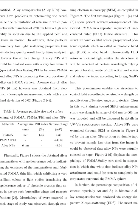
Physically, Figure 1 shows the obtained silver
nanoparticles with golden orange colour
indicat-ing the existence of the nanoparticles and
fabri-cated PMMA thin film which exhibiting a very
brilliant colour as light strikes translating the
opalescence colour of photonic crystals that
ex-ist in nature such butterflies wings and peacock
feathers [29]. Morphology of every material in
each stage of study was observed through
scan-ning electron microscopy (SEM) as compiled in
Figure 2. The first two images (Figure 1 (a) and
(b)) show perfect ordered arrangement of
fab-ricated PMMA in a repeated sequence of
face-centered cubic (FCC) lattice structure. This
structure could exhibit optical properties of
pho-tonic crystals which so called as phopho-tonic band
gap (PBG) or stop band. Theoretically PBG
arises as incident light strikes the structure, it
will be reflected at certain wavelength relying
on particles size, angle of diffraction and
mate-rial refractive index according to Bragg Snell0s
law.
This phenomenon enables the structure to
control light according to required wavelength by
modification of its size, angle or materials. Thus
in this work aiming toward SERS enhancement
study wavelength in range of 400 nm to 600 nm
was targeted and will be discussed in details in
UV-Vis spectroscopy section. Alloys NPs were
examined through SEM as shown in Figure 2
(c) by drying alloy NPs solution on double tape
to prevent sample loss thus from the image it
could be observed that alloy NPs were actually
stacked on tape. Figure 2 (d) displays the
mor-phology of PMMA@alloy core-shell in
suspen-sion in which tiny white dots indicate alloy NPs
attachment and could be seen to completely
in-corporates surround the PMMA sphere
In further, the percentage composition of
el-ements especially Au and Ag in bimetallic
al-loy nanoparticles was analysed via energy
im-Figure 1. Photo of opalescence PMMA thin
film (left) and alloy nanoparticles (right)
obtained in this work.
Figure 2. SEM image of a) and b) PMMA thin
film, (c) alloy NPs and (d) PMMA@alloy
core-shell dispersion.
age in Figure 2(d) illustrates the spectrum
ob-tain from the EDX analysis while Table 2
sum-marises each element with the indicating mass
percentage. 65.92 % of Au and 8.02% of Ag was
analysed in alloy NPs coated 17.43% of PMMA
polymer host
High percentage of Au in this bimetallic
structure could be explained according to their
nature of stability itself and due to strong light
resistance compared to Ag. In this work we are
using 1:1 ratio of Au and Ag precursor thus in
or-der to obtained equal percentage the ratio could
be tuned to 2:1 or 3:1 of Ag and Au volume
Table 2. EDS analysis for every element
constituent of PMMA@alloy core-shell.
Element (keV) Mass % K C K* 0.277 17.43 9.0891
O K* 8.63
Ag L* 2.983 8.02 8.8211 Au M* 2.121 65.92 82.0898
UV-Vis spectroscopy spectrum in Figure
3 demonstrates SPR peak of alloy NPs and
PMMA@alloy core-shell at the wavelength of 451
nm and 438 nm respectively. Slight shift and less
intense of PMMA@alloy core-shell as compared
to alloy NPs was observed due to the
assimila-tion of PMMA and alloy NPs dielectric
environ-ment which also contribute to the merge of SPR
and the photonic band gap (PBG) properties.
This SPR peak was believed could be increased
by the increasing the alloy NPs composition or
by decreasing polymer diameter which provides
higher surface area for the incorporation sites.
However for the actual usage PMMA@alloy
core-shell is in thin film form thus the hybrid
prop-erties of PBG and SPR could not be study
us-ing typical UV-Vis spectroscopy. Thus Raman
spectroscopy analysis will be carried out for final
detection studies to confirm the applicability.
Figure 3. UV-Vis absorption spectrum of Alloy
nanoparticles and PMMA@Alloy core shell.
MDPCs SERS substrate could be potentially
used as SERS sensitive substrate due to its
ex-cellent plasmonic properties. Furthermore the
holey structure of the core-shell particles itself
have a contribution in trapping the electron and
create a region of high electron density. In
ad-dition 3D MDPC SERS substrate also could be
fabricated in large area and would serve large
active area upon detection. Another remarkable
point is it was reproducible and highly stable to
be stored in long time.
IV. SUMMARY
In conclusion, the core-shell structure
con-sist of PMMA as core and alloy (Au-Ag) as the
shell was successfully synthesised in this work
in developing 3D SERS substrates referring to
both physical and optical properties. The
core-shell advantages with interior nanogap in
be-tween Au-Ag alloy nanoparticles on the
sur-face of PMMA sphere. Besides the
fabrica-tion of PMMA@alloy SERS substrate was
per-formed via self-assembly method which is low
cost and laboratory practise. Alloy NPs were
in-troduced onto PMMA surface with modification
using PEI. The uses of PEI successfully modified
negatively charge PMMA surface to enhance the
incorporation of alloy NPs. The average size of
6 nm alloy NPs was obtained with intense SPR
peak at the wavelength of 438 nm indicating the
improvement of plasmonic activity of alloy NPs
compared to Au NPs from our previous work
along with stability.
The results from the fabrication the
ob-tained SERS substrate consisted of two
prop-erties arising from photonic crystals and metal
nanoparticles that were from periodic
arrange-ment PMMA@alloy and alloy nanoparticles
re-spectively. These properties were so called as
PBG and SPR properties each from periodic
ar-rangement of PMMA@alloy and alloy
nanopar-ticles enables sensitive SERS substrate. The
enhancement of Raman signal was expected to
gradually increased and stable for a long time
period. Compared to conventional SERS
sub-strate , the PMMA@alloy coreshell could serve
the most versatile yet sensitive substrate due to
the ability of alloy NPs itself in gripping SERS
reporter molecule vertically possess better
per-formance in chemical and biological analysis.
V. ACKNOWLEDGMENT
We would like to express our
(MOHE) for research grant FRGS 59391 and
Universiti Malaysia Terengganu for research
sup-ports and facilities.
[1] Wan, F, Shi , H, Chen, W, Gu , Z, Du , L, Wang , P, Wang, J, Huang, Y, 2017, Charge Transfer Effect on Raman and Surface Enhanced Raman Spectroscopy of Furfural Molecules, Nanomate-rials, vol. 7, pp. 1-9.
[2] Marta , S, D, Novara , C, Giorgis , F, Bonifacio, A, Sergo, V, 2017, Optimization and Characteri-zation of Paper-Made Surface Enhanced Raman Scattering (SERS) Substrates with Au and Ag NPs for Quantitative Analysis, Materials, vol. 10, pp. 1-15.
[3] Lyandres, O, Yuen, M, Shah, N, C, VanDuyne, R, P, Walsh Jr, J, T, Glucksberg, M, R, 2008, Progress Toward an In Vivo Surface-Enhanced Raman Spectroscopy Glucose Sensor, Diabetes technology and therapy, vol. 10, pp. 257-265. [4] Tian, S, Neumann, O, McClain, M, J, Yang, X,
Zhou, L, Zhang, C, Nordlander, P, Halas, N, J, 2017, Aluminum Nanocrystals: a Sustainable Substrate for Quantitative SERS-based DNA Detection,Nano Letter, vol. 17, pp. 5071-5077. [5] Zhu, T, Hu, Y, Yang, K, Dong, N, Yu, M, Jiang,
N, 2018, A novel SERS nanoprobe based on the use of core-shell nanoparticles with embedded reporter molecule to detect E. coli O157:H7 with high sensitivity,Microchimica Acta, vol. 30, pp. 1-9.
[6] Gudun, K, Elemessova, Z, Khamkhash, L, Ralchenko, E, Bukasov, R, 2017, Commercial Gold Nanoparticles on Untreated Aluminum Foil: Versatile, Sensitive, and Cost-Effective SERS Substrate,Journal of Nanomaterials, Ar-ticle ID 9182025, pp. 1-8.
[7] Tan, K, H, Tajuddin, H, A, Ahmad, R, Shuhaimi, A, Johan, M, R, 2017, Fabrications of Nanocomposite Gold-Polymer Metamaterials Consisting of Periodic Microcavities with Tun-able Optical Properties Optik, vol. 150, pp. 54-61.
[8] Ghazali, N, Johan, M, R, 2016, Environmental modification of self-assembled plasmonic core-shell cluster (silica-gold nanoparticles) for sur-face enhanced Raman scattering (SERS), Opti-cal Society of America, vol. 6, pp. 1-7.
[9] Zhang, C, Jiang, S, Z, Yang, C, Li, C, H, Huo, Y, Y, Liu, X, Y, Liu, A, H, Wei, Q, Gao, S, S, Gao, X, G, Man, B, Y, 2016, Gold@silver bimetal nanoparticles/pyramidal silicon 3D substrate with high reproducibility for high-performance SERS,Scientific Reports, vol. 6, pp. 1-8 [10] Perez, N, P, Puebla, R, A, 2018,SPIE
Proceed-ings, vol. 10507, pp. 1-6.
[11] Kong, X, Squire, K, Wang, A, X, 2018, Fabri-cation of continuous and isolated 3D plasmonic micro-structured super-crystals arrays for SERS sensing,SPIE Proceedings, vol. 10510, pp. 1-6. [12] Kong, X, Xi, Y, Duff , P. L, Chong, X, Li, E,
Ren, F, Rorrer, G. L, Wang, A, X, 2017, Detect-ing explosive molecules from nanoliter solution: A new paradigm of SERS sensing on hydrophilic photonic crystal biosilica, Biosensors and Bio-electronics, vol. 88, pp. 6370.
Biosen-sors and Bioelectronics, vol. 72, pp. 268274.
[14] Gong, T, Cui, Y, Goh, D, Voon, K, K, Shum, P, P, Humbert, G, Auguste, J, L, Dinh, X, Q, Yong, K, T, Olivo, M, 2015, Highly sensitive SERS detection and quantification of sialic acid on single cell using photonic-crystal fiber with gold nanoparticles,Biosensors and Bioelectron-ics, vol. 64, pp. 227233.
[15] Mott, D, Thuy, N, T, B, Aoki, Y, Maenosono, S, 2010, Aqueous synthesis and characteriza-tion of Ag and AgAu nanoparticles: addressing challenges in size, monodispersity and structure, Phil. Trans. R. Soc. A, vol. 368, pp. 42754292.
[16] Verbruggen, S, W, Keulemans, M, Martens, J, A, Lenaerts, S, J, 2013, Physical Chemistry C, vol. 117, pp. 19142-19145.
[17] Link, S, Wang, Z, L, El-Sayed, M, A, J, 1999, Al-loy Formation of GoldSilver Nanoparticles and the Dependence of the Plasmon Absorption on Their Composition, Physical Chemistry B, vol. 103, pp. 3529-3533.
[18] Hazra, A, Hossain, S, M, Pramanick, A, K, Ray, M, 2017, Gold-silver nanostructures: Plasmon-plasmon interaction,Vacuum, vol. 146, pp. 437-443.
[19] Kamaruddin , N, N, Kassim, S, Harun, N, A, 2017, Volume effect of non-polar solvent towards the synthesis of hydrophilic polymer nanopar-ticles prepares via inverse miniemulsion poly-merization, AIP Conference Proceedings, 1885, 020056.
[20] Ismail, Z, Kassim, S, Harun, N, A, 2017, Devel-opment of hydrophilic poly(N-vinylpyrrolidone) nanoparticles via inverse miniemulsion polymer-ization technique,AIP Conference Proceedings, 1885, 020079.
[21] Kapeliouchko, V, Palamone, G, Poggio, T,
Zuc-cheri, G, Passeri, R, Sparnacci, K, Antonioli, D, Deregibus, S, Laus, M, 2009, PMMAbased core-shell nanoparticles with various PTFE cores, Journal of Polymer Science Part A: Polymer
Chemistry, vol. 47, pp. 29282937.
[22] Sa, Y, Yu, N, Wolke, J, G, C, Chanchareonsook, N, Goh, B, T, Wang, Y, Yang, F, Jansen, J, A, 2017, Bone Response to Porous Poly(methyl methacrylate) Cement Loaded with Hydroxya-patite Particles in a Rabbit Mandibular Model, Tissue Engineering: Part C, vol. 23, pp. 262-273.
[23] James, J, Thomas, G. V, Pramoda, K, P, Thomas, S, 2017, Transport behaviour of aromatic solvents through styrene bu-tadiene rubber/poly [methyl methacrylate] (SBR/PMMMA) interpenetrating polymer net-work (IPN) membranes, Polymer, vol. 116, pp. 76-88.
[24] Totu, E, E, Nechifor, A, C, Nechifor, G, Enein, H, Y, A, Cristache, C, M, 2017, Poly(methyl methacrylate) with TiO2 nanoparticles inclu-sion for stereolitographic complete denture man-ufacturing the fututre in dental care for elderly edentulous patients?, Journal of Dentistry, vol. 59, pp. 68-77.
[25] Esmaeil, S, Zakiyan, Aziz, H, Ghasemi, I, 2017, Composite Science Technology, vol. 142, pp.
10-19.
[26] Rodriguez, E, Shahbikian, S, Marcos, B, Huneault, M, A, 2018, Hydrolytic stability of polylactide and poly(methyl methacrylate) blends,Journal of Applied Polymer Science, vol. 45991, pp. 1-14.
emulsion polymerization of poly (methacrylic acid) nanoparticles, AIP Conference Proceed-ings, 1885 (1), 020032.
[28] Tahrin, R, A, A, Azman, N, S, Kassim, S, Harun, N, A, 2017, Preparation and properties of PMMA nanoparticles as 3 dimensional pho-tonic crystals and its thin film via Surfactant-free emulsion polymerization, AIP Conference
Proceedings, 1885 (1), 020092.