1P53 over expression in skin lesions: An immunohistochemical study
Received: 24/6/2014 Accepted: 24/12/2014
Abstract
* Department of pathology, college of medicine, Hawler Medical University, Erbil, Iraq.
Introduction
Mutation of the p53 gene represents the most common genetic alteration in human tumors. The accumulation of the protein can also occur as a result of epigenetic
changes, and therefore it is not an obligatory indicator of a gene mutation.1
P53 protein has an intracellular half-life of
only 20 minutes.1 It is present at very low concentration below the limit of Immunohistochemical detection in most untransformed cells and non neoplastic tissue.2 The altered protein product of the mutant gene has a much extended half-life and can be detected with immunohisto-chemical techniques.1 Epidemiological data strongly implicate sunlight as the principal
environmental cause of skin cancer.3 Sunlight acts as tumorigenic mutagen, it also acts as tumor promoter by favoring the clonal expansion of p53-mutated cells. These combined actions of sunlight result in normal individuals carrying a substantial burden of keratinocytes predisposed to cancer.4 The role of sunlight is supported
by the finding of sunlight-induced mutations in the p53 tumor suppressor
gene in actinic keratosis. The precancer-ous lesion for squamprecancer-ous cell carcinoma of the skin and other precancerous conditions issquamous-cell carcinoma in situ.4,5 It may progress to invasive disease if not treated completely.6,7 The thickness of the epidermis and skin color are known to
Background and objective: Immunohistochemistry is the application of immunologic principles and techniques to demonstrate molecules in cells and tissues. Gene p53 is a tumor-suppressor gene. The product of this gene is a nuclear protein thought to be involved in the control of the cell cycle, apoptosis, and the maintenance of genomic stability. Gene p53 is the most frequently mutated tumor suppressor gene found in human
cancer. The aim of this study was to investigate p53 protein over expression in skin cancer and compare it with benign skin lesion.
Methods: A retrospective study was conducted on paraffin block from skin biopsy of 36 patients with various skin lesions; eight benign cases and 28 malignant cases. The sample of patients was collected from the Pathology Laboratory in Rizgary Teaching Hospital in Erbil, Kurdistan region, Iraq from December 2011 to December 2012. The age of the patients ranged from 34 to 80 years. The p53 protein over expression was investigated by immunohistochemical staining. Sample sections were stained and scored.
Results: Nineteen out of 28 (76.8%) skin cancer showed over expression of p53 gene compared with benign skin lesion and there was a statistically significant difference. There was statistical significant difference in relation to the age group of patients with various skin cancers which was higher in patient above 40 years.
Conclusion: Increased expression of p53 a nuclear protein can be detected in human skin cancer compared with benign skin lesion and it may play an important role in pathogenesis of many types of skin cancers.
Keywords: Immunohistochemistry, Tumor Suppressor gene (p53) protein and skin cancers.
776 attenuate the amount of UV light that reaches the basal layer of the epidermis
and these factors may modify the distribution of p53 expression in the upper
and basal layers of the epidermis.8,9 The aim of the study is to investigate p53 protein over expression in malignant skin lesions and compare it with the benign skin lesions and to evaluate the frequency rate of p53 expression measured in biopsies from various skin cancers.
Methods
primary antibody was placed over the slide. All 10-12 other procedures were the same. The distribution of p53 immunoreactivity in epidermal cells in which the nucleus was stained
brownish-red, including keratinocytes and melanocytes, were interpreted as
p53-positive cells. The examination and interview were done by three pathologists.
P53 immunoreactivity were assessed by the proportion score (percentage) of p53-positive cells in the whole epidermis.
The results were quantified by expressing the number of positive keratinocytes as a percentage of 1000 cells counted in each section.11-13 Tumor cells with staining <10% were regarded as negative and >10% were considered positive.3 Tumor
staining pattern was divided into 5 categories (nil, <10%, 10-20%, 21-50%,
>50%). The proportional score of positively staining cells in each tumor was recorded as (10-20% = +), (21-50% = ++) and (>50% = +++) but was classified as either p53-positive or p53-negative for analysis.
Statistical Analysis:
Statistical parameters as mean, standard deviation, t-test, z-test and chi-square test were used in the study. P value ≤0.05 was regarded as significant, values <0.01 were regarded as highly significant.
This is a retrospective study conducted on paraffin block from skin biopsy of 36 patients with various skin lesions including 8 benign lesions and 28 malignant cases. The sample of patients was collected from the Pathology Laboratory in Rizgary
Teaching Hospital in Erbil, Kurdistan region, Iraq for the period from December
2011 to December 2012. The age of the patients ranged from 34 to 80 years. Ethical approval was obtained from the Ethical Committee of Hawler Medical College. Immunohistochemical staining method was carried out on these samples using the DO-7 monoclonal antibody (Novocastra Laboratories, Newcastle upon
Tyne, United Kingdom) at a 1:50 dilution using microwave antigen retrieval.
All slides were counterstained with hematoxylin and examined at 400 magnification. For each case, a negative
control section was examined to monitor
staining quality based on the level of nonspecific background staining. The
negative control section was prepared identical as the test section except that no
Results
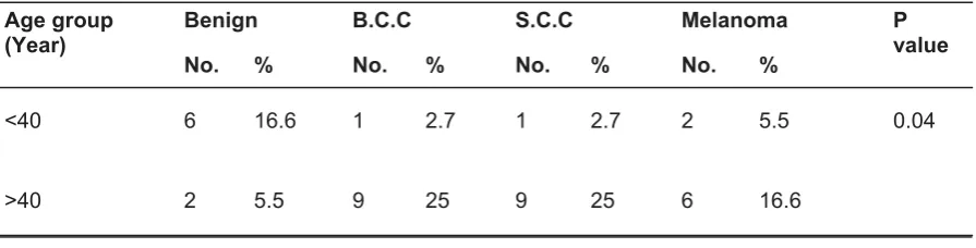
There was statistical significant value in relation to age group examined in patients with various skin cancers which was higher in patient aged more than 40 years versus less than 40 years as shown in Table 1.
Table 1: Age distribution in relation to various skin lesions.
Age group
(Year) Benign B.C.C S.C.C Melanoma P value
No. % No. % No. % No. %
<40 6 16.6 1 2.7 1 2.7 2 5.5 0.04
Regarding gender, there was no statistical
significant difference in different type of skin cancer (Table 2). The p53 immunoreactivity of the skin lesions of the total 36 collected sample from patients
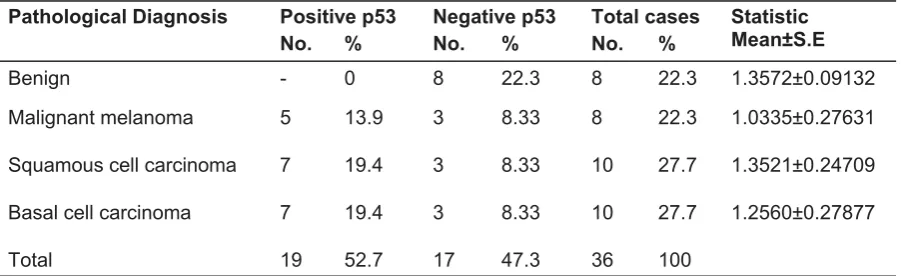
with various skin lesions is shown in Table 3. The IHC expression of p53 in malignant cases was positive in only 19 of total 28 cases (76.8%).The samples with most biopsies showed medium to intense p53-positive cells (Table 4); positive staining constituted 68.4% while negative 32.4%.
Discussion
Recently p53 mutation has been implicated in the pathogenesis of many skin cancer.14
Most of these mutations are acquired, not inherited.15 Assessment of p53 status in human cells can be performed by immunohistochemical detection of nuclear p53 accumulation giving a rapid preliminary indication of p53 status in tumors.16 Most skin cancers in the present study occur in old age group above 40 year. Age
is considered to be a determinant of the neoplastic process.17 Other studies agree that skin cancers typically arise in patients aged 50–70.4 Differences in the estimation of p53 cut off value level in different studies account for some of the variability as some studies consider benign skin lesion as being positive.18 In the current study there was scattered discrete immunreactivity of p53 staining in keratinicytes which are considered as negative (the cut off value in present study >10%) In the current study there was statistical significant difference in p53 expression in patients with skin cancer compared with benign skin lesion. One investigator found that higher p53 immunoreactivity in the malignant cases.19 The observed staining pattern in our study were scattered in basal layer of epidermis in benign skin, but in BCC, SCC, cutaneous melanoma there was intense diffuse staining throughout the lesion. P53 staining intensity among malignant cases, shows statistically significant association most biopsies showing medium to intense
Table 2: Gender factor in relation to various skin lesion.
Table 3: Number of positive and negative p53 immunoreactivity cases.
Gender Benign B.C.C SCC Melanoma P value
Male 3 3 8 4 0.103
Female 5 7 2 4
Pathological Diagnosis Positive p53 Negative p53 Total cases Statistic
Mean±S.E
No. % No. % No. %
Benign - 0 8 22.3 8 22.3 1.3572±0.09132
Malignant melanoma 5 13.9 3 8.33 8 22.3 1.0335±0.27631
Squamous cell carcinoma 7 19.4 3 8.33 10 27.7 1.3521±0.24709
Basal cell carcinoma 7 19.4 3 8.33 10 27.7 1.2560±0.27877
Total 19 52.7 17 47.3 36 100
Table 4: P 53 immunohistochemistry proportional scoring intensity.
P53 scoring 10-20%=+ 21-50=++ >50=+++
Number of cases 3(15.8%) 3(15.8%) 13(68.4%)
778 p53-positive cells, positive staining constituted 68.4%. In the present study p53 over expression was detected in 70% of BCC,70% of SCC and 83% in M.M. while variable results was found in other studies that 90 % of squamous cell carcinoma and approximately 50% of basal cell carcinomas, and 20% of melanomas.5-7 This variability is due to small sample size in this study. The prevalence of abnormal p53 expression in series of invasive melanomas from Queensland was 83%. Estimates of p53 expression vary widely, due to in part the differing characteristics
Conclusion
Increased expression of p53 protein can be detected in human skin cancer compared with benign lesion and mutation of p53 may form an important part of pathogenesis of many type of skin cancer.
of the antibodies used for detection and the criteria used for determining positive staining.17 In addition, individuals exhibit heterogeneity in their ability to repair DNA damage induced by UV radiation. This heterogeneity will affect the relationship between sun exposure and p53 mutation.20
Figure 1: Basal cell carcinoma of skin IHC staining for p53 magnifideX400.
Figure 2: Squamouse cell carcinoma of skin IHC staining for p53 magnifideX400.
Figure 3: Malignant melanoma of skin IHC staining for p53 magnifideX400.
Conflicts of interest
The authors report no conflicts of interest.
References
well-differentiated squamous cell carcinoma of the skin. Dermatol 1997; 195(3):224-7.
15. Ringer DP, Schniper LE. Principles of cancer biology. Gene mutation that can lead to cancer.
In: Lenhard RE, Osteen RT, Gansler T. Clinical Oncology. Atlanta, GA: American Cancer
Society; 2001: 25– National Cancer Institute. Understanding Gene Testing. Accessed at
16. Sidransky D, Hollstein M. Clinical implications
of the p53 Gene. Annu Rev Med 1996; 47(3): 285-301.
17. Whiteman DC, Parsons PG, Green AC. p53 ex-pression and risk factors for cutanous melanoma: A case control-study. Int J Cancer 1998; 77 (6):843-8.
18. Jonason AS1, Kunala S, Price GJ, Restifo RJ, Spinelli HM, Persing JA, et al. Frequent clones of p53-mutated keratinocytes in normal
human skin. Proc Natl Acad Sci USA 1996; 93 (24):14025-9.
19. Batinac PT, Zamolo G, Jonjic N, Gruber F, Petroveckj M. P53 protein expression and cell proliferation in non neoplastic and neoplastic proliferative skin diseases. Tumori 2004; 90 (6):120-7.
p53 mutation frequency in normal skin as a predictor of risk of basal cell carcinoma. J Natl Cancer Inst 1998; 90(7):523-31.
1. Chan J KC. P53 expression. Rosai and Ackerman's Surgical Pathology. 10th Edition.
New York, SA: Mosby Elsever; 2011. PP 58. 2. Purdie CA, Grady J, Piris J, Wylie AH, Biral. P53
expression in colorectal cancer. AJP 1991; 138 (4):22-7.
3. Ghaderi R, Haghighi F. Immunohistochemistry assessment of p53 protein in basal cell carcinoma. Iran J Allergy Asthma Immunol 2005; 4(4):167-71. 4. Kricker A, Armstrong B K, English DR, Heenan PJ.
Pigmentary and cutaneous risk factors for nonmelanocytic skin cancer a case control study.
Int J Cancer 1991; 48(5):650-62.
5. Marisa RN, Christopher PC. In Mills SE, Carter D, Greenson JK, Reuter VE and stoler MH, Editors. Sternberg. Non melanocytic cutaneous tumor. Diagnostic Surgical Pathology. 5th Edition. Baltimore USA: Lippinvott Williams and Wilkins; 2010. PP46-60.
6. Dinehart SM, Nelson-Adesokan P, Cockerell C, Russell S, Brown R. Metastatic cutaneous squamous cell carcinoma derived from actinic keratosis. Cancer 1997; 79(38):920-3.
7. Johnson TM, Rowe DE, Nelson BR, Swanson NA. Squamous cell carcinoma of the skin (excluding lip and oral mucosa).Journal American Acad Derma-tol 1992; 26(5):467-84.
8. Agar NS, Halliday GM, Bametson RS .The basal layer in human squamous tumors harbors more UVA than UVB fingerprint mutations: A role for UVA in human skin carcinogenesis. Proc Natl Acad Sci USA 2004; 101(16):4954-9.
protein in basal cell carcinomas of human skin. Am J Pathol 1992; 141(1):25-9.
10. SoiniY, Kamel D,Pääkkö PLethtoV-P,Oikarinen A, VähäkangasK. Aberrant accumulation of p53 associates with Ki67 and mitotic count in benign skin lesions. British J Dermatol 1994; 131(4): 514-20.
11. Brown DC, Gatter KC. Ki67 protein: The immaculate deception? Histopathol 2002; 40(1): 2-11.
12. Shimuzu T, Oga A, Murakami T, Muto M. Overexpression of p53 protein associated with proliferative activity and histological degree of malignancy in solar keratosis. Dermatol 1999; 28 (6):113-8.
13. Hermeking H, Eick D. Mediation of c-Myc-induced apoptosis by p53. Science 1994; (26) 5:2091-3.
14. Shimuitzu T, Muto M, Murakami T, Furumoto H, Mogami S, Asagami C. Over expression of p53 protein associated with proliterative activity