Original Article
P63 and Ki-67 Expression in Dentigerous Cyst and Ameloblastomas
Zohreh Jaafari-Ashkavandi a, Bita Geramizadeh b, Mohammad Ali Ranjbar a
a
Dept. of Oral and Maxillofacial Pathology, School of Dentistry, Shiraz University of Medical Sciences, Shiraz, Iran. b
Transplant Research Center, Dep. of Pathology, School of Medicine, Shiraz University of Medical Sciences, Shiraz, Iran.
KEY WORDS
P63; Ki-67;
Dentigerous Cyst; Ameloblastoma;
Unicystic Ameloblastoma
Received October 2014;
Received in revised form February 2015; Accepted April 2015;
ABSTRACT
Statement of the Problem: P63 gene is a member of TP53 and its homologous gene family. Its expression was observed in some odontogenic lesions, more ex-pression in aggressive lesions.
Purpose: This study aimed to investigate the possible diagnostic impact of P63 protein on dentigerous cysts and various types of ameloblastoma. Its expression with Ki-67 proliferation marker was also compared.
Materials and Method: This cross-sectional retrospective study was enrolled on 25 cases of dentigerous cyst including 21 unicystic ameloblastomas and 17 con-ventional ameloblastomas. The expression of P63 and Ki-67 was assessed by im-munohistochemical (IHC) examinations. Data were analyzed by employing Mann-Whitney and correlation coefficient tests.
Results: P63 expression was significantly higher in ameloblastoma than unicystic ameloblastoma and dentigerous cysts. There was no significant difference between unicystic ameloblastoma and dentigerous cyst in P63 expression. A 90% cut-off point was obtained for basal layer which gave 88% sensitivity and 78% specificity to distinguish more invasive lesions from others. There was not any correlation between P63 and Ki-67 immunostaining in the three study groups.
Conclusion: More aggressiveness and more invasiveness of odontogenic lesions depicted higher rate and also more intensive expression of P63. Moreover, the expression of P63 protein had not any correlation with Ki-67 protein in dentigerous cysts and ameloblastomas.
Corresponding Author: Jaafari Ashkavandi Z., Dept. of Oral and Maxillofacial Pathology, School of Dentistry, Shiraz University of Medical Sciences, Shiraz, Iran. Tel: +98-71-36263193-4
Fax: +98-71-36270325 Email: [email protected]
Cite this article as: Jaafari-Ashkavandi Z., Geramizadeh B., Ranjbar MA. P63 and Ki-67 Expression in Dentigerous Cyst and Ameloblastomas. Dent Shiraz Univ Med Sci., December
2015; 16(4): 323-328.
Introduction
Odontogenic cysts and tumours arise from the odonto-genic epithelium of tooth germ. Ameloblastoma is the most common odontogenic tumour with clinical signifi-cance. This neoplasm appears in three forms: conven-tional, unicystic and peripheral. These lesions present different clinical and histopathological features which need different managements. Conventional type is a locally invasive benign tumour with high recurrence rate. [1-2] Unicystic ameloblastoma mimics the dentig-erous cyst in clinical, radiographical, and even
histo-pathological features. Unicystic ameloblastoma may arise from a dentigerous cyst, but these odontogenic lesions have different clinical behaviours and treatment managements. Therefore, accurate diagnosis and identi-fying the processes which explain the tumour growth and invasion are the matter of concern.
is discovered in skin, oesophagus, oral mucosa, prostate, breast, lung, salivary glands, and odontogenic epitheli-um of tooth germ as well as dental follicle of impacted teeth. [5-7] Studies showed that P63 is an essential pro-tein for epithelial stratification [8] and various isoforms of P63 have different roles. ∆NP63 proteins contribute to cell proliferation; while, TAP63 isoforms induce cell differentiation. [9]
There are some researches on expression of P63 in odontogenic cysts and tumours. [10-14] They reported that more aggressive tumours such as keratocystic odon-togenic tumour (KCOT) and ameloblastoma have more expression of P63. [10-12] However, most of these studies demonstrated semi-quantitative data and did not present any cut-off point to help diagnosis. Ameloblas-tomas, unicystic and solid types, may arise from a den-tigerous cyst and may show transitional changes from a non-aggressive cyst to a locally invasive tumour. There-fore, we analyzed the comparative expression of P63 in these lesions to evaluate this protein as a marker in early diagnosis which consequently helps selecting accurate management. Ki-67 is the most frequently applied pro-liferation marker for evaluating proliferative activity and biologic behaviour of many pathologic lesions, in-cluding odontogenic cysts and tumours. [15-16] Regard-ing the role of P63 protein in epithelial cell proliferation, we also evaluated the correlation of Ki-67 and P63 in those odontogenic lesions.
Materials and Method
In this retrospective cross-sectional analytical study, 25 cases of dentigerous cyst including 21 cases of unicystic ameloblastoma and 17 ameloblastomas were selected from the archive of oral pathology department, School of Dentistry, Shiraz University of Medical Sciences. The samples had adequate epithelial component to eval-uate the quantity of stained cells. Clinical data including
lesion site, age and sex of patients were obtained from patients’ registered medical documents. Cases with un-certain diagnosis, severe inflammation, and small size lesions were excluded.
Immunohistochemistry
After reviewing and confirming the proposed diagnosis of tumours, two 4µm sections of formalin-fixed and paraffin-embedded specimens were deparaffinised in xylene, then rehydrated by using various concentrations of alcohol, and finally washed with distilled water. Im-munohistochemistry (IHC) was performed by using envision-labelledperoxidasesystem(DAKOCarpinteria; CA, USA). Antigen retrieval was performed by DAKO Cytomation target retrieval solution (PH=9) for 20 minutes. Endogenous peroxidase activity was blocked by 0.3% H2O2. Antigen-antibody reaction was detected by mouse monoclonal anti-P63 antibody (clone 7JUL; Ready to Use, Novocastra, Newcastle, UK) for one hour at room temperature. Then, 3, 3 di-aminobenzidine (DAB Liquid K3467; DAKO Corporation, Denmark) was used as chromogen. Sections were counterstained with Harris' Hematoxylin, washed with tap water and covered by glass coverslips. The positive control was normal oral mucosa for both antibodies. As a negative control, primary antibody was replaced by phosphate-buffered solution. The mean percentage of P63- and Ki-67- positive cells was calculated in 200 cells selected from at least three random fields (at 400x magnifica-tion). In dentigerous cysts and luminal unicystic amelo-blastoma, brown nuclei in basal layer and suprabasal layers (100 cells for each) and in mural and convention-al ameloblastomas, peripherconvention-al and centrconvention-al cells of ame-loblastic islands (100 cells for each) were considered to be scrutinized. Intensity of P63-staining wasalso evalu-atedin three groups:I (mild,light- brown), II (moder-ate), III (severe, dark-brown).
Table 1: P63 expression (mean±SD) in various cell layers and Ki-67 Labelling Index (LI) in the three study groups
P63 expression
Ki-67 LI Basal/Peripheral cells Suprabasal/Central cells
Dentigerous Cyst (n=25) 71.8 ± 23.1 55.8± 25.8 2.4± 2.3
Unicystic Ameloblastoma(n=21) 75.8± 9.9 71.5± 15.9 2.9± 2.5
Luminal(4) 71.25± 32.3 64.7± 14 -
Mural(17) 77 ± 30.2 73.2 ± 16.3 -
Ameloblastoma(n=17) 97.7 ± 5 80.3± 16.6 5.4± 4.4
Statistical analysis
Data was analysed by using SPSS software (version 11). T-test, Mann-Witney, and correlation tests were used as appropriated. P-value<0.05 was considered as signifi-cant. A receiver operating characteristic (ROC) curve was obtained to distinguish more invasive lesions (mu-ral and solid ameloblastoma) from other non-aggressive lesions.
Results
The patients were 33 men and 30 women with the mean±SD age of 27±15.2 years. The brown nuclei were indicative of P63 and Ki-67 expression.
P63 expression
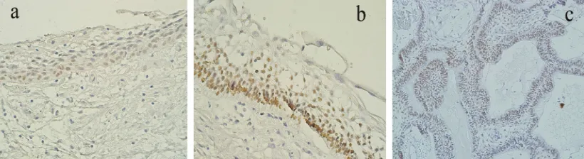
In dentigerous cyst group; P63- positive cells were mainly located in basal cell layer of epithelial lining and the layers above this area had a lower mean of expres-sion (Figure 1a); however, Mann-Whitney test showed that this difference was not statistically significant (p= 0.3). Superficial cells did not show any staining. The mean of P63 expression and its intensity are summa-rized in Tables 1 and 2. There were 5 cases with mild to moderate inflammation that showed a slightly lower P63-expression, but it was not significantly different with non-inflamed cysts (p> 0.05).
Table 2: Intensity of P63 expression in study groups
Mild N (%)
Moderate N (%)
Severe
N (%) Total Dentigerous cyst 10 (40) 13 (52) 2(8) 25 (100) Unicystic A. 2 (9.5) 11(52.4) 8 (38.1) 21 (100) Ameloblastoma 1 (5.9) 5(29.4) 11 (64.7) 17(100) Total 13(20.6) 29(46) 21(33.3) 63(100)
A: Ameloblastoma
The unicystic ameloblastomas consisted of 4 cases of luminal and 17 cases of mural types. The pattern of P63 expression was similar to dentigerous cysts. Basal layer showed more positive cells; however, the intensity of staining was higher (Table 2, Figure 1b, c). There was no significant difference between the cystic lining
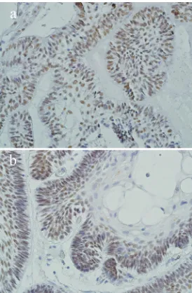
of luminal and mural ameloblastomas (p= 0.38 by Mann-Whitney test). The ameloblastoma group includ-ed 10 follicular and 7 plexiform subtypes. P63 positive cells were found in most peripheral and central cells of ameloblastic nests and in both histological subtypes (Table 1, Figure 2a). However, squamous metaplasia and lining of microcysts were not labeled (Figure 2b). Follicular and plexiform ameloblastomas did not show significant difference in P63 expression.
Figure 2a: P63 expression in ameloblastoma (×400 magnifica-tion). b: Squamous metaplasia does not show P63 expression.
There was no significant difference in overall P63 expression betweendentigerous cyst and unicystic ame-loblastoma; however, T-test analysis showed a statisti-cally significant difference between P63 expression in dentigerous cyst and luminal unicystic ameloblastoma in suprabasal (p= 0.02), but not in basal layers (p= 0.5). Also, peripheral cells of ameloblastic nests in solid and mural ameloblastomas revealed a significant difference in labelling P63 (p= 0.01), but not in central cells.
using Chi-square test (Table 2) and the results showed that intensity was significantly different between the three groups (p= 0.001); however, there was no differ-ence between various tumour subtypes.
Ki-67 expression
Ki-67-positive staining was detected in 40 cases includ-ing 13 cases of dentigerous cyst, 13 cases of unicystic ameloblastomas, and 14 cases of solid ameloblastoma. Ki-67 was labelled heterogeneously in basal and su-prabasal layers of cystic samples and in peripheral and central cells of ameloblastic nests. Ki-67-labelling index (LI) was 2.4±2.3 in dentigerous cysts, 2.9±2.5 in uni-cystic, and 5.2±4.4 in solid ameloblastoma (Table 1). Solid ameloblastoma had significantly higher LI than unicystic ameloblastoma and dentigerous cyst. The dif-ference in Ki-67 LI between dentigerous cyst and uni-cystic ameloblastomas was not statistically significant. The correlation test revealed no correlation between P63 and Ki-67 expression in the study groups.
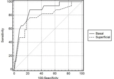
Figure 3: ROC curve for basal and superficial layers: evaluation of basal layer is better for differentiation of aggressive lesions
According to the receiver operating characteristic (ROC) curve analysis, we obtained a 90% cut-off point for basal layer which gave 88% sensitivity and 78% specificity to distinguish more invasive lesions includ-ing mural and solid ameloblast from others (dentigerous and luminal). The area under ROC curve was 0.84 with 95% confidence interval (CI). Cut-off point for supraba-sal layers was 77% with 72% sensitivity and 77% speci-ficity. The area under this curve was 0.75. This analysis demonstrated that basal layer was a better area than suprabasal layers to differentiate aggressiveness of the lesions (Figure 3).
Discussion
In this study, the expression of P63 protein was evaluat-ed since previous studies reportevaluat-ed its effects on
aggres-sive behaviour of odontogenic cysts and tumours. We also assessed the correlation of P63 expression with Ki-67 proliferation marker. The expression of P63 with mild intensity was found in basal and parabasal layers of cystic samples, but not in superficial layers. Earlier studies surveyed P63 protein in odontogenic cysts and reported that staining was not observed in superficial layers of the cysts other than odontogenic keratocyst (currently known as keratocystic odontogenic tumour, KCOT) and that non-aggressive cysts showed mild ex-pression of P63. [10-11] Immunoreactivity for this pro-tein in luminal lining of unicystic ameloblastoma was similar to that in dentigerous cyst; however, the intensi-ty of staining was higher and suprabasal layers showed greater percentage of stained cells. Unicystic ameloblas-toma in many cases shows transitional changes from dentigerous cyst to an ameloblastoma. Moreover, some researchers demonstrated that in the epithelial linings of KCOT, unlike dentigerous and radicular cysts, P63-positive cells were present from basal to upper cell lay-ers with more intensity. [10, 12-13] Therefore, pattern and intensity of P63-staining might be a diagnostic aid for more aggressive cysts.
In the present study, the immune reaction of P63 was slightly lower than that in inflamed cysts, though not statistically significant. Gonçalves et al. found this finding in the cases of severe inflamed radicular cyst. [12] Our study excluded severe inflamed cysts.
hy-pothesis which construed that P63 protein may contrib-ute to the tumour genesis of odontogenic structures. [13]
In the present study, less differentiated cells that were located in basal cell layer of cystic lesions and in the tumoral nests displayed extensive P63- immunore-activity; whereas, terminal differentiated cells like squamous cells and the lining of microcysts did not show staining. It seems that during the transformation of a cyst to a tumour, the upper cell layers lose their differ-entiation and express P63. This figure was also reported by Kumamoto et al. in keratinized and granular cells of ameloblastoma. [6] These features support anti-differen-tiation activity P63 in odontogenic cyst and tumours.
Some authors stated that P63 was in association with epithelial cell proliferation due to the expression pattern of this protein in basal and parabasal layers in epithelial component of mucosa and cysts. [10-11, 17-18] In the present study, we analysed Ki-67 prolifera-tion marker in the samples and evaluated its LI in com-parison with P63-expression. Ki67-positive cells were found in basal and parabasal layers of cystic lesions, and peripheral and central cells of ameloblastomas. Ki-67 LI did not show any significant different between dentiger-ous cyst and unicystic ameloblastoma. It may be at-tributed to the slow growth of unicystic ameloblastoma and its lower aggressive behaviour in comparison with solid ameloblastoma. According to the results of the present and previous studies, P63 protein is expressed in proliferative compartment of odontogenic lesions. [10-11] Nevertheless, our statistical analysis did not show any correlation between expression of Ki-67 and P63 markers. In contrast with these results, Vered et al.
found a correlation between Ki-67 and P63 immunore-action in epithelial dysplasia and oral squamous cell carcinoma. [19] In agreement with our findings, Takada
et al. have found an increasing Ki-67 LI with
progres-sion of dysplasia, but P63 expresprogres-sion has not risen. They included that P63-positive cells could provide stem cell features rather than direct correlation with carcinogenesis. [20] Because of the presence of P63-positive cells in the suprabasal layers of cystic lining and central cells in ameloblastic nests, this reaction probably was indicator of amplifying cells in addition to the stem cells. Takeda et al. indicated that only intense-stained P63 positive cells were true stem cells. [20] P63 expression was also displayed in basal and parabasal
cell layers of oral epithelium and epidermis, where the amplifying or transient amplifying cells were found. [21-22] Also, it seemed that other stem cell markers such as CK19 did not have any particular relation with P63-positive cells. [20] P63 positive cells are necessary for proliferation; however, their presence is not indica-tor of proliferation and anti-differentiation activity is among their important roles, as well.
Various P63 isoforms have different functions and the balance between these isoforms varies during for-mation and progression of the tumour. Therefore, to determine the major role of P63 in tumour genesis of a given tumour, the type of dominant isoform is im-portant. [23] Therefore, PCR technique was required to determine the dominant type.
Conclusion
P63 and Ki-67 had higher expression in more aggres-sive ameloblastic lesions. Therefore, over expression of P63 and Ki-67 in combination with histomorphological examination may provide useful diagnostic aid for ag-gressive odontogenic epithelial cysts and tumours, with a 90% cut-off point for P63 staining in basal layer (88% sensitivity and 78% specificity). Also, evaluation of basal layer was more precise than suprabasal layers. P63 positive cells were present in basal and suprabasal lay-ers. The pattern of immunoreactions seemed to be relat-ed to anti-differentiation and proliferation activity of this protein. We suggest further studies to assess various isoforms of P63 in the odontogenic lesions and their correlation with Ki-67 positive cells.
Acknowledgments
The authors are grateful to Dr. Shahram Hamedani (DDS, MSc) for his suggestions and English editorial assistance. Appreciations are also expressed to the Vice-Chancellery of Shiraz University of Medical Science for supporting this research (Grant#88-4795). This manu-script was mostly based on the DMD thesis of Dr. Rana Ahmadi Mahmood-Abadi and Dr. Shahla Amirsalari.
Conflict of interest
There was no conflict of interest to declare.
References
RS, Mendenhall NP. Ameloblastoma. Am J Clin Oncol.
2007; 30: 645-648.
[2] Ide F, Mishima K, Yamada H, Kikuchi K, Saito I,
Kusa-ma K. Intraosseous ameloblastoKusa-ma with a prominent
ex-traosseous component: pitfalls in diagnosis. Head Neck
Pathol. 2010; 4: 192-197.
[3] Yang A, McKeon F. P63 and P73: P53 mimics, menaces
and more. Nat Rev Mol Cell Biol. 2000; 1: 199-207.
[4] Yang A, Kaghad M, Caput D, McKeon F. On the
shoul-ders of giants: p63, p73 and the rise of p53. Trends
Genet. 2002; 18: 90-95.
[5] Yang A, Kaghad M, Wang Y, Gillett E, Fleming MD,
Dötsch V, et al. p63, a p53 homolog at 3q27-29, encodes
multiple products with transactivating, death-inducing,
and dominant-negative activities. Mol Cell. 1998; 2:
305-316.
[6] Di Como CJ, Urist MJ, Babayan I, Drobnjak M, Hedvat
CV, Teruya-Feldstein J, et al. p63 expression profiles in
human normal and tumor tissues. Clin Cancer Res. 2002;
8: 494-501.
[7] Brkić A, Mutlu S, Koçak-Berberoğlu H, Olgaç V.
Patho-logical changes and immunoexpression of p63 gene in
dental follicles of asymptomaticimpacted lower third
mo-lars: an immunohistochemical study. J Craniofac Surg.
2010; 21: 854-857.
[8] Koster MI, Kim S, Mills AA, DeMayo FJ, Roop DR. p63
is the molecular switch for initiation of an epithelial
strat-ification program. Genes Dev. 2004; 18: 126-131.
[9] Kumamoto H, Ohki K, Ooya K. Expression of p63 and
p73 in ameloblastomas. J Oral Pathol Med. 2005; 34:
220-226.
[10]Lo Muzio L, Santarelli A, Caltabiano R, Rubini C,
Pieramici T, Fior A, et al. p63 expression in odontogenic
cysts. Int J Oral Maxillofac Surg. 2005; 34: 668-673.
[11]Seyedmajidi M, Shafaee S, Shafigh E, Bijani A, Hamidi
H. p63 expression in randomized odontogenic cysts.
Saudi Med J. 2011; 32: 463-466.
[12]Gonçalves CK, Fregnani ER, Leon JE, Silva-Sousa YT,
Perez DE. Immunohistochemical expression of p63,
epi-dermal growth factor receptor (EGFR) and notch-1 in
radicular cysts, dentigerous cysts and keratocystic odon
togenic tumors. Braz Dent J. 2012; 23: 337-343.
[13]Gurgel CA, Ramos EA, Azevedo RA, Sarmento VA, da
Silva Carvalho AM, dos Santos JN. Expression of Ki-67,
p53 and p63 proteins in keratocyst odontogenic tumours:
animmunohistochemical study. J Mol Histol. 2008; 39:
311-316.
[14]Kim YS, Lee SK. Different Protein Expressions between
Peripheral Ameloblastoma and Oral Basal Cell
Carcino-maOccurred at the Same Mandibular Molar Area. Korean
J Pathol. 2014; 48: 151-158.
[15]Ashkavandi ZJ, Najvani AD, Tadbir AA, Pardis S,
Ranjbar MA, Ashraf MJ. MCM3 as a novel diagnostic
marker in benign and malignant salivary gland tumors.
Asian Pac J Cancer Prev. 2013; 14: 3479-3482.
[16]Güler N, Comunoğlu N, Cabbar F. Ki-67 and MCM-2 in
dental follicle and odontogenic cysts: the effects of
in-flammation onproliferative markers. Scientific World
Journal. 2012; 2012: 946060.
[17]Melino G, Lu X, Gasco M, Crook T, Knight RA.
Func-tional regulation of p73 and p63: development and
can-cer. Trends Biochem Sci. 2003; 28: 663-670.
[18]Atarbashi Moghadam S, Atarbashi Moghadam F,
Mokh-tari S, Eini E. Immunohistochemical analysis of P63
ex-pression in odontogenic lesions. Biomed Res Int. 2013;
2013: 624176.
[19]Vered M, Allon I, Dayan D. Maspin, p53, p63, and Ki-67
in epithelial lesions of the tongue: from hyperplasia
through dysplasia to carcinoma. J Oral Pathol Med. 2009;
38: 314-320.
[20]Takeda T, Sugihara K, Hirayama Y, Hirano M, Tanuma
JI, Semba I. Immunohistological evaluation of Ki-67,
p63, CK19 and p53 expression in oral epithelial
dyspla-sias. J Oral Pathol Med. 2006; 35: 369-375.
[21]Suzuki D, Senoo M. Expansion of epidermal progenitors
with high p63 phosphorylation during wound healing of
mouse epidermis. Exp Dermatol. 2013; 22: 374-376.
[22]Crivelini MM, de Araújo VC, de Sousa SO, de Araújo
NS. Cytokeratins in epithelia of odontogenic neoplasms.
Oral Dis. 2003; 9: 1-6.
[23]de Oliveira LR, Ribeiro-Silva A, Zucoloto S. Prognostic
impact of p53 and p63 immunoexpression in oral
squa-mous cell carcinoma. J Oral Pathol Med. 2007; 36:
191-197.
[24]Nylander K, Vojtesek B, Nenutil R, Lindgren B, Roos G,
Zhanxiang W, et al. Differential expression of p63
isoforms in normal tissues and neoplastic cells. J Pathol.