R E S E A R C H A R T I C L E
Open Access
Spectral domain optical coherence
tomography and B-scan ultrasonography in
the evaluation of retinal tears in acute,
incomplete posterior vitreous detachment
Solmaz Abdolrahimzadeh
1*, Domenica Carmen Piraino
2, Vittorio Scavella
2, Barmak Abdolrahimzadeh
3,
Filippo Cruciani
2, Magda Gharbiya
2and Santi Maria Recupero
4Abstract
Background:The purpose of this study was to evaluate the extension and traction effects of posterior vitreous detachment (PVD) complicated with retinal tears using spectral domain optical coherence tomography (OCT) and B-scan ultrasonography.
Methods:Complete ophthalmological examination, B-scan ultrasonography and spectral domain OCT were performed in patients with acute PVD and retinal tears. Vitreous detachment was classified as complete or incomplete, based on extent of posterior pole or peripheral vitreous detachment. Retinal tear location and persistent traction on the retinal flap was evaluated with B-scan ultrasonography and OCT. Categorical data were evaluated with Fisher’s exact test. Statistical significance was considered asP< 0.05.
Results:Twenty-six eyes of 25 patients were assessed. Four eyes (15 %) presented complete PVD with detachment at the posterior pole and periphery. 22 eyes (85 %) presented incomplete PVD with detachment in the periphery. Twenty eyes presented retinal tears in the superior quadrants with respect to only 6 in the inferior quadrants (p= 0.006). There was a higher incidence of retinal tears in the pre with respect to post-equatorial areas (19 vs 7 eyes,p= 0.019). B-scan ultrasonography and OCT revealed persistent traction on the retinal tear flap in 19 and 15 eyes, respectively. Conclusions:In acute PVD, retinal tears are prevalently associated with peripheral vitreous detachment. The impact of complete or incomplete PVD can be of clinical value when evaluating patients with retinal tears.
Keywords:B-scan ultrasonography, Complete posterior vitreous detachment, Peripheral vitreous detachment, Retinal tears, Spectral domain optical coherence tomography
Background
Posterior vitreous detachment (PVD) is the separation of the posterior hyaloid membrane from the surface of the retina. According to the literature, age-related PVD begins in the form of a limited separation of the vitre-ous from the perifoveal retina and gradually advances to culminate in detachment of the vitreous from the optic disc in a variable period of time, which can be
months or years [1]. However, partial PVD is more common than once speculated and acute symptomatic PVD does not always suggest complete detachment of the vitreous [2–5]. Binocular indirect ophthalmoscopy and 3-mirror lens biomicroscopy are the cardinal diag-nostic procedures in the evaluation of acute PVD. How-ever, the presence of a Weiss ring does not necessarily indicate complete PVD and the posterior hyaloid face may still be attached [6]. Furthermore, the Weiss ring can have morphological diversity as it can be divided or lost during the process of vitreous separation. The clin-ical detection of the Weiss ring can indicate that the posterior vitreous is detached from the optic disc but
* Correspondence:[email protected] 1
Ophthalmology Unit, DAI Head/Neck, Azienda Policlinico Umberto I, University of Rome“Sapienza”, viale del Policlinico 155, Rome 00161, Italy Full list of author information is available at the end of the article
cannot give information on the status of the peripheral vitreous. Optical coherence tomography (OCT) has provided new perspectives with high–resolution im-aging of the retina and vitreoretinal interface and can identify fine linear signals, indicating vitreous attach-ment, on the peripapillary, foveal, perifoveal and mid-peripheral areas [1, 7–9]. B-scan ultrasonography is fundamental in establishing PVD in the peripheral ret-ina or in cases with opaque optical media like haemor-rhage and cataract [10–12].Therefore, the extent of PVD can be better established with a combination of both optical coherence tomography (OCT) and B-scan ultrasonography [1, 7, 8, 13].
Retinal tears are among the complications of ir-regular detachment of the posterior vitreous from the retinal surface [4]. Some authors have reported that they occur as a complication of age-related PVD following acute symptomatic vitreopapillary separ-ation [14–16]. Retinal tears are frequently located on the anterior limit of the PVD, also called the vitreous base, which can range from the equator to the ora
serrata. They have been reported in 8–22 % of pa-tients [17–20].
Some studies in the literature have evaluated the risk of delayed retinal breaks following PVD, in order to establish guidelines for the follow up of patients. How-ever, there are no studies where the extension and traction effects of vitreous detachment as shown by B-scan ultrabiomicroscopy and OCT are evaluated. The scope of this study was to investigate if vitreous de-tachment is complete or incomplete in the acute phase of PVD complicated with retinal tear formation and whether the detachment involves the posterior or peri-pheral vitreous.
Methods
Patients with acute, symptomatic PVD and retinal tears were examined at the retina center of the Oph-thalmology Unit of the University of Rome,“Sapienza”. All patients gave informed consent to inclusion in the research, which was given institutional review board
approval, and the study was conducted in accordance with Tenets of the Declaration of Helsinki.
A careful history of the patients was obtained to es-tablish whether the symptoms (visual floaters and/or photopsia) were of recent onset. Exclusion criteria were: symptoms of more than 1 week duration, his-tory of recent eye trauma, eye surgery, eye disease, high myopia, media opacity due to corneal or lens opacity and narrow pupil dilatation. The comprehen-sive ophthalmological examination undertaken by the retina specialist included visual acuity measurement with Snellen charts, slit lamp examination, vitreous bio-microscopy, indirect ophthalmoscopy and 3-mirror lens biomicroscopy of the retina. All patients underwent B-scan ultrasonography and spectral domain OCT in order to establish the location and extension of vitreous
detachment. PVD was classified as complete or incom-plete and as involving the posterior pole or periphery. Furthermore, traction effects of PVD on retinal tear flaps were evaluated with both instruments.
B-scan ultrasonography was performed by one expe-rienced investigator, D.C.P., using the Cinescan S (Quantel Medical, Clermont-Ferrand, France) with a 10 MHz and 20 MHz probe. The examination tech-nique was adopted from a method previously reported by Josè Lorenzo Carrero [4, 13]. The patients were placed supine on a reclining chair. Examination was performed with the 10MHZ probe on the eyelids. Then following topical anesthesia, the 20MHZ probe was positioned on the ocular surface through 2.5 % methyl-cellulose. The dB gain was adjusted when using each probe to give the finest images of the ocular structures.
The quality of the images obtained using each probe was assessed with particular reference to the vitreoretinal interface at the posterior pole, the peripheral vitreous and the retinal tears. Kinetic examination was performed by evaluating vitreous movement during voluntary motion of
the eye while the probe was kept immobile according to the method previously described [4].
Spectral domain OCT (Spectralis Family Acquisition Module, V 5.1.3.0; Heidelberg Engineering) with Hei-delberg Eye Explorer (V 1.6.2.0; HeiHei-delberg) was used
Fig. 3B-scan ultrasonography showing extent of vitreous detachment. Retinal tear with vitreo-retinal traction (a); retinal tear (arrow) (b); anterior vitreous detachment (arrowhead) (c); and posterior vitreous attachment (d)
to obtain images by one experienced investigator, V.S. All patients were dilated with tropicamide 1 % (Visu-farma, Italy) and phenylephrine hydrochloride 0.5 % (Visufarma, Italy). Single-line vertical and horizontal 100-frame line scans, 50-frame (20° x 15° 19- line) raster scans, and 60-frame radial scans were taken of the posterior pole to obtain an optimal view of the vitreous.
Retinal tears were studied by instructing patients to gaze in the direction of the tear. The operators were aware of the results of the fundus examination before performing the test. 100-frame line scans and, where possible according to the localization of the tear, 50-frame raster and radial scans were performed along the retinal tear and its borders.
Retinal laser treatment for retinal tears was per-formed in all eyes. Fundus examination to evaluate laser treatment was carried out at 1 and 3 months from treatment.
Categorical data were evaluated with Fisher’s exact test. Statistical significance was considered asP< 0.05.
Results
Twenty-six eyes of 25 patients, 9 male and 16 fe-male, aged 45 to 71 years (mean 60.5 +/− 7.58) were included in the study. The mean spherical equivalent of subjective refraction error was −0.37 diopters (range −3.25 to + 2.50). Best-corrected visual acuity of patients was 20/20 in 23 eyes and 20/40 in 3 eyes where there was vitreous cellularity.
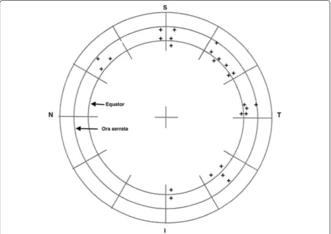
The location of the retinal tears was post-equatorial in 7 eyes, pre-equatorial in 13 eyes and in the ex-treme pre-equatorial periphery in 6 eyes. The distri-bution of retinal tears in the retinal quadrants, in general agreement with previous reports on retinal tears, is shown in Fig. 1 [17–21]. There was a higher in-cidence of retinal tears in the superior quadrants and
those localised in the prequatorial region (p= 0.006 and
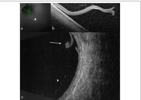
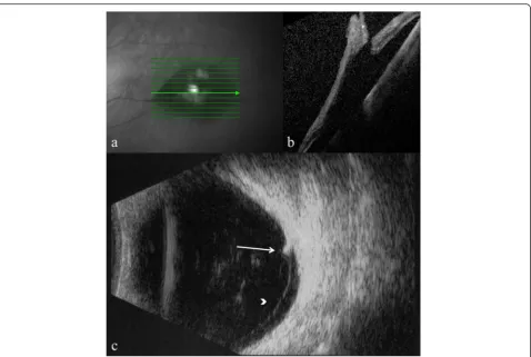
p= 0.019, respectively). There were no cases of frank vitreous hemorrhage, however, in five eyes the vitreous body presented cellularity and in these cases the retinal tears were localized in the pre-equtorial region. Com-plete PVD was observed with B-scan ultrasonography in only four eyes (15 %), which showed both posterior pole PVD and peripheral PVD. However, in three of these cases OCT did not show posterior pole PVD due to the technical limitation of the instrument as the posterior border of the detached hyaloid had moved very anterior to the retinal plane and could not be visualized. Ultrasonography showed that vitreous sep-aration was only peripheral in 22 eyes (85 %) and the posterior pole was still attached. Fine details of residual perifoveal and peripapillary vitreous adher-ence in 2 eyes were shown with OCT, which had not been evidenced with B-scan ultrasonography (Figs. 2, 3 and 4).
Persistent vitreous traction on the flap was observed in 19 eyes with ultrasonography and in 15 eyes with OCT, where scans could be obtained (Figs. 5, 6 and 7).
Fundus examination at 1, and 3 months from treat-ment did not show further retinal tears. In one patient with a pre-equatorial retinal tear and vitreous cellularity, retinal detachment occurred at month 1 and required surgical management.
Discussion
The present study showed that in 85 % of cases of acute symptomatic PVD with retinal tears, vitreous detachment was incomplete and retinal tears were as-sociated with peripheral PVD.
Various studies in the literature describe PVD based on ophthalmoscopic evidence, however, OCT allows a more precise classification of vitreous detachment of the posterior pole, which has been divided in stages based on location in the peripapillary, foveal, perifoveal, and midperipheral areas [7]. B-scan ultrasonography has been used to study the peripheral vitreous, precisely because the ophthalmoscopy finding of the Weiss ring indicates the separation of the posterior vitreous from the optic disc, but not necessarily from the peripheral retina [4]. To the best of our knowledge there are no
studies performed with the concurrent use of ultrason-ography and OCT to study both the posterior and per-ipheral vitreous in acute PVD complicated with retinal tears.
Josè Lorenzo Carrero recently reported on incom-plete PVD using kinetic ultrasound examination [4]. He suggested that incomplete PVD may accommo-date different forms like incomplete posterior or in-complete peripheral PVD and this may modify the common progression of vitreous detachment leading to different clinical complications [4]. Indeed, retinal tears were observed in 85 % of our cases with in-complete PVD where the detachment was chiefly peripheral and did not involve the posterior pole.
The presumed pathogenesis of retinal tears is be-lieved to be excessive traction of the posterior vitre-ous cortex on areas of firm vitreoretinal adhesion. Anomalous PVD, where vitreous liquefaction takes place before adequate reduction of vitreoretinal ad-hesions leading to retinal tears, has been described by Sebag [22]. Byer reported that in some eyes, dis-tinct, microscopical, pathological vitreoretinal adhe-sions exist which are not clinically visible and retinal tears can occur following sudden traction on these
areas [23]. In the present study, persistence of fine vitreous attachment on the retinal tear flap was evi-denced in 19 and 15 eyes with B-scan ultrasonog-raphy and OCT, respectively. It is thought that retinal tears result from dynamic forces created dur-ing ocular movement causdur-ing opposdur-ing tractional forces on the minute vitreoretinal adhesions. It is reasonable to hypothesize that in incomplete periph-eral PVD, the vitreous could be pulled posteriorly to the plane of detachment at the peripheral site of vit-reous separation, causing traction on vitreoretinal adhesions, which lead to retinal tear formation.
The limit of our study is the small number of eyes, thus, our results must be interpreted with caution. Furthermore, with the advent of swept source OCT the interface of the vitreous can be studied from the macula up to the periphery [24, 25]. The present study evaluated the vitreous with B-scan ultrasonog-raphy and OCT only in the acute stage of retinal tear formation but the extension of PVD and the presence of persistent vitreous traction on the retina over time can be clinically important and future longitudinal studies may establish if eyes with tenacious attach-ment at the posterior pole are a liability for new tears
[26, 27]. This could give further clinical information on the impact of complete or incomplete PVD when evaluating risk in patients with retinal tears. Further-more, studies on larger patient populations, treated with laser therapy for retinal tears, could indicate ret-inal detachment risk after specific treatment in rela-tion to vitreous attachment/detachment morphology in the area of the retinal tear and indicate appropriate intervals for follow up. Moreover, with the advent of enzymatic vitreolysis for focal vitreo-macular traction, [28, 29] increased awareness of traction mechanisms might lead to new therapeutic options in specific cases. Further studies on larger patient populations are warranted.
Conclusions
In this study we investigated the extension of acute posterior vitreous detachment in 26 eyes with retinal tears evaluated with B-scan ultrasonography and optical coherence tomography. In the acute phase of vitreous detachment, retinal tears are prevalently associated with incomplete, peripheral vitreous detachment. The impact of complete or incomplete PVD can be of clinical value when evaluating patients with retinal tears.
Abbreviations
OCT, optical coherence tomography; PVD, posterior vitreous detachment
Funding
The authors declare that they did not receive any funding for the present study.
Availability of data and materials
Data supporting our findings is contained in the manuscript.
Authors’contributions
SA made substantial contributions to the design and conception of the study, analysis and interpretation of images, drafting and critical revision of the manuscript for important intellectual content. DCP contributed to the acquisition and interpretation of B-scan ultrasonography images, preparation of figures, and drafting of the manuscript. VS contributed to the acquisition and interpretation of OCT images and drafting of the manuscript. FC and BA contributed to the conception of the study, analysis of images and drafting of the manuscript. MG contributed to analysis and interpretation of images and critical revision of the manuscript for important intellectual content. SMR contributed to conception and design of the study and critical revision of the manuscript for important intellectual content. All authors read and approved the final version of the manuscript for publication.
Competing interests
The authors declare that they have no competing interests.
Consent to publish Not applicable.
Ethics and consent to participation
All patients gave informed consent to inclusion in the research, which was given Institutional Review Board approval by the Ophthalmology Department, University of Rome“Sapienza”. The study was conducted in accordance with Tenets of the Declaration of Helsinki.
Author details
1Ophthalmology Unit, DAI Head/Neck, Azienda Policlinico Umberto I,
University of Rome“Sapienza”, viale del Policlinico 155, Rome 00161, Italy.
2
Ophthalmology Unit, Department of Sense Organs, University of Rome
“Sapienza”, viale del Policlinico 155, Rome 00161, Italy.3Polimed Beltramelli
Medical Centre, via I.C. Falbo 9, Roma 00154, Italy.4Opthalmology Unit,
NESMOS Department, University of Rome“Sapienza”, St. Andrea Hospital, via di Grottarossa 1035-1039, Rome 00189, Italy.
Received: 10 October 2015 Accepted: 19 May 2016
References
1. Johnson MW. Posterior vitreous detachment: evolution and complications of its early stages. Am J Ophthalmol. 2010;149:371–82.
2. Kakehashi A, Kado M, Akiba J, Hirokawa H. Variations of posterior vitreous detachment. Br J Ophthalmol. 1997;81:527–32.
3. Weber-Krause B, Eckardt C. Incidence of posterior vitreous detachment in the elderly. Ophthalmology. 1997;94:619–23.
4. Carrero JL. Incomplete posterior vitreous detachment: prevalence and clinical relevance. Am J Ophthalmol. 2012;153:497–503.
5. Puliafito CA, Hee MR, Schuman JS, et al. Optical coherence tomography of ocular diseases. Thorafare, NJ: SLACK Inc; 1996.
6. Kakehashi A, Inoda S, Shimuzu Y, Makino S, Shimzu H. Predictive value of floaters in the diagnosis of PVD. Am J Ophthalmol. 1998;125:113–5. 7. Uchino E, Uemara A, Ohba N. Initial stages of posterior vitreous detachment
in healthy eyes of older persons evaluated by optical coherence tomography. Arch Ophthalmol. 2001;119:1475–9.
8. Hohn F, Mirshahi A, Hattenback LO. Optical coherence tomography for diagnosis of posterior vitreous detachment at the macular region. Eur J Ophthalmol. 2009;19:442–7.
9. Gharbiya M, Cruciani F, Cuozzo G, et al. Macular thickness changes evaluated with spectral domain optical coherence tomography after uncomplicated phacoemulsification. Eye (Lond). 2013;27:605–11. 10. Arzabe CW, Akiba J, Jalkh AE, et al. Comparative study of vitreoretinal
relationships using biomicroscopy and US. Graefes Arch Clin Exp Opthalmol. 1991;229:66–8.
11. Dugel PU, Smiddy WE, Byrne SF, et al. Echographic findings with clinical correlation. Ophthalmology. 1994;101:815–21.
12. Van Newkirk MR, Johnson MW, Hughes JR, et al. B-scan ultrasonographic findings in the stages of idiopathic macular hole. Trans Am Ophtalmol Soc. 2000;98:163–9.
13. Carrero JL, Perez-Flores I, Cid-Galano M, et al. B-scan ultrasonography to screen for retinal tears in acute symptomatic age-related posterior vitreous detachment. Ophthalmology. 2009;116:94–9.
14. Jaffe NS. Complications of acute posterior vitreous detachment. Arch Ophthalmol. 1968;79:568–71.
15. Lindner B. Acute posterior vitreous detachment and its retinal complications. Acta Ophthalmol. 1966;87(suppl):1–108.
16. Novack MA, Welch RB. Complications of acute sumptomatic posterior vitreous detachment. Am J Ophthalmol. 1984;97:308–14.
17. Byer NE. Natural history of posterior vitreous detachment with early management as the premier line of defence against retinal detachment. Ophthalmology. 1994;101:1503–13.
18. Sharma S, Walker R, Brown G, Cruess A. The importance of qualitative vitreous examination in patients with acute posterior vitreous detachment. Arch Ophthalmol. 1999;117:343–6.
19. Murakami K, Jalkh AE, Avila MP, et al. Vitreous floaters. Ophthalmology. 1983;90:1271–6.
20. Boldrey EE. Risk of retinal tears in patients with visual floaters. Am J Ophthalmol. 1983;96:783–7.
21. Sarrafizadeh R, Hassan TS, Ruby AJ, et al. Incidence of retinal detachment and visual outcome in eyes presenting with posterior vitreous separation and dense fundus-obscuring hemorrhage. Ophthalmology. 2001;108: 2273–8.
22. Sebag J. Anomalous posterior vitreous detachment: a unifying concept in vitreo-retinal disease. Graefes Arch Clin Exp Ophthalmol. 2004;242:690–8. 23. Byer NE. What happens to untreated asymptomatic retinal breaks, and are
24. Mori K, Kanno J, Gehlbach PL, Yoneya S. Montage images of spectral-domain optical coherence tomography in eyes with idiopathic macular holes. Ophthalmology. 2012;119:2600–8.
25. Itakura H, Kishi S, Li D, Nitta K, Akiyama H. Vitreous changes in high myopia observed by swept-source optical coherence tomography. Invest Ophthalmol Vis Sci. 2014;55:1447–52.
26. Coffee R, Westfall AC, Davis GH, et al. Symptomatic posterior vitreous detachment and the incidence of delayed retinal breaks: case series and meta-analysis. Am J Ophtalmol. 2007;144:409–13.
27. Richardson PSR, Benson MT, Kirby GR. The posterior vitreous detachment clinic: do new retinal breaks develop in the six weeks following an isolated symptomatic posterior vitreous detachment? Eye. 1999;13:237–40. 28. Salmans P, Delaey C, De Smet MD, et al. Intravitreal injection of
microplasmin for treatment of vitreomacular adesion. Results of a prospective, randomized, sham-controlled Phase II Trial (The MIVI-IIT Trial). Retina. 2010;30:1122–7.
29. Benz MS, Packo KH, Gonzales V, et al. A placebo-controlled trial of microplasmin intravitreous injection to facilitate posterior vitreous detachment prior to vitrectomy. Ophthalmology. 2010;117:791–7.
• We accept pre-submission inquiries
• Our selector tool helps you to find the most relevant journal
• We provide round the clock customer support
• Convenient online submission
• Thorough peer review
• Inclusion in PubMed and all major indexing services
• Maximum visibility for your research
Submit your manuscript at www.biomedcentral.com/submit