1
Abstract:Medical image processing is the most challenging and emerging field nowadays. To solve various problems in medical imaging such as medical image segmentation, object extraction and image classification etc this work presents a performance of the rough set based approaches. Other computational intelligence technologies that include neural networks, particle swarm optimization, support vector machines and Genetic Algorithm are also presented in this paper. There are so many people affected by brain tumour and it is one of the most harmful diseases. The intention of feature selection approach is to select a small subset of features that minimize redundancy and maximize relevance to the target such as the class labels in classification.
I. INTRODUCTION
1.1 Brain tumour:
An abnormal growth of the tissues is known as tumour. It is an abnormal mass of tissue in which cells grow and multiply uncontrollably. Brain tumours can be ant type like primary or metastatic, and either malignant or benign. A metastatic brain tumour also called a cancer. In this, cancer can spread from anywhere in the body to the brain. Electrochemical impulses are generated by neurons that affect the glands, muscles and other neurons and produces human thoughts, actions and feelings. The normal pattern of neuronal activity becomes disturbed, causing strange sensations, emotions, and behaviour in epilepsy. To produce high quality images of the parts contained in the human body there is an advanced medical imaging technique called Magnetic Resonance Imaging (MRI). When treating brain tumors, ankle, and foot, then often used MRI imaging. From MRI imaging, we get high resolution images. These highly resolution images helps us to drive the detail or information to examine human brain development and discover abnormalities. There are fuzzy methods, neural networks, atlas methods, knowledge based
techniques, shape methods, variation segmentation methodology for classifying MR images.
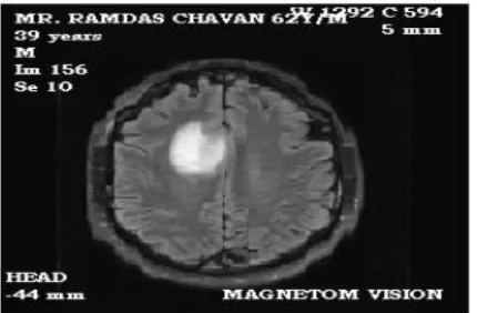
The primary step in image analysis is Pre-processing of MRI images. It performs image enhancement and noise reduction techniques. By using these techniques, we increase the image quality. After this, to detect the tumour in the images some morphological operations are applied. Basically to find out the size and shape of the tumour, morphological operations are applied and in the end, to make visible the tumour in the image, the tumour is mapped onto the original gray scale image with 255 intensities. On a number of patients the algorithm has been tried, MRI data of brain tumour images in Figure 1.1 shows an MRI image of tumour affected brain.
Fig.1.1 MRI image of tumour affected brain 1.2 Methodology:
Pre-processing of given MRI image is the first stage of the algorithm. After that segmentation and then performs morphological operations. Steps of algorithm are as following: 1) Give MRI image of brain as input.
2) Convert it to gray scale image.
3) Apply high pass filter for noise removal.
4) Apply median filter to enhance the quality of image.
SHIV SHAKTI
International Journal in Multidisciplinary and
Academic Research (SSIJMAR)
Vol. 6, No. 5, October 2017 (ISSN 2278 – 5973)
Brain Tumor detection using RST-PSO
* Monika, **Deepti Ahlawat *M.Tech scholar, N.C College, Israna
5) Compute threshold segmentation. 6) Compute watershed segmentation. 7) Compute morphological operation. 8) Finally output will be a tumour region
.
Keeping these points in consideration following will be our objectives:
To extract features from MRI images. A feature vector consists of different features like statistical features, GLCM features with 0,45,90,135 degree orientation and to use the supervised learning algorithm RST-PSO to get the best features set. The evaluation parameters will be sensitivity,
specificity and accuracy.
II. LITERATURE REVIEW
This paper permits an efficient and absolutely computerized brain tumor segmentation technique [1]. The proposed process includes a fuzzy C-way (FCM) based techniques to enhance the first-rate of T1-weighted coronal MR photos, a fast bounding field (FBB) detection algorithm to locate a rectangle round tumour. Medical imaging is advantageous in analysis of the disease. Many humans suffer from brain tumor; it is a severe and hazardous disease [2]. Scientific imaging provides right analysis of brain tumor. There are lots of systems to become aware of mind tumor from MRI image. In this paper [3] our method supplies as a minimum comparable outcome to the wave detection in view of the discrimination of subjects being at hazard for CAPD and topics being no longer at hazard for CAPD. Brain snapshot Segmentation is an elaborate and challenging part in the medical image Processing. This paper [4] describes two new techniques for mind tumor detection making use of Meta heuristic algorithms. Many photograph threshold system uses the histogram of the photograph. In this paper, the target operate of Otsu method which is a statistical approach, Particle Swarm Optimization with an intuitive algorithm (PSO) by way of maximizing, the top-rated threshold values on a medical image had been studied to seek out [5]. This paper [6] presents a novel premiere algorithm for MRI brain tumor recognition. To do this, we use the newly developed meta-heuristic MSFLA (Modified Shuffled Frog Leaping Algorithm).
In proposed algorithm [7] every particle is encoded in a average number vector and an effective process is developed to move particles within the solution house. In this paper [8], a two-degree CAD device has been evolved for automatic detection and type of mind tumor via magnetic resonance pictures (MRIs). Basic homes of hybrid multi-granularity difficult set of variable precision are discussed, which provides a new approach to cope with the incomplete data machine [9]. Rough set theory has been used to define the necessity of functions in literature. We propose a new hard set based function choice approach known as Parameterized Average Support Heuristic (PASH) [10]. In this paper [11] three novel strategies are proposed specifically Binary Artificial Bee Colony (BABC), horizontal feature extraction and function gallery expansion. BABC is a binary model of Artificial Bee Colony (ABC) which is hired as function choice
method for green reduction in selected features. In this paper [12] we suggest different approach to discover appropriate reducts: RS Reduct this method combines numerous factors of hard set concept [12. In this paper [13] documented the set-up and results of the Multimodal Brain Tumor Image Segmentation Benchmark (BRATS) organized in conjunction with the MICCAI 2012 and 2013 conferences. Brain tumor detection in an early stage is a difficult assignment, because the imaging is quite doubtful [14]. The necessity of automated mind tumor segmentation and detection is high. To attain an accurate MRI image of the mind tumor is hard. The approach proposed here is a segmentation procedure of 2D MRI picture the use of numerous filtering techniques. Experimental research based on a practical state of affairs concerning terrorist sports and also artificial random records are performed, demonstrating the ability and efficacy of the proposed work [15]. Several approaches to feature selection based totally on rough set concept are experimentally in comparison. Additionally, a new vicinity in feature choice, feature grouping, is highlighted and a difficult set-based totally feature grouping method is designated. In this paper, we proposed a conventional information analyzed techniques, this thesis studied the essential position of discernibility matrix in finding the middle of attributes then proposed an improved attribute discount set of rules for incomplete records structures based at the limited tolerance relation [17].
III. Proposed work
Medical image processing is the most challenging and emerging field nowadays. Processing of MRI images is one of the parts of this field. This work presents a performance of the rough set based approaches to solve various problems in medical imaging such as medical image segmentation, object extraction and image classification. Rough set frameworks hybridized with other computational intelligence technologies that include neural networks, particle swarm optimization, support vector machines and Genetic Algorithm are also presented.
3
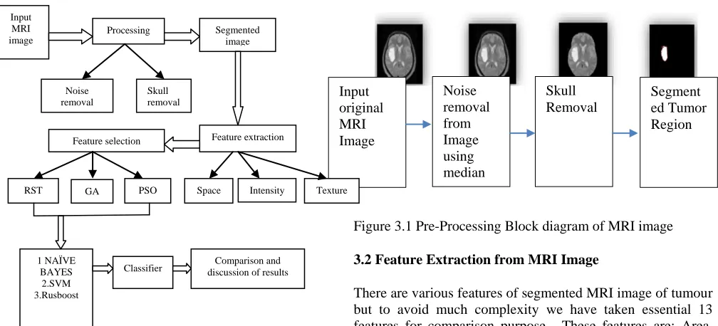
Figure 3.1: Proposed framework for RST, GA and RST-PSO based supervised feature selection
Overall working can be divided in following points: 1. Pre-processing of MRI Images
2. Feature extraction from MRI images 3. Feature reduction using RST
4. Feature reduction using GA and Particle Swarm Optimization (PSO)
5. Classification of feature extracted from RST, GA and PSO using SVM, Naive Bayes and Rusboost classifiers.
6. Comparison and discussion.
3.1 Pre-processing of MRI Images: The MRI images under test acquired by the MRI scanner are always susceptible by the environment. The image restoration tries to minimize the effects of degradations, by means of a filter. It is a fundamental problem in the image processing to improve quality through the reduction of the noise. A wide variety of techniques dedicated to carry out this task already exist. In the MRI image compression system, significant tumor detection depends on the regions of interest which are usually of noisy nature and low contrast. Hence an image enhancement and de-noising may be required to preserve the image quality, highlighting the image features and suppressing the noise. Noise in image not only lowers its quality but also can cause feature extraction algorithms to be unreliable. The de-noising and feature enhancement techniques presented in this work will improve the reliability of image processing.
Figure 3.1 Pre-Processing Block diagram of MRI image 3.2 Feature Extraction from MRI Image
There are various features of segmented MRI image of tumour but to avoid much complexity we have taken essential 13 features for comparison purpose. These features are: Area, Perimeter, Circularity, Mean, Variance, Standard Deviation, Skewness, Kurtosis, Contrast, Correlation, Energy, Entropy, and Homogeneity.
3.3 Feature reduction using Rough Set Theory
RST is powerful tool to minimize no. of features from larger database and those reduced feature produce almost same results as compared to all the features combined used.
3.4 Feature reduction using RST and Particle Swarm Optimization (PSO)
Our work mainly focuses on combining RST and PSO, because a larger database like MRI image feature database can be efficiently and speedily reduced using RST with PSO. By using Rough Set Theory we used concept of equivalence class set, lower approximation, and positive region boundary. To find re-duct from given attributes we used indiscernibility definition from RST.
3.5 Classification and Performance evaluation:
In order to evaluate performance of RST, PSO and RST-GA, we have chosen three classifiers.
1. SVM (Support Vector Machine) 2. Naïve Bayes
3.
RusboostBefore applying classifier first, we divided our reduced feature data in 80:20 ratio and 80% data is used as training data and remaining 20% is used as testing data. The parameters such as Regression, Performance plot, Confusion Matrix and ROC Values are used to analyze the performance. The performance of the feature selection is increased after applying PSO. Input
original MRI Image
Noise removal from Image using median filter
Skull Removal
Segment ed Tumor Region Input
MRI image
Processing Segmented image
Noise removal
Skull removal
Feature extraction Feature selection
Space Intensity Texture RST GA PSO
1 NAÏVE BAYES 2.SVM 3.Rusboost
IV. RESULTS
In our work we have proposed combination of Rough Set Theory (RST) using Particle Swarm Optimization (PSO) algorithm to reduced no. of attributes to successfully analyse type of brain tumour from given MRI image. The proposed work is implemented in MATLAB R 2016a.
We have divided our work in six sub cases which are 1. Pre-processing of MRI Images0
2. Feature extraction from MRI images 3. Feature reduction using RST
4. Feature reduction using Genetic Algorithm (GA) and Particle Swarm Optimization (PSO)
5. Classification of feature extracted from RST, GA and PSO using SVM, Naïve Bayes and Rusboost classifiers.
6. Comparison and discussion. 4.1 Pre-processing of MRI Image:
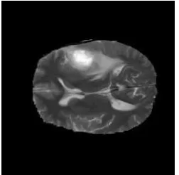
Here first we read MRI images, second noise is removed using median filter third skull is removed and last brain tumour is segmented from image. Image segmentation is the process of dividing an image into several parts. This is used to identify objects or other information in digital images. There are many ways to perform image segmentation. First, we have used edge and the Sobel operator to calculate the threshold value. We then tuned the threshold value and use edge again to obtain a binary mask that contains the segmented cell. So segmentation of image is achieved by this way.
Figure 4.1 Input MRI Image
Figure 4.2 MRI Image after noise removal using median filter
Figure 4.3 Brain tumour segmented image
By the process of image segmentation, all images of tumour are extracted from dataset of tumour. Now using feature extraction, all the features are extracted from each and every segmented tumour image.
4.2 Rough Set Theory using PSO based feature reduction:
In this Rough set theory with PSO is used for feature reduction. Confusion matrix is plotted for this case. Confusion matrix shows correctness of predicted labels by true labels figure 4.4 shows comparison of confusion matrix for different classifier
.
(a)
5
Figure 4.4 Confusion Matrices using three classifiers (a) SVM (b) Naïve Bayes (c) RUS Boost.
It is observed that when we apply RST using PSO, to reduce no. of features, having three decision labels or classes, true positive for label 1,2,3 using different classifier are given here
.
Figure 4.5 ROC curve for RST using PSO method
It is observed from ROC curve that proposed method of feature reduction using RST with PSO fits better in comparison to other two cases. ROC curve is better when it is near to 1 and upward. It means more labels are matching with testing data.
4.3 Cumulative Results and Comparison
Figure 4.6 Accuracy Curve Comparison
Observation is that average values of accuracy in PSO is higher than other two for all three classifiers
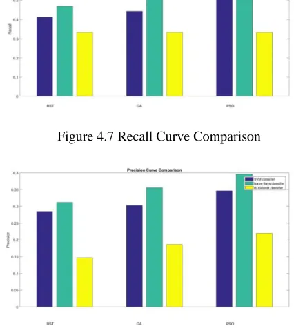
Figure 4.7 Recall Curve Comparison
Figure 4.8 Precision Curve comparison
We have implemented RST using PSO to reduce no. of attributes in a database and compare performance of our method with simple RST method and RST using GA. We have taken three classifiers SVM, Naive Bayes and RUS Boost for performance comparison. Parameters chosen to compare performance are accuracy, precision, recall. We also analyse performance with the help of ROC curve and Confusion matrix built using classifiers. Figures show comparison between three methods on the basis of accuracy, recall and precision. We have taken mean value of accuracy, precision and recall from all three output decision values. RST using PSO is showing better performance in comparison to other two methodologies. By analysing all results for a database of 3064 MRI images , it can be concluded that RST using PSO gives better results than simple RST and RST using GA.
V. CONCLUSION
Rough set theory can be used in medical domain and for importance of features, patterns in sample data, and feature space dimensionality reduction. Classification and dimensionality reduction is the main focusing area in most of the literature on rough set theory. it is obvious that the rough set approach provides a promising means of solving a number of medical imaging problems. It should be observed that rough set or near set by themselves or in combination with other computational intelligence technologies work remarkably well in separating medical images into approximation regions that facilitate automated image segmentation and object recognition.
knowledge discovery and model interpretation. The field of medical diagnosis and monitoring using medical images faces several technological, scientific and societal challenges. From this study, it can be concluded that the RST PSO based core re-duct algorithm can be effectively used for real time medical image analysis due to its simplicity, consistency and robustness. In removing the redundant features the proposed algorithm performs well. From the experimental results, it has been found that the proposed RST PSO based core re-duct algorithm efficiently selects useful features which are of significant quality.
REFERENCES
1. T. Xu and M. Mandal, "Automatic brain tumor extraction from T1-weighted coronal MRI using fast bounding box and dynamic snake," 2012 Annual International Conference of the IEEE Engineering in Medicine and Biology Society, San Diego, CA, 2012, pp. 444-447.
2. Deepa and A. Singh, "Review of brain tumor detection from MRI images," 2016 3rd International Conference on Computing
for Sustainable Global Development (INDIACom), New Delhi,
2016, pp. 3997-4000.
3. D. J. Strauss, W. Delb and P. K. Plinkert, "Objective detection of the central auditory processing disorder:A new machine learning approach," in IEEE Transactions on Biomedical
Engineering, vol. 51, no. 7, pp. 1147-1155, July 2004.
4. M. Karnan and K. Selvanayaki, "Improved implementation of brain MR image segmentation using Meta heuristic algorithms," 2010 IEEE International Conference on
Computational Intelligence and Computing Research,
Coimbatore, 2010, pp. 1-4.
5. M. Ü. Özıç, Y. Özbay and Ö. K. Baykan, "Detection of tumor with Otsu-PSO method on brain MR image," 2014 22nd Signal Processing and Communications Applications Conference
(SIU), Trabzon, 2014, pp. 1999-2002.
6. A. Ladgham, A. Sakly and A. Mtibaa, "MRI brain tumor recognition using Modified Shuffled Frog Leaping Algorithm," 2014 15th International Conference on Sciences and Techniques of Automatic Control and Computer
Engineering (STA), Hammamet, 2014, pp. 504-507
7. Q. Kang, H. He, H. Wang and C. Jiang, "A Novel Discrete Particle Swarm Optimization Algorithm for Job Scheduling in Grids," 2008 Fourth International Conference on Natural
Computation, Jinan, 2008, pp. 401-405.
8. M. K. Abd-Ellah, A. I. Awad, A. A. M. Khalaf and H. F. A. Hamed, "Design and implementation of a computer-aided diagnosis system for brain tumor classification," 2016 28th
International Conference on Microelectronics (ICM), Giza,
2016, pp. 73-76
9. He Lin, Qianyi Wang, Xin Lu and Haibo Li, "Hybrid multi-granulation rough sets of variable precision based on tolerance," 2015 12th International Conference on Fuzzy
Systems and Knowledge Discovery (FSKD), Zhangjiajie, 2015,
pp. 231-235
10. M. Zhang and J. T. Yao, "A rough sets based approach to feature selection," Fuzzy Information, 2004. Processing NAFIPS '04. IEEE Annual Meeting of the, 2004, pp. 434-439 Vol.1. doi: 10.1109/NAFIPS.2004.1336322
11. M. B. S. S. Akhil, P. Aashish and K. Manikantan, "Feature selection using Binary-ABC algorithm for DWT-based face recognition," 2015 IEEE International Conference on
Computational Intelligence and Computing Research (ICCIC),
Madurai, 2015, pp. 1-7
12. Y. Caballero, D. Alvarez, R. Bello and M. M. Garcia, "Feature Selection Algorithms Using Rough Set Theory," Seventh International Conference on Intelligent Systems Design and
Applications (ISDA 2007), Rio de Janeiro, 2007, pp. 407-411
13. B. H. Menze et al., "The Multimodal Brain Tumor Image Segmentation Benchmark (BRATS)," in IEEE Transactions on Medical Imaging, vol. 34, no. 10, pp. 1993-2024, Oct. 2015. doi: 10.1109/TMI.2014.2377694.
14. R. Noureddine, K. Tarhini and S. Saleh, "Segmentation and extraction of brain injury lesions from MRI images: Matlab implementation," 2015 International Conference on Advances
in Biomedical Engineering (ICABME), Beirut, 2015, pp. 45-48.
15. R. Sun and R. Han, "Data mining based on fuzzy rough set theory and its application in the glass identification," 2009
International Conference on Information and Automation,
Zhuhai, Macau, 2009, pp. 154-157
16. M. Samantaray, M. Panigrahi, K. C. Patra, A. S. Panda and R. Mahakud, "An adaptive filtering technique for brain tumor analysis and detection," 2016 10th International Conference on
Intelligent Systems and Control (ISCO), Coimbatore, 2016, pp.
1-5
17. R. Diao, S. Jin and Q. Shen, "Antecedent selection in fuzzy rule interpolation using feature selection techniques," 2014 IEEE
International Conference on Fuzzy Systems (FUZZ-IEEE),
Beijing, 2014, pp. 2206-2213.
18. R. Jensen and Q. Shen, "Semantics-preserving dimensionality reduction: rough and fuzzy-rough-based approaches," in IEEE
Transactions on Knowledge and Data Engineering, vol. 16, no.
12, pp. 1457-1471, Dec. 2004.
19. M. Liu, X. Zhang and B. Wang, "Research on agricultural decision support system based on rough set theory," 2009 International Conference on Future BioMedical Information
Engineering (FBIE), Sanya, 2009, pp. 102-109.
20. R. Li, "Medical Image Segmentation Based on Watershed Transformation and Rough Sets," 2010 4th International
Conference on Bioinformatics and Biomedical Engineering,
Chengdu, 2010, pp. 1-5.