Abeba T. Giorgis, Addisu Worku. Ethiop Med J, 2019, Vol. 57, No. 2
ORIGINAL ARTICLE
OUTCOMES OF COMBINED MANUAL SMALL INCISION CATARACT SURGERY
AND TRABECULECTOMY
Abeba T. Giorgis, MD1*, Addisu Worku, MD1
ABSTRACT
Background: Within an aging population, coexistence of glaucoma and cataract is frequent. The treatment of
ei-ther condition can influence the course of the oei-ther. Cataract extraction, glaucoma filtration surgery or combined procedures are the management options. The choice of procedures depends on the level of visual impairment, se-verity of glaucoma, level of intraocular pressure and other factors.
Objective: The study was aimed at assessing the outcomes of combined manual small incision cataract surgery and
trabeculectomy in lowering intraocular pressure and restoration of vision.
Methods: A retrospective chart review of one-year follow-up of patients who under went combined manual small
incision cataract surgery and trabeculectomy at Menelik II referral hospital, Addis Ababa, Ethiopia.
Results: Operations performed on Forty-nine eyes of 43 patients were included in the study. Intraocular pressure
decreased from mean 27.04 mmHg to 13.41 mmHg at 12 months postoperative follow-up, with mean reduction of 13.63 mmHg. Hypotensive medication was not required in 71.4% of the operated eyes. Visual acuity, which was less than 6/18 in all eyes on Snellen chart improved to greater than 6/18 (71.4%) in 35 eyes.
Conclusion: Combined manual small incision cataract surgery and trabeculectomy is effective in terms of IOP control and vision restoration in treating patients with coexisting cataract and glaucoma.
Key words: Cataract, Glaucoma, Manual small incision cataract surgery and trabeculectomy.
INTRODUCTION
Coexistence of glaucoma and cataract is common among elderly individuals. The management decision to treat glaucoma varies between developed and de-veloping countries based on the availability of hy-potensive medications, laser therapy and surgical management. Phacoemulsification over the last 2 dec-ades and the recently introduced femtosecond cataract surgery have been the standard for cataract extraction in the developed world (1,2), while manual small inci-sion cataract surgery (MSICS), which is inexpensive procedure with comparable outcome, has remained the standard in many developing countries, including Ethiopia (3).
As far as glaucoma surgery is concerned, trabeculec-tomy has remained the gold standard filtration proce-dure since its introduction by Cairns and Watson in 1968 (4) while recently minimally invasive glaucoma surgery (MIGS) using new devices has been practiced mainly in the developed countries (5) and newer de-vices are also on the horizon (6).
However, the management decision has been chal-lenging and remained debatable when cataract and glaucoma coexist. Bleb failure rate increases even after cataract surgery with a small incision size, and on the other hand cataract either develops or accelerates after filtration surgery (7,8,9) There-fore, managing the two coexisting eye diseases at the same time may be justified to avoid the short comings of two separate surgeries in addition to being more economical and time saving for both the patient and the health care team. It is less stressful to the patients.
Since the introduction of combined surgery for coexisting glaucoma and cataract, there has been number of available options of treatment. These include 1) Combined extracapsular cataract extrac-tion - trabeculectomy (ECCE-Trab), 2) Manual small-incision cataract extraction -_trabeculectomy (MSICS-Trab), 3) Phaco-trabeculectomy (Phaco-Trab) and 4) Phaco-minimally invasive glaucoma surgery (Phaco-MIGS), using the latest micro-invasive glaucoma devices. (10-14).
1Department of Ophthalmology, School of Medicine, College of Health Sciences, Addis Ababa University.
The main purpose of combining the procedures is to get the eye pressure controlled, preferable without hypotensive drug (s) while restoring vision of patients with surgery that has minimal surgery related compli-cations.
Combined MSICS-Trab has been practiced in places where MSICS is a surgical procedure for cataract ex-traction. Comparable outcome of combined MSICS-Trab and Phaco-MSICS-Trab in terms of intraocular pressure (IOP) control and visual improvement has been re-ported from studies in India (15, 16).
To our knowledge, there is no study that analyzed the efficacy of the combined procedure (manual small incision cataract surgery and trabeculectomy) among Ethiopia patients. Therefore, this retrospective study was designed to assess IOP lowering effect and vision restoration of the procedure.
PATIENTS AND METHODS
The study was a one-year retrospective chart review of consecutive patients who underwent combined manual small incision cataract surgery and trabeculec-tomy at the Department of Ophthalmology, Menelik II tertiary referral hospital in 2016, Addis Ababa, Ethio-pia.Patients with a diagnosis of combined vision impair-ing cataract and preoperative glaucoma (includimpair-ing primary open-angle glaucoma (POAG), pseudo-exfoliative glaucoma (PXFG), chronic angle closure glaucoma, phacomorphic glaucoma, steroid induced glaucoma or ocular hypertension), and who had post-operative follow-up visits for a year and beyond were included in the study. Patients with previous failed filtration surgery, uveitic glaucoma and neovascular glaucoma were excluded. Accordingly, among 63 patients (75 eyes) that were operated during the year, 43 patients (49 eyes) were included in the study.
Glaucoma was diagnosed based on gonioscopy and the presence of characteristic optic disc changes (thinning, excavation or focal notch of the neuro-sensory rim, or asymmetrical cupping between the eyes of > 0.2) and persistence high intraocular pres-sure (IOP) of 21 mmHg and above meapres-sured on more than one visit. In eye with dense cataract that did not allow examination of the funds, the diagnosis was based on previous record and presence of risk factors including persistently elevated IOP, glaucoma in the other eye, and presence of pseudo-exfoliative material either in the operated or the second eye.
Ocular hypertension was diagnosed based on the presence non-excavated optic nerve head, while IOP was persistently high and absence of other risk factors for glaucoma. Postoperative posterior seg-ment examination was used to identify the status of the optic nerve head in eyes that had dense cata-ract.
The surgical steps were similar in all patients: it was performed under retrobulbar anesthesia and the conventional steps of MSICS were followed. Mitomycin C (MMC) 0.2mg/ml was the anti-fibrotic agent used and applied under the conjunc-tiva or Tenon’s capsule for 2-3 minutes on the sur-gical area before half-thickness scleral tunnel con-struction. Can-opener or capsulorrhexis technique through side port was the type of anterior capsu-lotomy done and PMMA IOL was used in all eyes. After completion of the cataract extraction and IOL implantation, posterior sclerostomy was per-formed at the middle of the floor of scleral tunnel by punching towards the limbal cornea-scleral junction using a Kelly’s Descement punch. The size of the sclerotomy was around 1.5 mm hori-zontally and 3 mm vertically, while the whole tun-nel length vertically at the center was around 4 mm. Peripheral Iridectomy was not routinely done.
Two 9-0 or 10-0 nylon sutures, one noted as per-manent and the other as releasable were applied to approximate the scleral tunnel close to the scle-rostomy site. The conjunctival peritomy was repo-sitioned back and sutured using the same suture material, two simple wing and one mattress at the middle of the limbal peritomy. Deeping of the an-terior chamber and bleb formation with solution through side port was done routinely before com-pletion of the procedure. Steroid and antibiotic were injected sub-conjunctively and applied topi-cally before patching the eye overnight. Postopera-tive eye drops medications included antibiotic for 1-2 weeks and steroids 10-12 weeks.
Data collection: The surgical registration book of the hospital eye operation theater was used to generate the list of patients that underwent the procedure during the one year period and to retrieve their respective charts. After identifying the patients fulfilling the in-clusion criteria, relevant data were reviewed and en-tered into a computer.
Preoperative data: Patients’ age, sex, number of hy-potensive medications that had been used, Snellen visual acuity (best corrected distance vision, i.e., taken either with eye glasses or pinhole), intraocular pres-sure, (measured with either Goldmann or I-care tono-meter at the time of surgery decision) and the type of glaucoma diagnosis were included.
Postoperative data: Visual acuity, IOP, prescribed
number of hypotensive medications and complications were documented at first week, and at 1, 3, 6 and 12 months.
Data Analysis: The data was analyzed using
Statisti-cal Package for Social Sciences (SPSS) version 20. Data of intraocular pressure and number of hypoten-sive medications used are presented in mean with standard deviation. Paired Student’s t-test was used to compare mean preoperative and postoperative in-traocular pressure change and independent Student t-test was used to compare subgroups. Chi-squared t-test was used to test associations between categorical vari-ables. Statistical significance P- values of <0.05 were considered significant.
Outcome Measures: success rate of IOP control,
visual acuity, number of hypotensive drugs used and complications.
Success of intraocular pressure control was graded as follows:
I. Complete success: IOP in the range of 5 to 20 mm Hg without hypotensive medi-cation.
II. Qualified success: IOP in the range of 5 to 20 mm Hg with hypotensive medica-tion(s).
III. Failure: IOP > 21 mm Hg with hy-potensive medication(s) at the end of fol-low up.
Good vision: Snellen Visual acuity 6/18 and above.
Ethical clearance: The study was approved by the
research and publication committee of the Depart-ment of ophthalmology and the institutional review board of the College of Health Sciences, Addis Ababa University.
RESULTS
Forty-three patients were included in the study. Females were larger in number, 25 (58.2%), than males, 18 (41.9%). Their age ranged from 48-82 years while the mean age was 66.10 + 9.87 years. Majority of the patients, 30 (69.8%), were age above 60 years (See Table 1). Eighteen patients were residents of Addis Ababa, while the rest live in the different regions.
Table 1: Age and sex distribution of patients underwent Manual Small Incision Cataract Surgery Combined with
Trabeculectomy, January – December 2016.
Age
Sex
Total Female Male
40 -50 3 1 4
51 - 60 7 2 9
61 - 70 11 7 18
> 70 4 8 12
The preoperative ocular characteristics of the oper-ated 49 eyes are depicted in Table 2. The Snellen visual acuity was less than 6/18 in all eyes, and 36 (73.5%) eyes had vision 6/60 and below. Pseudoex-foliative glaucoma and Primary open-angle glaucoma were the common diagnosis made in 22 (44.9%) and 12 (24.5%) eyes, respectively. Cataract that was dense enough not to allow assessment of the optic nerve head was found in 25 eyes (51%).
Advanced stage glaucoma diagnosis was made based on glaucomatous optic neuropathy (vertical cup-disc ration 0.9 and above) in 16 (32.7%) and 5 (10.2%) eyes preoperative and postoperative, re-spectively. The median duration of glaucoma diag-nosis was 14 months ranging 1 to 36 months. The mean number of hypotensive medications used (39 eyes) was 2.65 +0.63, while 24 (49%) had two drops and 15 patients were taking additional oral acetazolamide tablets.
Table 2: Preoperative ocular characteristics of eyes underwent Manual Small Incision Cataract Surgery Combined
with Trabeculectomy, January – December 2016
Preoperative ocular characteristics of patients No of eyes (%)
Laterality
Right eye 29 (59.2)
Left eye 20 (40.8)
Visual acuity
<6/18 – 6/60 13 (26.5)
<6/60 36 (73.5)
Diagnosis
Pseudoexfoliative glaucoma 22 (44.9)
Primary open angle glaucoma 12 (24.5)
Ocular hypertension 7 (14.2)
Angle closure glaucoma 4 (8.2)
Phacomorphic glaucoma 2 (4.1)
Steroid induced glaucoma 2 (4.1)
Glaucomatous optic neuropathy
Non glaucomatous (CDR: 0, 0.1, 0.2 and 0.3) 5 (10.2)
Early (CDR: 0.4 and 0.65) 1 (2.0)
Moderate (CDR: 0.7, 0.8 and 0.85) 2 (4.1)
Severe (CDR: > 0.9) 16 (32.7)
Age related macular degeneration, high myopia and diabetic retinopathy were the co-existing ocular pa-thology in 8 (16.3%), 6 (12.2%) and 2 (4.1%) eyes respectively.
After treatment, the preoperative visual acuity that was less than 6/18 in all the eyes improved to 6/18 and above in 28 (57.1%), 38 (77.6%), 37 (75.5%) and 35 (71.4%) eyes at first week and 3, 6 and 12 months, respectively (Table 3).
Advanced stage glaucoma, age related macular degeneration, posterior capsular opacity, de-centered intraocular lens, and corneal opacity were the causes documented for eyes with low visual acuity, less than 6/18.
Table 3: Preoperative and post-operative visual acuity of eyes underwent Manual Small Incision Cataract Surgery
Combined with Trabeculectomy, n = 49.
Time Number of eyes (%)
>6/18 <6/18 – 6/60 <6/60
Preoperative - 13 (26.5) 36 (73.5)
In the first week 28 (57.1) 9 (18.4) 12 (24.5%)
3 months 38 (77.6) 3 (6.1) 8 (16.3)
6 months 37 (75.5) 6 (12.2) 6 (12.2)
1 year 35 (71.4) 5 (10.2) 9 (18.4)
Postoperatively, the preoperative mean intraocular pressure, 27.04 mmHg, decreased statistically signifi-cantly (P < 0.0001) to 13.00, 13.10, 14.38 and 13.41 mmHg at one week, 3, 6 and 12 months, respectively (Table 4).
Table 4: Intra-ocular pressure of preoperative and during postoperative follow-up visits, n= 49 eyes.
Visit Mean IOP SD P-value
Preoperative 27.04 1.08 -
1 week 14.40 0.42 < 0.001
3 months 12.29 1.07 < 0.001
6 months 13.01 1.76 < 0.001
12 months 14.32 1.17 < 0.001
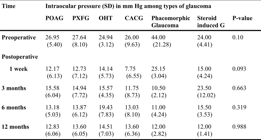
Table 5: Comparison of preoperative and postoperative mean intraocular pressure among the different types of glaucoma, n=49 eyes.
Time Intraocular pressure (SD) in mm Hg among types of glaucoma
POAG PXFG OHT CACG Phacomorphic
Glaucoma Steroid induced G P-value
Preoperative 26.95
(5.40)
27.64 (8.10)
24.94 (3.12)
26.00 (9.63)
44.00 (21.28)
24.00 (4.41)
0.10
Postoperative
1 week 12.17
(6.13) 12.73 (7.12) 14.14 (5.73) 7.75 (6.55) 25.15 (3.04) 15.00 (4.24) 0.093
3 months 15.58
(6.04) 14.94 (7.72) 15.57 (4.35) 11.75 (8.73) 10.50 (2.12) 23.50 (12.02) 0.663
6 months 13.18
(5.03) 13.87 (6.12) 19.43 (7.83) 13.03 (8.10) 11.00 (4.24) 15.50 (3.53) 0.319
12 months 12.83
(6.06) 13.60 (6.05) 14.51 (7.03) 13.60 (6.36) 12.00 (2.82) 12.00 (1.41) 0.988 POAG: primary open angle glaucoma, PXFG: psuedoexfolation glaucoma, OHT: ocular hypertension, CACG: chronic angle closure glaucoma, IOP: intraocular pressure
Table 6: Preoperative and postoperative number of anti glaucoma medications used to control intraocular pressure.
The overall success rate of IOP control at the comple-tion of 12 months follow-up was 87.7% (43 eyes), Complete 71.4% (35 eyes) and qualified 16.3% (8 eyes). The pressure remained above 21 mm Hg in six eyes while they were on hypotensive medications. The analysis of success rate showed no statistically signifi-cant difference either in complete or qualified success rate, or in failure among the different types of glau-coma.
In the first three months of postoperative period, 43 (87.3%) of the operated eyes were without hypoten-sive medication. And at one-year follow-up, 35 (71.4%) of the eyes were free of medication. The mean number of hypotensive medication lowered from 2.65 + 0.63 preoperative to 1.49 +0.82 at 12 months follow up (Table 6).
Visit Mean
num-ber of medi-cations
Standard deviation
P- value
Preoperative 2.65 0.63 -
3 months 1.10 0.05 <0.001
6 months 1.16 0.75 <0.001
Zonular dehiscence, posterior capsule tear and vitre-ous loss were the types of intra-operative complica-tions recorded in 6 eyes, among them 5 eyes were with pseudoexfoliative glaucoma. The types of post-operative complications recorded in the first one month follow-up period were hyphema (1 eye), pupil-lary inflammatory membrane (2 eyes), shallow ante-rior chamber (2 eyes) and choroidal effusion with hypotony, i.e. IOP < 5 mm Hg, (1 eye).
At the last visit, filtering bleb was present in 75.5% and flat bleb was recorded in 12 (24.5%) eyes.
DISCUSSION
The study has assessed the outcome of combined cata-ract extcata-raction with glaucoma surgery in terms of vi-sion restoration, intraocular pressure control, need of hypotensive drug to control the IOP and complica-tions. Cataract and glaucoma are the two common eye diseases that occur among the elderly population. This was the fact in this study that has identified mean age 66.10 years and the majority to be age above 60 (69.8%). Other studies have also reported similar mean age (61.3, 64.13, and 66.20, years) among pa-tients underwent combined procedure either Phaco-Trab or MSICS-Phaco-Trab (15, 17, 18). Our result also also shown a similar finding with a mean age of 66.10 years and the majority to our patients were aged above 60 (69.8%).
Pseudoexfoliative glaucoma was the commonest type of glaucoma diagnosis in this study, 22 eyes (44.9%). And this can be explained by the fact that pseudoexfo-liation and pseudoexfoliative glaucoma are common conditions among Ethiopians (19-21). The high and fluctuating nature of IOP in pseudoexfoliative glau-coma requires keeping the IOP at low and stable level, which achievable with filtering surgery as was indi-cated in this study.
The visual acuity of our patients improved to > 6/18 in 28 eyes (57.1%) and 35 eyes (71.4%) in the first one week and at 12 month follow-up. The number of eye with visual acuity > 6/18 during the first one week postoperative follow-up was less as compared the subsequent visits. This can be explained by the pres-ence of wound inflammatory reaction of the anterior segment and wound healing process. The visual acuity of > 6/18 that was achieved at the last follow-up is lower when compared to > 6/12 in 43 eyes (78.2%) at 3 years in India (15). On the other hand, visual acuity > 6/18 that was achieved in 38 eyes (77.8%) at 3 months, is better as compared to other study report at 8 weeks that achieved the same level of vision im-provement in 23 eye (65.7%), (3). Fourteen eyes (28.6%) remained visual impaired, less than 6/18 vis-ual acuity; which can be explained by the glaucoma-tous optic nerve head damage and other preexisting ocular pathology documented in their charts.
In this study, the mean intraocular pressure was significantly reduced from the baseline mean level (27.04 + 6.04) and remained below 15 mmHg throughout the year (P < 0.001), with mean reduc-tion 13.63 mmHg at last follow-up. This level of IOP is comparable to 13.9 + 3.81mmHg reported at 3 years follow-up by Mittal S et al (15); and to an eight weeks mean IOP reduction of 12.52 + 35 mmHg reported by Khurana AK et al (17). And it is better than 17.1 + 10mm Hg reported at 6 months follow-up by Thomas R et al (16). Singh P et al (18) reported mean IOP reduction form baseline 23.93 + 0.75 mm Hg to 11.2 + 1.5 mm Hg at 6 weeks post-operative follow-up in 45 patients, which is lower as compared to the 3 months mean IOP level of this study, 12.29 + 1.07 mmHg, but the time different should be taken in to consideration.
In the first postoperative week, the IOP remained low in all types of glaucoma and ocular hyperten-sion, except in phachomorphic glaucoma, which can be explained by the presence of anterior chamber reaction before, during and after surgery, which is the nature of the disease.
Among the pseudoexfoliative glaucoma cases, the preoperative 27.64 mm Hg mean IOP remained less than 15 mm Hg during the one-year follow-up pe-riod, which is advantageous to keep the IOP low and stable to halt the damaging nature of fluctuating IOP in this form of glaucoma. Additionally, cataract extraction lowers IOP in all types of glaucoma due to widening of the anterior chamber angel and the possibility of remodeling of the trabecular mesh-work (22,23).
The two patients with phacomorphic glaucoma un-derwent the surgery because of delayed presenta-tion, more than a month, and consideration of the possibility of persistence angle closure; otherwise, they could have been managed by cataract extrac-tion alone.
The 87.7% success of IOP control (IOP < 21 mmHg) with and without medication, at the last follow up, is comparable with that of retrospective comparative study of MSICS-Trab (a similar proce-dure with this study) and Phaco-Trab done in India, that achieved 89.0% and 92.3% success at 3 years follow-up, respectively. On the other hand, it is bet-ter than other study report from the same country with mean 6 months follow up that achieved IOP control in 73% MSICS-Trab group and in 75.6% Phaco-Trab group (15,16).
Forty-three (87.7%) and 35 (71.4%) eyes required no hypotensive drugs after the surgery during the first three months and the last follow up period, which is beneficial to the patients in terms of cost, being free of drug side effects and the physiological impact of having and applying medications. It is also beneficial and encouraging to the treating physician to have his/ her patient being managed with less frequent follow-up visit.
The procedure enabled to reduce the number of hy-potensive drugs that had been used prior to the sur-gery. The preoperative 2.65 + 0.63 mean number of hypotensive drugs reduced to 1.49 + 0.82 at one-year postoperative follow-up. The other studies, men-tioned above, have also reported postoperative reduc-tion of hypotensive drugs need (14,15).
The type and frequency of occurrence of both intra-operative and postintra-operative complications varies among studies reports, including this study (15,16, 18).
Pupillary fibrinous inflammatory membrane, the type of early postoperative complication, docu-mented in three eyes, could be related to the surgi-cal manipulation to the anterior chamber structures during the surgical procedure. Besides, two of the three eyes were with pseudeoexfoliation, which by itself is associated with more wound reaction than the other types of glaucoma. Postoperative flat bleb was recorded in 6 (12.2%) eyes, which could be related to healing or fibrosis of the scleral tunnel.
The possibility of non-detailed documentation of clinical information during the preoperative, intra-operative and postintra-operative periods is believed to be the limitation of this retrospective study.
Conclusion: Combined manual small incision
cata-ract surgery and trabeculectomy is effective in terms of IOP control and vision restoration in treating patients with coexisting cataract and glaucoma.
Recommendation: The procedure is doable and cost
effective for eye care professionals who handle glaucoma patients and practicing manual small inci-sion cataract surgery.
REFERENCES
1. Linebarger EJ,, Hardten DR, Shah GK, Lindstrom RL. Phacoemulsification and modern cataract surgery.
Surv Ophthalmol. 1999; 44(2): 123-47.
2. Donaldson EK, Braga-Mele R , Cabot F, et al. Femtosecond laser–assisted cataract surgery. J Cataract Refract Surg 2013; 39:1753–1763.
3. Singh K, Misbah A, Saluja P, Singh A. Review of manual small-incision cataract surgery. Indian J Oph-thalmol. 2017; 65(12): 1281–1288.
4. David R, Freedman J, Luntz MH. Comparative study of Watson's and Cairns's trabeculectomies in a Black population with open angle glaucoma. Br J Ophthalmol. 1977; 61(2): 117–119.
5. SooHoo JR, Seibold LK, Radcliffe NM, Kahook MY. Minimally invasive glaucoma surgery: Current implants and future innovations. Can J Ophthalmol. 2014; 49(6): 528-33.
6. Lee JH, Amoozgar B, Han Y. Minimally Invasive Modalities for Treatment of Glaucoma: An Update. J Clin Exp Ophthalmol 2017; 8(4). https://www.omicsonline.org/open-access/minimally-invasive-modalities-for-treatment-of-glaucoma-an-update-2155-9570-1000666.php?aid=93378, Accessed 05/03/2018.
7. Husain R, Liang S, Foster PJ, et al. Cataract surgery after trabeculectomy: the effect on trabeculectomy function. Arch Ophthalmol. 2012; 130(2): 165-7.
8. Longo A, Uva MG, Reibaldi A, Avitabile T, Reibaldi M. Long-term effect of phacoemulsification on tra-beculectomy function. Eye (Lond). 2015; 29(10): 1347-52.
9. Patel HY, Danesh-Meyer HV. Incidence and management of cataract after glaucoma surgery. . Curr Opin Ophthalmol. 2013; 24(1): 15-20.
10. Friedman DS, Jampel HD, Lubomski LH, et al. Surgical strategies for coexisting glaucoma and cataract: an evidence-based update. Ophthalmology. 2002; 109(10): 1902-13.
11. Allingham RR, Damji KF, Freedman S, Moroi SE, Rhee D eds. Surgical approaches for coexisting glau-coma and cataract. Shields Textbook of Glauglau-coma. 6th ed. Philadelphia, Lippincott Williams & Wilkins, 2011: 578-588.
12. Marchini G, Ceruti P, Vizzari G, Berzaghi D, Zampieri A. Management of Concomitant Cataract and Glaucoma. Dev Ophthalmol. 2017;59:155-164.
14. Richter MR, Coleman LA. Minimally invasive glaucoma surgery: current status and future prospects. Clin Ophthalmol. 2016; 10: 189–206.
15. Mittal S, Mittal A, Ramakrishnan. Safety and efficacy of manual small-incision cataract surgery combined with trabeclectomy: Comparison with phacotrabeculectomy. Asian J Ophthalmol. 2008; 10: 221-9. 16. Thomas R, Parikh R, Muliyil. Comparison between Phacoemulsification and the Blumenthal Technique
of Manual Small-Incision Cataract Surgery Combined with Trabeculectomy. Journal of Glaucoma, 2003; 12:333–339.
17. Khurana AK, Chawla U, Passi N, et al. Combined SICS and trabeculectomy. Nepal J Ophthalmol. 2011; 3 (5): 13- 18.
18. Singh P, Kunhammad S. Visual rehabilitation and intraocular pressure control after combined manual small incision cataract surgery and mitomycin-C augmented trabeculectomy. Nepal Med Coll J. 2014; 16 (2-4): 177-181.
19. Olawoye OO, Pasquale L, Ritch R. Exfoliation syndrome in sub-Saharan Africa. Int Ophthamol. Published online 2014. DOL: 10.1007/s10792-014-9953-5.
20. Tenkir A, Solomon B, Deribew A. Glaucoma Subtypes in Ethiopia Clinical Patients. J Glaucoma. 2013; 22:110–116.
21. Giorgis AT, Mulugeta A, Aga A, Deyassa N. The spectrum of glaucoma presentation at Menelik II Hospi-tal, Addis Ababa. Ethiop Med J. 2012; 50(3): 259-64.
22. Berdahl PJ. Cataract Surgery to Lower Intraocular Pressure. Middle East Afr J Ophthalmol. 2009; 16(3): 119–122.