4-23-2007
Analysis of the Cellular Proteins, TIA-1 and TIAR,
and their Interaction with the West Nile Virus
(WNV) 3' SL Minus-Strand RNA
Mohamed Maged EmaraGeorgia State University
Follow this and additional works at:https://scholarworks.gsu.edu/biology_diss
Part of theBiology Commons
This Dissertation is brought to you for free and open access by the Department of Biology at ScholarWorks @ Georgia State University. It has been accepted for inclusion in Biology Dissertations by an authorized administrator of ScholarWorks @ Georgia State University. For more information, please [email protected].
Recommended Citation
Emara, Mohamed Maged, "Analysis of the Cellular Proteins, TIA-1 and TIAR, and their Interaction with the West Nile Virus (WNV) 3' SL Minus-Strand RNA." Dissertation, Georgia State University, 2007.
by
Mohamed Emara
Under the Direction of Margo A. Brinton
ABSTRACT
The 3’ terminal stem loop of the WNV minus-strand [WNV3’(-) SL] RNA was
previously shown to bindthe cell protein, T-cell intracellular antigen-1 (TIA-1), and the
related protein, TIAR. These two proteins are known to bind AU-rich sequences in the
3’UTRs of some cellular mRNAs. AU stretches are located in three single-stranded
loops (L1, L2, and L3) of the WNV3’(-) SL RNA. The RNA binding activity of both
proteins was reduced when L1 or L2, but not L3, AU sequences were deleted or
substituted with Cs. Deletion or substitution with Cs of the entire AU-rich sequence in
either L1 or L2 in a WNV infectious clone was lethal for the virus while mutation of
some of these nt decreased the efficiency of virus replication. Mutant viral RNAs with
small plaque or lethal phenotypes had similar translational efficiencies to wildtype RNA,
but showed decreased levels of plus-strand RNA synthesis. These results correlated well
with the efficiency of TIA-1 and/or TIAR binding in in vitro assays.
In normal cells, TIA-1 and TIAR are evenly distributed in the cytoplasm and
nucleus. Between 6 and 24 hr after WNV infection, TIAR concentrated in the perinuclear
region and TIA-1 localization to this region began by 24 hr. Similar observations were
proteins colocalized with dsRNA, a marker for viral replication complexes, and with viral
non-structural proteins. Anti-TIAR or anti-TIA-1 antibody coimmunoprecipitated viral
NS3 and possibly other viral nonstructural proteins. In response to different types stress,
TIA-1 and TIAR recruit cell mRNA poly(A)+ into cytoplasmic stress granules (SG)
leading to general translational arrest in these cells. SG were not induced by flavivirus
infection and cells became increasingly resistant to arsenite induction of SG with time
after infection. Processing Body (PB) assembly was also decreased beginning at 24 hr.
These data suggest that the sequestration of first TIAR and then TIA-1 via their
interaction with viral components in flavivirus infected cells inhibits SG formation and
prevents the shutoff of host translation.
INDEX WORDS: West Nile virus, minus-strand RNA, 3’stem loop RNA, protein
binding sites, TIAR, TIA-1, NS3, processing body, stress granules, virus RNA
Analysis of the cellular proteins, TIA-1 and TIAR, and their interaction with the
West Nile virus (WNV) 3’ SL minus-strand RNA
by
Mohamed Emara
A Dissertation Submitted in Partial Fulfillment of the Requirements for the Degree
of Doctor of Philosophy
in the College of Arts and Sciences
Georgia State University
Copyright by
Mohamed M. Emara and Margo A. Brinton
Analysis of the cellular proteins, TIA-1 and TIAR, and their interaction with the
West Nile virus (WNV) 3’ SL minus-strand RNA
by
Mohamed Emara
Major Professor: Margo A.Brinton Committee Members: Teryl K. Frey Irene T. Weber
Electronic Version Approved by:
DEDICATION AND ACKNOWLEDGEMENTS
This dissertation is dedicated to the soul of my father Mr. Maged Emara, who taught
me that with hard and honest work I would be able to accomplish my goals. I would like
to express my deepest gratitude and appreciation to Dr. Margo A. Brinton for her
guidance, encouragement, and thoughtfulness. She was an exceptional advisor. I would
also like to express my appreciation to my committee members, Dr. Teryl K. Frey and
Dr. Irene T. Weber for their interest, advice and critical review of this dissertation. Also, I
would especially like to thank Dr. Svetlana V. Scherbik and William G. Davis for their
technical advice, discussions, and encouragment throughout my Ph.D study. Thanks to all
my colleagues in Dr. Brinton lab, Gretrud Radu, Slava Stockman, Husni ElBahesh, Sean
Courtney, Joanna Pulit-Penaloza, Dr. Andrey Perelygin, Dr. Mausumi Basu, Dr. Natalia
Astrakova, and Dr. Taronna Maines, for their friendship and assistance as well as
stimulating and interesting conversations. Thanks to my Egyptian colleagues at GSU, Dr.
Hassan Wally, Dr. Mahmoud Ghanem, and Dr. Hosam Ewis for their friendship and
support. I would like to thank my Mom and my sister for their constant love and
encouragement. My deepest thanks, appreciation, and love to my wife Samah Mahdy for
her unconditional support, encouragement, and love throughout my Ph.D. study. Finally,
TABLE OF CONTENTS
DEDICATION AND ACKNOWLEDGEMENTS... iv
Table of Contents... v
List of figuers... ix
Chapter I... 1
INTRODUCTION ... 1
Classification and Medical Importance of Flaviviruses... 1
Virion Morphology and Composition... 2
Flavivirus replication cycle... 3
Flavivirus genome organization... 5
Structural proteins... 9
The C protein. ... 9
The M protein. ... 9
The E protein... 10
Nonstructural proteins... 11
NS1. ... 11
NS2A, NS2B, NS4A, and NS4B. ... 12
NS2A... 13
NS2B... 13
NS3. ... 14
NS5. ... 15
Stress granules. ... 19
GOALS OF THIS DISSERTATION... 23
REFERENCES ... 26
CHAPTER II... 40
Mutation of mapped TIA-1/TIAR binding sites within the West Nile virus 3’ terminal minus-strand RNA sequence in an infectious clone negatively affects viral plus-strand synthesis... 40
ABSTRACT... 40
INTRODUCTION ... 41
RESULTS ... 45
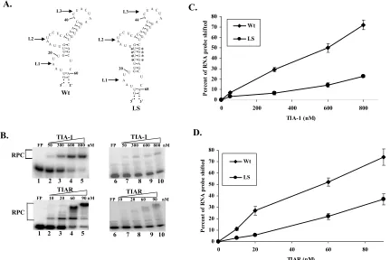
Expression, purification, and RNA binding activities of recombinant TIA-1 and TIAR. ... 45
Analysis of the WNV3’(-)SL RNA structure. ... 46
Mapping the binding sites for the TIA-1 and TIAR proteins within the WNV3’ (-)SL RNA. ... 50
Effect of substitution of L1 and L2 with Cs on virus production. ... 54
Effect of deletion of A and U nt in the mapped WNV3’(-)SL RNA TIA-1 and TIAR binding sites in a WNV infectious clone... 59
Effect of mutations in the WNV3’(-)SL RNA on the viral RNA translation. ... 60
Relative quantification of viral RNA replication by real-time RT-PCR. ... 62
DISCUSSION ... 65
Cells. ... 72
Cloning, expression, and purification of recombinant TIA-1 and TIAR from E. coli... 73
DNA constructs used as templates for RNA synthesis... 74
In vitro transcription of 32P-labelled and unlabeled RNA. ... 74
Transfection of in vitro transcribed full-length WNV genomic RNA... 75
Site directed mutagenesis of the infectious clone... 76
Gel mobility shift assays... 77
Quantitative Real-time RT-PCR of the intracellular viral RNA... 78
Detection of intracellular viral antigen. ... 79
RNA secondary structure prediction... 80
ACKNOWLEDGMENTS ... 80
REFERENCES ... 81
CHAPTER III ... 88
ADDITIONAL DATA... 88
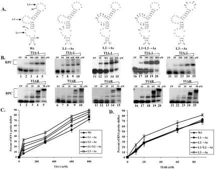
Effect of substitution of L1, L2, or L3 with As or Us... 88
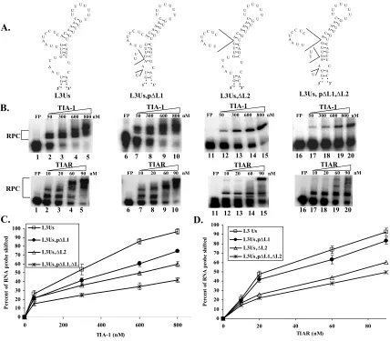
Effect of increasing the distance between L1 and L2 on TIA-1 and TIAR binding activity... 90
DISCUSSION ... 94
REFERENCES ... 97
Interaction of TIA-1/TIAR with West Nile and dengue virus products in infected cells
interferes with SG formation and PB assembly... 99
ABSTRACT... 99
INTRODUCTION ... 100
RESULTS ... 102
TIA-1 and TIAR colocalize with WNV and DV2 proteins in infected BHK cells. ... 102
TIA-1 and TIAR interact with sites of WNV and DV2 RNA replication. ... 107
WNV and DV2 interfere with SG formation in infected BHK cells. ... 113
WNV and DV2 infections interfere with PB assembly. ... 117
DISCUSSION ... 120
MATERIALS AND METHODS... 126
Cells. ... 126
Viruses. ... 126
Antibodies... 127
Western blotting... 127
Coimmunoprecipitation assays. ... 128
Detection of intracellular and cellular viral proteins by immunofluorescence... 128
Laser confocal imaging settings... 129
ACKNOWNLEDGMENTS ... 129
LIST OF FIGUERS
Figure 1.1: Flavivirus replication cycle. ... 6
Figure 1.2: The WNV genome... 8
Figure 1.3: Schematic structure of TIA-1 and TIAR... 20
Figure 1.4: Translation initiation in the absence or presence of stress factors. ... 22
Figure 2.1. Purification and RNA binding activities of recombinant TIA-1 and TIAR proteins... 48
Figure 2.2. Effect of C substitutions in L1, L2, and L3 of the WNV3’(-)SL RNA on in vitro rTIA-1 and rTIAR binding activity. ... 52
Figure 2.3. Effect of deletions in L1, L2, or L3 of the WNV3’(-)SL RNA on in vitro rTIA-1 and rTIAR binding activity... 53
Figure 2.4. Effect of sequential restoration of deleted nt in L1 and L2 on in vitro rTIA-1 and rTIAR binding activity... 55
Figure 2.5. Effect of C substitutions in L1, L2, or L3 of the WNV3’(-)SL RNA on virus production. ... 58
Figure 2.6. Effect of deletion of A and U nucleotides in L1, L2, or L3 of the WNV3’ (-)SL RNA on virus production... 61
Figure 2.7. Effect of the introduced mutations in L1 or L2 within the WNV3’(-)SL RNA on viral RNA transcription and/or translation... 64
Figure 3.2: Effect of deletions in L1 and/or L2 of the L3→Us mutant RNA on rTIA-1
and rTIAR binding activity... 92
Figure 3.3: Effect of increasing the distance between L1 and L2 within the WNV3’(-)SL RNA on rTIA-1 and rTIAR binding activity. ... 93
Figure 4.1. Colocalization of TIA-1 and TIAR with WNV proteins in infected BHK cells. ... 105
Figure 4.2. Colocalization of TIA-1 with WNV proteins in infected TIAR-/- MEFs. ... 106
Figure 4.3. Colocalization of TIA-1 and TIAR with DV2 proteins in infected BHK cells. ... 108
Figure 4.4. Interaction of TIA-1 and TIAR with WNV replication complex components. ... 111
Figure 4.5. Interaction of TIA-1 and TIAR with DV2 replication complex components. ... 112
Figure 4.6. WNV infection interferes with SG formation. ... 115
Figure 4.7. DV2 infection interferes with SG formation. ... 116
Figure 4.8. WNV infection interferes with PB assembly. ... 118
CHAPTER I
INTRODUCTION
Classification and Medical Importance of Flaviviruses.
Flaviviruses and alphaviruses were previously classified as members of the family
Togaviridae. However, due to differences in the replication and assembly strategies
(Westaway et al., 1980), as well as the genome structures (Rice et al., 1985) of
flaviviruses and alphaviruses, flaviviruses were reclassified as members of the genus
flavivirus within the family Flaviviridae. The family Flaviviridae currently includes two
other genera, the pestiviruses and the hepaciviruses (Lindenbach and Rice, 2007). The
genus flaviviruscontains more than 68 members that are separated into twelve antigenic
serogroups (Heinz et al., 2000). Some flaviviruses such as dengue virus (DV), Japanese
encephalitis virus (JEV), yellow fever virus (YFV), West Nile virus (WNV), St. Louis
encephalitis virus (SLEV), Murray Valley encephalitis virus (MVEV), and tick-borne
encephalitis virus (TBEV), are important human pathogens.
Most of the flaviviruses are transmitted to vertebrates via infected blood-sucking
arthropods, and are therefore called arthropod-borne viruses or “arboviruses”
(Lindenbach and Rice, 2007). Approximately 65% of the flaviviruses are transmitted by
mosquitoes, 22% by ticks, and the remaining 13% have no known arthropod vector
(Porterfield, 1996). All of the members of the YFV, DV, and JE serogroups are
mosquito-borne. Neuroinvasiveness is a common feature of those flaviviruses that cause
encephalitis. Although YFV can be prevented by vaccine, outbreaks and epidemics
caused by YFV have risen dramatically in the recent years in South America and
sub-Saharan countries in Africa, because of the difficulties of vaccinating many at-risk
populations (Monath, 1994). YFV infections have a mortality rate that can be as high as
20%. DV infects 50-100 million humans annually in tropical and subtropical regions
(Gubler, 2002). Symptoms range from a self-limiting febrile illness to the life threatening
dengue hemorrhagic fever and dengue shock syndrome (DHF-DSS), with a mortality rate
of ~10%. Members of the JEV serogroup such as JEV, WNV, SLEV, and MVEV, are
responsible for periodic epidemics and scattered cases of human central nervous system
(CNS) disease (Lindenbach and Rice, 2007). WNV is endemic in Africa,the Middle East,
Europe, central Asia,and most recently, North America. WNV infections in the USA first
occurred in the summer of 1999 (Asnis et al., 2000). In 2006, the Centers for Disease
Control and Prevention (CDC) reported 4261 positive human WNV cases in the US.
Also, the tick-borne flaviviruses, such as Powassan, louping ill, Kyasanur Forest Disease,
Russian Spring-Summer encephalitis viruses, and TBEV cause human meningitis and/or
encephalitis with a case fatality ranging from 1-30% (Shope, 1980).
Virion Morphology and Composition.
The flavivirus virion is spherical, enveloped and has a diameter of 40-60 nm
(Murphy, 1980). The virion is composed of three structural proteins, the envelope (E)
protein, the membrane (M) protein, and the capsid (C) protein; the E and M proteins are
(Lindenbach and Rice, 2007). Cryo-electron microscopy data suggested that the virions
have icosahedral symmetry (Heinz and Allison, 2000). Recent studies indicated that this
symmetry is imposed by interactions between E surface proteins, rather than by
interactions between capsid monomers (Kuhn et al., 2002).
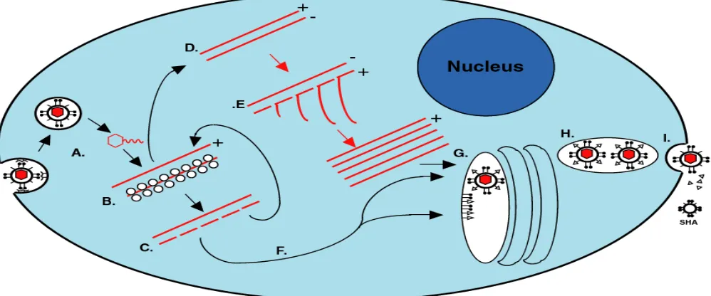
Flavivirus replication cycle.
Flavivirus surface protein binds to unknown receptor(s) on the host cell surface
followed by virus entry via receptor-mediated endocytosis (Fig. 1.1A). The viral
nucleocapsid is subsequently released into the cytoplasm of the infected cells by
acidic-pH fusion of the virion envelop with the endosomal membrane (Gollins and Porterfield,
1985; Gollins and Porterfield, 1986; Nawa, 1998). After uncoating of the nucleocapsid by
an unknown process, the viral genome is released into the cell cytoplasm and is then
translated into a single polyprotein (Fig. 1.1B). The poly protein is processed by viral and
cellular proteases into the mature viral structural and nonstructural proteins (Fig. 1.1C)
(Heinz et al., 1994). The genome RNA also serves as the template for transcription of the
complementary minus strand RNA. The minus-strandRNA in turn serves as a template
for the synthesis of genomicRNA. The minus-strand RNA is always present in infected
cells in association with the plus-strand RNA in a replicative form (RF) (Fig. 1.1D) or a
replicative intermediate (RI) (Fig. 1.1E) complex (Lindenbach and Rice, 2007).
Flavivirus RNA synthesis is postulated to occur via asymmetric and semiconservative
mode of replication (Chu and Westaway, 1985; Cleaves, Ryan, and Schlesinger, 1981).
time forming the RF, while the minus-strand template is efficiently reinitiated and
simultaneously synthesizes multiple copies of the plus strand RNA forming the RI. The
synthesis of plus and minus-strand viral RNA is disproportionate; about 10 times more
plus strand RNA than the minus-strand RNA is produced (Chu and Westaway, 1985).
Flavivirus replication takes place in viral replication complexes located on membranes in
the perinuclear region of the cytoplasm of infected cells.
Flavivirus infection causes extensive rearrangement and proliferation of the
cytoplasmic membranes present in the perinuclear region to provide an environment that
enhances virus replication (Brinton, 2002; Lindenbach and Rice, 2007). Later in the
infection cycle, these rearrangements induce the formation of smooth membrane vesicle
clusters known as vesicle packets (VP), that are found associated with smooth
endoplasmic reticulum (SER) or Golgi-like membranes (Mackenzie, Jones, and
Westaway, 1999). VP appear to be associated with another randomly folded membrane
structure, called convoluted membranes (CM) or paracrystalline arrays (PC) (Mackenzie
et al., 1998). Previous studies showed that the viral RNA as well as the viral replication
proteins are localized within the VP, suggesting that these structures might be sites of
RNA replication (Mackenzie et al., 1998; Westaway, Khromykh, and Mackenzie, 1999).
The viral serine protease NS3 and cofactor NS2B colocalize in the CM/PC structures
indicating that these are sites of proteolytic processing (Westaway et al., 1997b). It is
noteworthy that VP have only been observed at later times after infection, thus the site(s)
Although flavivirus packaging and release mechanisms are not well understood,
electron microscopy studies demonstrated that progeny virion assemblies formed in the
lumen of the ER (Lindenbach and Rice, 2007) (Fig. 1.1 G). Immature virions containing
prM-E heterodimers are found in cytoplasmic vesicles (Fig. 1.1 G and H). Mature virions
are generated in the trans-Golgi network via cleavage of the N-terminal portion of prM to
M (Stadler et al., 1997; Wengler and Wengler, 1989) and mature virions are released
from the infected cell by exocytosis after fusion of virion-containing cytoplasmic vesicles
with the plasma membrane (Stollar, Stevens, and Schlesinger, 1966) (Fig. 1.1I). The
release of the flavivirus particles from the infected cells starts at about 12 hr after
infection. For WNV and JEV, maximal virus titers are produced by 24 hr after infection
(Trent, Swensen, and Qureshi, 1969).
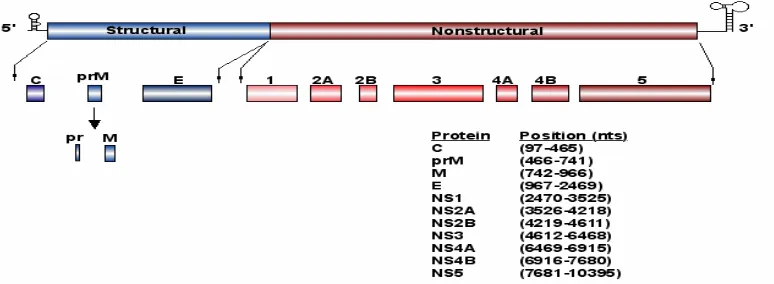
Flavivirus genome organization.
The WNV genome is a single-stranded RNA with positive polarity that is
approximately 11 Kb in length (Rice et al., 1985). It contains a type I cap at the 5’ end,
but lacks a poly (A) tract at the 3’ end. The genome contains a single, long open reading
frame and serves as the only viral mRNA in WNV infected cells. The viral mRNA
encodes a single polyprotein, which is translated and co- and posttranslationally
seven nonstructural (NS1, NS2a,NS2b, NS3, NS4a,NS4b and NS5) proteins (Fig. 1.2)
(Chambers et al., 1990).
The terminal 5’and 3’noncoding regions (NCRs) of the WNV genome RNA are 96
and 631 nucleotides (nts) long, respectively (Chambers et al., 1990). Previous studies
have indicated that both the 3’ and 5’ terminal nucleotides of the flavivirus genome can
form conserved stem-loop (SL) structures (Fig. 1.2) (Brinton and Dispoto, 1988; Brinton,
Fernandez, and Dispoto, 1986; Chambers et al., 1990). The SL structure formed by the
3’-terminal nts is more stable than the structure formed by the 5’-terminal nts. The
conservation of the terminal structures as well as of short sequence elements within these
structures among divergent flaviviruses suggest that they may function as cis-acting
elements (Brinton, 2002). Evidence supporting the formation of a SL structure by the
3’terminal nt of the complementary minus-strand (Shi, Li, and Brinton, 1996) was
previously obtained by RNase structure probing. Deletion of either the 3’or 5’ terminal
SL in flavivirus infectious clones was lethal strongly suggesting that these regions
contain essential cis-acting elements for virus replication (Bredenbeek et al., 2003;
Cahour et al., 1995; Men et al., 1996). Particular host cell proteins have previously been
reported to bind specifically to regions within the 3’ and 5’ NCRs of a number of animal
RNA virus genomes (Lai, 1998). The 3’-terminal structure of the WNV complementary
minus strand RNA was previously reported to bind specifically to cell proteins.
UV-induced cross-linkingstudies indicated that the molecular masses of these RNA binding
proteins were about 42, 50, 60, and 108 kDa. One of these proteins,p42, was identified as
Figure 1.2: The WNV genome. Noncoding regions (terminal black lines), structural
Structural proteins.
The C protein.
Mature C protein is produced by the cleavage of a hydrophobic region from the C
protein precursor, designated anchored C (anch C), by the viral serine protease NS3
(Amberg et al., 1994; Yamshchikov and Compans, 1994). Peptides in the hydrophobic
region of anch C were reported to serve as signals for the translocation of prM into the
endoplasmic reticulum (ER) lumen (Nowak et al., 1989). The mature C protein is small
(~11 kDa) and contains highly basic residues in its N- and C-termini (Rice et al., 1985),
that may facilitate its interaction with the genomic RNA during the formation of the
ribonucleoprotein complex. The middle portion of mature C contains a hydrophobic
region that facilities membrane association and this may play a role in virus assembly
(Markoff, Falgout, and Chang, 1997). At late times after infection, the C protein was
found in the cytoplasm as well as the nucleus of infected cells (Westaway et al., 1997a).
The M protein.
The M protein is the smallest protein (~8 kDa) in the virus particle. It is initially
expressed as the precursor protein M (prM). prM is incorporated into immature virions.
During egress of virions, prM is cleaved by the Golgi-network enzyme furin to generate
the mature structural protein M (Stadler et al., 1997). The mature M protein consists of an
ectodomain (~40 aa long) followed by two transmembrane domains (Chambers et al.,
1990; Lindenbach and Rice, 2007). Although M protein is not defined as the viral protein
induce neutralizing antibody response (Bray and Lai, 1991; Vazquez et al., 2002),
suggesting that it can maintain protective immunity.
The E protein.
E is a type I integral membrane protein of ~ 53 kDa that is located on the outer
surface of the virion. The E protein is thought to facilitate the binding and entrance of the
virus particle into the cell by mediating fusion of the viral envelope with cellular
membrane. In immature virions, the E protein forms a heterodimer with the prM protein
to maintain the correct folding and stabilization of E until prM is cleaved by cellular
protease to yield the mature virion (Heinz and Allison, 2000). The E protein contains six
intramolecular disulfide bonds that are conserved among all flaviviruses (Rey et al.,
1995). A high-resolution crystal structure of a soluble fragment of TBE E protein showed
that the mature E protein forms elongated head-to-tail homodimers that were prepared to
lie fairly parallel with the virus envelope with the distal end of each monomer anchored
in the membrane (Heinz and Allison, 2000). Each subunit of the E protein is divided into
three domains (I, II, and III) that corresponded to previously defined antigenic domains
(Mandl et al., 2000; Mandl et al., 1989). Several studies have demonstrated that the E
protein contains the major determinants that elect neutralizing antibodies and generate a
Nonstructural proteins.
The flavivirus nonstructural proteins are multifunctional proteins (Mackenzie et al.,
1998). All seven of the nonstructural proteins were previously shown to be components
of viral RNA replication complexes (Kapoor et al., 1995; Uchil and Satchidanandam,
2003; Vasudevan et al., 2001). However, exactly how each of these viral nonstructural
proteins is involved in the formation and functioning of the flavivirus replication
complexes is not well understood.
NS1.
NS1 is a ~ 46 kDa glycoprotein, which is found both in the cytoplasm as well as on
the surface of infected cells and is slowly secreted by mammalian cells (Smith and
Wright, 1985). The NS1-E junction is cleaved by host signal peptidase after NS1 has
been translocated into the lumen of the ER (Chambers et al., 1990). NS2A is cleaved
from the C-terminal end of NS1 by an unknown host protease associated with the ER
(Falgout and Markoff, 1995). Although, NS1 forms homodimers that are hydrophobic
and membrane-associated (Smith and Wright, 1985; Winkler et al., 1989; Winkler et al.,
1988), it is not clear how NS1 associates with cell membranes. A Kunjin NS1 mutation
that completely inhibited NS1 dimer formation (Hall et al., 1999), was shown to reduce
the level of virus growth by 100-fold compared to wild-type virus, but had no effect on
NS1 secretion (Hall et al., 1999). These observations indicate that NS1 dimerization
A role for NS1 in viral RNA replication (Lindenbach and Rice, 1997; Muylaert,
Galler, and Rice, 1997) has also been reported. Studies with dual labeled cryosections
showed that DV NS1 colocalized with viral dsRNA in VP, the most likely sites of RNA
replication, in infected Vero cells (Mackenzie, Jones, and Young, 1996; Westaway et al.,
1997b). Moreover, the accumulation of both the positive- and the negative-strand RNA
during the first phase of viral RNA synthesis was significantly reduced in cells infected
with a mutant YFV that contained a large in frame deletion in the NS1 gene (Lindenbach
and Rice, 1997). Providing wildtype NS1 in trans successfully complemented this mutant
virus RNA and restored viral RNA replication to wildtype levels, suggesting that NS1
plays functions prior to or early in minus-strand synthesis. However, the YFV NS1
deletion mutant RNA could not be trans-complemented by a DV NS1 suggesting that the
function of NS1 may be mediated by an interaction with another viral protein. Mutation
in NS4A suppressed this defect, indicating that an interaction between NS1 and NS4A is
required for viral RNA synthesis (Lindenbach and Rice, 1999).
NS2A, NS2B, NS4A, and NS4B.
The four small hydrophobic, nonstructural proteins (NS2A, NS2B, NS4A, and NS4B)
are tightly associated with cytoplasmic membranes and together with NS3, NS5, and NS1
were previously shown to be components of membrane bound viral RNA replication
complexes located in the perinuclear region of infected cells. Little is known about the
NS2A.
NS2A is found in infected cells in two forms, the full-length protein of ~ 22 kDa and
a smaller 20 kDa C-terminally truncated form (Chambers et al., 1990; Nestorowicz,
Chambers, and Rice, 1994). It was previously shown that mutagenesis of the C-terminus
of either full length or truncated NS2A was lethal for YFV replication (Nestorowicz,
Chambers, and Rice, 1994). Cryo-immunogold staining studies showed that NS2A
localized in VP, which is thought to be the site for virus RNA replication (Mackenzie et
al., 1998). Also, a recombinant NS2A protein was previously reported to bind in vitro to
NS3 and NS5 as well as to regions within the 3’ NCR of Kunjin virus genomic RNA
(Mackenzie et al., 1998). Mutational analysis of NS2A in a Kunjin virus infectious clone
indicated an essential role for this protein in virus assembly (Liu, Chen, and Khromykh,
2003). A recent study using Kunjin virus subgenomic replicons, demonstrated that a
single amino acid substitution in NS2A significantly reduced the viral inhibitory effect of
interferon beta (Liu et al., 2004). Together these data suggest that NS2A may be involved
in RNA packaging, RNA replication, and interference with the host cell immune
response.
NS2B.
NS2B is ~ 14 kDa protein that has a conserved central hydrophilic domain flanked by
two hydrophobic domains. It forms a stable complex with the viral serine protease (NS3),
and functions as a cofactor for the serine protease activity (Chambers et al., 1993;
the central hydrophilic region of NS2B and the N-terminal region of NS3 (Chambers et
al., 1993; Droll, Krishna Murthy, and Chambers, 2000; Falgout, Miller, and Lai, 1993).
This viral protein complex mediates cleavage at the junctions between anch C/virion C,
NS2A-NS2B, NS2B-NS3, NS3-NS4A, and NS4B-NS5 (Amberg et al., 1994; Cahour,
Falgout, and Lai, 1992; Chambers, Grakoui, and Rice, 1991; Falgout et al., 1991; Zhang,
Mohan, and Padmanabhan, 1992). Deletion analysis indicates that the 40 aa hydrophilic
domain of NS2B is required for NS3 serine protease activity. Mutations within the
hydrophilic region that disrupt the NS2B-NS3 interaction also abolish serine protease
activity (Chambers et al., 1993; Clum, Ebner, and Padmanabhan, 1997; Droll, Krishna
Murthy, and Chambers, 2000; Falgout, Miller, and Lai, 1993). The hydrophobic regions
of NS2B are involved in anchoring NS2B to ER membranes (Clum, Ebner, and
Padmanabhan, 1997).
NS3.
NS3 is a large (~70 kDa) multifunctional protein that has several enzymatic activities,
which are required for polyprotein processing, viral RNA replication, and RNA capping
(Westaway, Mackenzie, and Khromykh, 2003). The N-terminus of NS3 has serine
protease activity (Lindenbach and Rice, 2007), which is activated only after interacting
with NS2B. It was previously reported that the NS3-NS2B complex is associated with
membranes and is required for efficient polyprotein processing. The C-terminal portion
of NS3 contains helicase and NTPase activities (Borowski et al., 2001). The precise
the 3’ terminal secondary structures, which are involved in template recognition. NS3
not only has these two activities, but the C-terminal region also encodes RNA
triphosphatase (RTPase) (Wengler and Wengler, 1993), which is postulated to
dephosphorylate the 5’end of the viral RNA before its cap methylation by the
polymerase NS5. In a previous study using coimmunoprecipitation and immunoblotting
experiments a protein-protein interaction between NS3 and NS5 was demonstrated
(Kapoor et al., 1995) and the NTPase (Cui et al., 1998; Yon et al., 2005) as well as
RTPase (Yon et al., 2005) activities of a recombinant dengue NS3 were enhanced by the
presence of NS5. These observations indicate that the interaction of NS3 and NS5 might
regulate the activity of NS3 during virus replication.
NS5.
NS5 is the largest (103 kDa) and most highly conserved flavivirus protein. The
C-terminal portion of NS5 contains the eight RNA dependent RNA polymerase (RdRp)
motifs, that are highly homologues to the sequences of the RdRps of other positive-strand
RNA viruses (Koonin, 1993). Previous in vitro studies using purified recombinant NS5
protein (Ackermann and Padmanabhan, 2001; Nomaguchi et al., 2004; Tan et al., 1996)
confirmed the polymerase activity of NS5. Previous studies indicated that flavirus NS5
performs de novo RNA synthesis (Ackermann and Padmanabhan, 2001; Selisko et al.,
2006). As concluded from comparison of the kinetics of the de novo RNA synthesis by
different Flavivirdae members RdRps, DV RdRP showed a higher degree of
initiation to elongation (Selisko et al., 2006). In a recent study, the crystal structure of the
WNV RdRp was determined and it was shown that this structure is similar to that of two
other members of the family Flaviviridae, hepatitis C virus (HCV) and bovine virus
diarrhea virus (BVDV) (Malet et al., 2007). The overall structure has the classic fold of
an RdRP, which consists of palm, thumb, and finger domains as well as specific
flavivirus features such as the priming loop that provides a platform which stabilizes the
viral RNA initiation complexes during the de novo initiation of flavivirus RNA syntheisis
(Malet et al., 2007). The N-terminal part of this protein contains a methyltransferase that
is predicted to function as an RNA capping enzyme (Koonin, 1993). NS5 was shown to
have both guanine-N-7 and ribose 2’-O methylation activities (Ray et al., 2006). A
recent mutational analysis of the terminal 74 nt WNV5’(+)SL structure of WNV showed
that the second and third nt of the viral genome as well as the bottom two helices of the
WNV5’(+)SL RNA structure are required for N-7 methylation. Also, a minimum
sequence of 20 nt in the 5’(+)SL that includes the first and the second nt was essential
for 2’-OH ribose methylation (Dong et al., 2007). Although the presence of the MTase
domain did not influence RdRp activity (Selisko et al., 2006), mutagenesis studies
confirmed that both RdRp (Khromykh, Sedlak, and Westaway, 1999) and MTase (Zhou
et al., 2007) are required for WNV life cycle.
TIA-1 and TIAR cellular proteins.
T-cell intracellular antigen-1 (TIA-1) and T-cell intracellular antigen related protein
to regulate alternative splicing of particular pre-mRNAs, translationally silence some
mRNAs, and sequester cytoplasmic mRNAs in stress granules (SG) (Kedersha and
Anderson, 2002). Although first discovered in T cells, TIA-1 and TIAR are expressed in
most types of cells, including brain, spleen, and macrophages, which are sites of
flavivirus replication in vivo. Both proteins are found in the cytoplasm and the nucleus
and shuttle between the two compartments (Beck et al., 1996). Both proteins were also
shown to be essential for embryonic development (Beck et al., 1998; Piecyk et al., 2000).
For homozygous TIAR knock outs, embryo lethality was 100% in BALB/c mice and
90% in C57BL/6 mice. For TIA-1 knock outs in both strains, the rate of embryonic
lethality was 50% (Piecyk et al., 2000). Attempts to knock out both proteins in mice were
unsuccessful (Piecyk et al., 2000) and suppression of both TIA-1 and TIAR in DT40
chicken cells resulted in cell death (Le Guiner, Gesnel, and Breathnach, 2003).
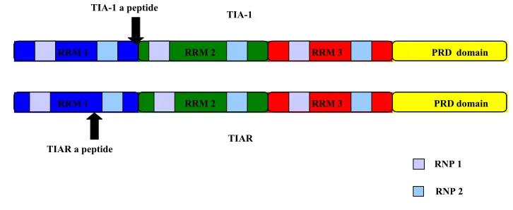
Two isoforms of both TIA-1 and TIAR have been reported and are translated from
alternatively spliced mRNAs (Fig. 1.3). The two isoforms of TIA-1 differ from each
other by the presence (TIA-1a) or absence (TIA-1b) of a mini exon encoding an
11–amino acid peptide within the RRM1. TIARa includes a mini exon that encodes a
17–amino acid peptide within RRM1, whereas TIARb lacks this exon. The long isoforms
of both proteins have molecular weights of ~ 42 kDa, whereas the short isoforms have
molecular weights of ~ 40 kDa. It was previously shown that the two TIA-1 isoforms are
equally abundant in cells, whereas the short isoform of TIAR is 6 times more abundant in
TIA-1 and TIAR are members of the RNA recognition motif (RRM) family of RNA
binding proteins (Anderson, 1995). Both proteins contain three RRM domains near their
amino termini and a glutamine rich domain at their carboxyl termini (Fig. 1.3). The
glutamine rich domain is also called prion related domain (PRD), which shares structural
and functional characteristics with the aggregation domains of mammalian and yeast
prion proteins (Gilks et al., 2004). Each RNA-binding domain contains
two-ribonucleoprotein sequences (RNP 1 and RNP2) that are conserved among all RNA
binding proteins. TIA-1 and TIAR proteins share 80% overall amino acid identity with
the highest degree of similarity (91% identity) in the RRM domain 3 and the lowest
degree of similarity (51% identity) in the carboxyl terminal domain (Kawakami et al.,
1992). Although RRM2 is sufficient for the specific binding of either protein to
uridylate-rich RNA, RRM1 and RRM3 enhance RRM2 RNA binding (Dember et al., 1996). In
somatic cells, RRM2 and the first half of the C-terminal domain mediate TIA-1 and
TIAR nuclear accumulation, whereas RRM3 facilitates nuclear export (Zhang et al.,
2005).
TIA-1 and TIAR protein binding sites were previously mapped to AU-rich regions
within the 3’UTRs of a subset of cellular mRNAs, that included TNF-α (Gueydan et al.,
1999; Piecyk et al., 2000), cyclooxgenase-2 (COX-2) (Cok et al., 2004; Dixon et al.,
2003), and β2-adrenegic receptor (β2-AR) (Kandasamy et al., 2005) mRNAs. For the
TNF-α mRNA, TIAR binding was mapped to a 39 nt region within the 3’UTR that
contained a 35 nt AU tract (Gueydan et al., 1999; Lewis et al., 1998). However, the
3’UTRs. The 3’terminal region of the WNV complementary minus strand RNA differs
from RNAs previously reported to bind to TIA-1 and TIAR in having AU rich sequences
within loops of a secondary structure. This binding is mediated through the RRM2
domain of TIA-1 and TIAR proteins. Kinetic studies showed that the binding activity of
TIA-1 RRM2 for the WNV3’(-)SL RNA was 10 times lower than that of the TIAR
RRM2 for this RNA (Li et al., 2002) .
Stress granules.
Mammalian stress granules (SG) are ribonucleoprotein aggregates or discrete
cytoplasmic foci, which are formed in response to environmental stresses such as
oxidative phosphorylation ER stress, and some viral infections (Fig. 1.4) (Anderson and
Kedersha, 2006). Several cell proteins, including TIA-1, TIAR, and Ras-Gap-SH3
domain-binding protein (G3BP) are involved in SG assembly (Anderson and Kedersha,
2006). SG are sites at which transient storage of untranslated mRNAs takes place in
stressed cells. The sequestration of these mRNAs in SG is initiated by the
phosphorylation of eukaryotic translation initiation factor 2 α (eIF2α), a key regulatory
factor in translation initiation, and this inhibits translation by preventing the assembly of
active preinitiation complexes. After eIF2α phosphorylation occurs, TIA-1 and TIAR
bind to the translationally inactive initiation complexes as well as to the mRNA poly(A)+
and self-aggregate forming SG (Kedersha and Anderson, 2002). Both the RRM domains
Figure 1.3: Schematic structure of TIA-1 and TIAR. RRM1 (blue), RRM2 (green), RRM3 (red), and the C-terminal glutamine rich prion related domain (yellow). Each RRM domain contains two ribonucleoprotein sequences (RNP 1 and RNP2). The positions of the additional mini peptides in the long isoforms of TIA-1 and TIAR are indicated by arrows.
RRM 1 RRM 2 RRM 3 PRD domain
TIA-1
TIAR
RRM 1 RRM 2 RRM 3 PRD domain
TIA-1 a peptide
TIAR a peptide
RNP 1
al., 2004; Kedersha et al., 1999). A recombinant TIA-1 protein lacking the three RNA
binding domains was unable to bind poly(A)+ RNA and recruit it to SG (Kedersha et al.,
1999). Deletion of the TIA-1 PRD inhibited protein aggregation and SG assembly (Gilks
et al., 2004). In mammalian cells SG were found to be physically associated with another
type of foci, known as processing bodies (PB) (Kedersha et al., 2005). PB are spheroid
particles (Cougot, Babajko, and Seraphin, 2004; Kedersha et al., 2005; Wilczynska et al.,
2005) that were identified in the cytoplasm of eukaryotic cells (Bashkirov et al., 1997;
Eystathioy et al., 2002; Eystathioy et al., 2003; Sheth and Parker, 2003) and were
reported to contain components of the 5’-3’ mRNA degradation pathway such as
GOALS OF THIS DISSERTATION
1. Mapping the binding site(s) of the TIA-1 and TIAR proteins on the WNV3’(-)SL
RNA.
Data from previous in vitro selection/amplification (SELEX) experiments showed
that the two cellular proteins, TIA-1 and TIAR, bind to AU stretches in RNAs. However,
the optimal RNA sequences recognized by TIA-1 in SELEX assays were not the same as
those recognized by TIAR (Dember et al., 1996). In another previous study, the binding
sites for both TIA-1 and TIAR in the 3’NCR of the tumor necrosis factor alpha (TNF-α)
mRNA were mapped to a large fragment of AU-rich sequence containing clustered
AUUUA pentamers (Gueydan et al., 1999). These cell mRNA binding sites for TIA-1
and TIAR were considered to be linear. In the WNV3’(-)SL RNA, the AU rich
sequences are located in single stranded loops (L1, L2, and L3) within a secondary
structure. Because TIA- and TIAR are ARE-binding proteins, the AU sequences will be
the initial mutation targets in experiments designed to identify nucleotides in the
WNV3’(-)SL RNA required forTIA-1 and TIAR protein binding.
Recombinant TIA-1 and TIAR proteins will be overexpressed in bacteria, purified,
and used for the analysis of RNA-protein interactions in gel mobility shift assay
experiments. The binding sites of both proteins will be mapped by measuring the relative
binding activities of each purified protein for different WNV 3’(-)SL RNA mutant
probes containing mutations and/or deletions in the three predicted single stranded loops.
essential for binding by deleting or replacing each loop separately or in combination with
a track of C residues. Subsequently, fine mapping of the binding sites will be done by
mutating A and U nt in the loops.
2. Determine whether the mapped binding site nt are cis-acting in the context of a
virus infection.
Deletion of either the 3’or 5’ terminal SL in various flavivirus infectious clones was
lethal, which strongly suggested that these regions contain essential cis-acting elements
for virus replication (Bredenbeek et al., 2003; Cahour et al., 1995; Men et al., 1996). In a
previous study, the growth of WNV in cells lacking TIAR was six to eight fold lower
than in wild type cells. In TIA-1 knock out cells, WNV grew to peak virus titers 6 hours
later than in the wild type cells (Li et al., 2002). These results suggested that both
proteins may play a role in the WNV replication cycle. To test this hypothesis more
directly, mutations and/or deletions in the mapped TIA-1 and TIAR binding sites within
the WNV3’(-)SL will be introduced into the a WNV infectious clone to test the effect of
these mutations on virus production. Those mutations and/or deletions found to have a
negative effect on virus production will be also tested for their effect on the translational
efficiency of the viral RNA in transfected BHK cells by detecting viral antigen
production by immunoflourescence. The effect of mutations and/or deletions found to
have a negative effect on virus production on the efficiency of viral plus-strand and
3. Determine whether the TIA-1/TIAR-viral RNA interactions occur in vivo and test
the effect of WNV infection on cellular SG formation.
In previous in vitro RNA-protein interaction assays, the TIA-1/TIAR proteins were
shown to bind specifically to the WNV3’(-)SL RNA (Li et al., 2002). The minus-strand
RNA of flaviviruses is present in infected cells only in RNA replication complexes
(Lindenbach and Rice, 2007). An antibody to dsRNA that does not detect either cellular
ribosomal RNA or tRNA was previously utilized to detect flavivirus replication
complexes in infected cells (Miller, Sparacio, and Bartenschlager, 2006). To test whether
TIA-1/TIAR colocalize with viral dsRNA and/or viral protein(s) in infected cells, BHK
cells will be infected with WNV and incubated with anti-WNV hyperimmune serum or
anti-dsRNA and anti-TIA-1 or anti-TIAR antibody and colocalization of cell and viral
proteins will be analyzed by confocal microscopy.
A number of detailed studies have shown that TIA-1 and TIAR are core components
of SG and that they recruit cell mRNAs into cytoplasmic foci formed in response to stress
factors (Kedersha et al., 2002). Sequestration of cell mRNAs in SG leads to repression of
their translation until the cells recover from stress or die (Kedersha et al., 2002). To
determine the effect of WNV infection on cellular SG formation, BHK infected with
WNV will be treated with sodium arsenite to induce SG at different times after infection
REFERENCES
Ackermann, M., and Padmanabhan, R. (2001). De novo synthesis of RNA by the dengue virus RNA-dependent RNA polymerase exhibits temperature dependence at the initiation but not elongation phase. J Biol Chem276(43), 39926-37.
Amberg, S. M., Nestorowicz, A., McCourt, D. W., and Rice, C. M. (1994). NS2B-3 proteinase-mediated processing in the yellow fever virus structural region: in vitro and in vivo studies. J Virol68(6), 3794-802.
Anderson, P. (1995). TIA-1: structural and functional studies on a new class of cytolytic effector molecule. Curr Top Microbiol Immunol198, 131-43.
Anderson, P., and Kedersha, N. (2006). RNA granules. J Cell Biol172(6), 803-8.
Asnis, D. S., Conetta, R., Teixeira, A. A., Waldman, G., and Sampson, B. A. (2000). The West Nile Virus outbreak of 1999 in New York: the Flushing Hospital experience. Clin Infect Dis30(3), 413-8.
Bashkirov, V. I., Scherthan, H., Solinger, J. A., Buerstedde, J. M., and Heyer, W. D. (1997). A mouse cytoplasmic exoribonuclease (mXRN1p) with preference for G4 tetraplex substrates. J Cell Biol136(4), 761-73.
Beck, A. R., Medley, Q. G., O'Brien, S., Anderson, P., and Streuli, M. (1996). Structure, tissue distribution and genomic organization of the murine RRM-type RNA binding proteins TIA-1 and TIAR. Nucleic Acids Res24(19), 3829-35.
Beck, A. R., Miller, I. J., Anderson, P., and Streuli, M. (1998). RNA-binding protein TIAR is essential for primordial germ cell development. Proc Natl Acad Sci U S A95(5), 2331-6.
nucleoside triphosphatase (NTPase)/helicase: evidence for dissociation of the NTPase and helicase activities of the enzyme. J Virol75(7), 3220-9.
Bray, M., and Lai, C. J. (1991). Dengue virus premembrane and membrane proteins elicit a protective immune response. Virology185(1), 505-8.
Bredenbeek, P. J., Kooi, E. A., Lindenbach, B., Huijkman, N., Rice, C. M., and Spaan, W. J. (2003). A stable full-length yellow fever virus cDNA clone and the role of conserved RNA elements in flavivirus replication. J Gen Virol84(Pt 5), 1261-8.
Brinton, M. A. (2002). The molecular biology of West Nile Virus: a new invader of the western hemisphere. Annu Rev Microbiol56, 371-402.
Brinton, M. A., and Dispoto, J. H. (1988). Sequence and secondary structure analysis of the 5'-terminal region of flavivirus genome RNA. Virology162(2), 290-9.
Brinton, M. A., Fernandez, A. V., and Dispoto, J. H. (1986). The 3'-nucleotides of flavivirus genomic RNA form a conserved secondary structure. Virology 153(1),
113-21.
Cahour, A., Falgout, B., and Lai, C. J. (1992). Cleavage of the dengue virus polyprotein at the NS3/NS4A and NS4B/NS5 junctions is mediated by viral protease NS2B-NS3, whereas NS4A/NS4B may be processed by a cellular protease. J Virol
66(3), 1535-42.
Cahour, A., Pletnev, A., Vazielle-Falcoz, M., Rosen, L., and Lai, C. J. (1995). Growth-restricted dengue virus mutants containing deletions in the 5' noncoding region of the RNA genome. Virology207(1), 68-76.
Chambers, T. J., Hahn, C. S., Galler, R., and Rice, C. M. (1990). Flavivirus genome organization, expression, and replication. Annu Rev Microbiol44, 649-88.
Chambers, T. J., Nestorowicz, A., Amberg, S. M., and Rice, C. M. (1993). Mutagenesis of the yellow fever virus NS2B protein: effects on proteolytic processing, NS2B-NS3 complex formation, and viral replication. J Virol67(11), 6797-807.
Chu, P. W., and Westaway, E. G. (1985). Replication strategy of Kunjin virus: evidence for recycling role of replicative form RNA as template in semiconservative and asymmetric replication. Virology140(1), 68-79.
Cleaves, G. R., Ryan, T. E., and Schlesinger, R. W. (1981). Identification and characterization of type 2 dengue virus replicative intermediate and replicative form RNAs. Virology111(1), 73-83.
Clum, S., Ebner, K. E., and Padmanabhan, R. (1997). Cotranslational membrane insertion of the serine proteinase precursor NS2B-NS3(Pro) of dengue virus type 2 is required for efficient in vitro processing and is mediated through the hydrophobic regions of NS2B. J Biol Chem272(49), 30715-23.
Cok, S. J., Acton, S. J., Sexton, A. E., and Morrison, A. R. (2004). Identification of RNA-binding proteins in RAW 264.7 cells that recognize a lipopolysaccharide-responsive element in the 3-untranslated region of the murine cyclooxygenase-2 mRNA. J Biol Chem279(9), 8196-205.
Cougot, N., Babajko, S., and Seraphin, B. (2004). Cytoplasmic foci are sites of mRNA decay in human cells. J Cell Biol165(1), 31-40.
Cui, T., Sugrue, R. J., Xu, Q., Lee, A. K., Chan, Y. C., and Fu, J. (1998). Recombinant dengue virus type 1 NS3 protein exhibits specific viral RNA binding and NTPase activity regulated by the NS5 protein. Virology246(2), 409-17.
Dixon, D. A., Balch, G. C., Kedersha, N., Anderson, P., Zimmerman, G. A., Beauchamp, R. D., and Prescott, S. M. (2003). Regulation of cyclooxygenase-2 expression by the translational silencer TIA-1. J Exp Med198(3), 475-81.
Dong, H., Ray, D., Ren, S., Zhang, B., Puig-Basagoiti, F., Takagi, Y., Ho, C. K., Li, H., and Shi, P. Y. (2007). Distinct RNA elements confer specificity to flavivirus RNA cap methylation events. J Virol.
Droll, D. A., Krishna Murthy, H. M., and Chambers, T. J. (2000). Yellow fever virus NS2B-NS3 protease: charged-to-alanine mutagenesis and deletion analysis define regions important for protease complex formation and function. Virology 275(2),
335-47.
Eystathioy, T., Chan, E. K., Tenenbaum, S. A., Keene, J. D., Griffith, K., and Fritzler, M. J. (2002). A phosphorylated cytoplasmic autoantigen, GW182, associates with a unique population of human mRNAs within novel cytoplasmic speckles. Mol Biol Cell13(4), 1338-51.
Eystathioy, T., Jakymiw, A., Chan, E. K., Seraphin, B., Cougot, N., and Fritzler, M. J. (2003). The GW182 protein colocalizes with mRNA degradation associated proteins hDcp1 and hLSm4 in cytoplasmic GW bodies. Rna9(10), 1171-3.
Falgout, B., and Markoff, L. (1995). Evidence that flavivirus NS1-NS2A cleavage is mediated by a membrane-bound host protease in the endoplasmic reticulum. J Virol69(11), 7232-43.
Falgout, B., Miller, R. H., and Lai, C. J. (1993). Deletion analysis of dengue virus type 4 nonstructural protein NS2B: identification of a domain required for NS2B-NS3 protease activity. J Virol67(4), 2034-42.
Gilks, N., Kedersha, N., Ayodele, M., Shen, L., Stoecklin, G., Dember, L. M., and Anderson, P. (2004). Stress granule assembly is mediated by prion-like aggregation of TIA-1. Mol Biol Cell15(12), 5383-98.
Gollins, S. W., and Porterfield, J. S. (1985). Flavivirus infection enhancement in macrophages: an electron microscopic study of viral cellular entry. J Gen Virol66
( Pt 9), 1969-82.
Gollins, S. W., and Porterfield, J. S. (1986). pH-dependent fusion between the flavivirus West Nile and liposomal model membranes. J Gen Virol67 ( Pt 1), 157-66.
Gubler, D. J. (2002). Epidemic dengue/dengue hemorrhagic fever as a public health, social and economic problem in the 21st century. Trends Microbiol10(2), 100-3.
Gueydan, C., Droogmans, L., Chalon, P., Huez, G., Caput, D., and Kruys, V. (1999). Identification of TIAR as a protein binding to the translational regulatory AU-rich element of tumor necrosis factor alpha mRNA. J Biol Chem274(4), 2322-6.
Hall, R. A., Khromykh, A. A., Mackenzie, J. M., Scherret, J. H., Khromykh, T. I., and Mackenzie, J. S. (1999). Loss of dimerisation of the nonstructural protein NS1 of Kunjin virus delays viral replication and reduces virulence in mice, but still allows secretion of NS1. Virology264(1), 66-75.
Heinz, F. X., and Allison, S. L. (2000). Structures and mechanisms in flavivirus fusion.
Adv Virus Res55, 231-69.
Heinz, F. X., Auer, G., Stiasny, K., Holzmann, H., Mandl, C., Guirakhoo, F., and Kunz, C. (1994). The interactions of the flavivirus envelope proteins: implications for virus entry and release. Arch Virol Suppl9, 339-48.
Kandasamy, K., Joseph, K., Subramaniam, K., Raymond, J. R., and Tholanikunnel, B. G. (2005). Translational control of beta2-adrenergic receptor mRNA by T-cell-restricted intracellular antigen-related protein. J Biol Chem280(3), 1931-43.
Kapoor, M., Zhang, L., Ramachandra, M., Kusukawa, J., Ebner, K. E., and Padmanabhan, R. (1995). Association between NS3 and NS5 proteins of dengue virus type 2 in the putative RNA replicase is linked to differential phosphorylation of NS5. J Biol Chem270(32), 19100-6.
Kawakami, A., Tian, Q., Duan, X., Streuli, M., Schlossman, S. F., and Anderson, P. (1992). Identification and functional characterization of a TIA-1-related nucleolysin. Proc Natl Acad Sci U S A89(18), 8681-5.
Kedersha, N., and Anderson, P. (2002). Stress granules: sites of mRNA triage that regulate mRNA stability and translatability. Biochem Soc Trans30(Pt 6), 963-9.
Kedersha, N., Chen, S., Gilks, N., Li, W., Miller, I. J., Stahl, J., and Anderson, P. (2002). Evidence that ternary complex (eIF2-GTP-tRNA(i)(Met))-deficient preinitiation complexes are core constituents of mammalian stress granules. Mol Biol Cell
13(1), 195-210.
Kedersha, N., Stoecklin, G., Ayodele, M., Yacono, P., Lykke-Andersen, J., Fritzler, M. J., Scheuner, D., Kaufman, R. J., Golan, D. E., and Anderson, P. (2005). Stress granules and processing bodies are dynamically linked sites of mRNP remodeling. J Cell Biol169(6), 871-84.
Kedersha, N. L., Gupta, M., Li, W., Miller, I., and Anderson, P. (1999). RNA-binding proteins TIA-1 and TIAR link the phosphorylation of eIF-2 alpha to the assembly of mammalian stress granules. J Cell Biol147(7), 1431-42.
Koonin, E. V. (1993). Computer-assisted identification of a putative methyltransferase domain in NS5 protein of flaviviruses and lambda 2 protein of reovirus. J Gen Virol74 ( Pt 4), 733-40.
Kuhn, R. J., Zhang, W., Rossmann, M. G., Pletnev, S. V., Corver, J., Lenches, E., Jones, C. T., Mukhopadhyay, S., Chipman, P. R., Strauss, E. G., Baker, T. S., and Strauss, J. H. (2002). Structure of dengue virus: implications for flavivirus organization, maturation, and fusion. Cell108(5), 717-25.
Lai, M. M. (1998). Cellular factors in the transcription and replication of viral RNA genomes: a parallel to DNA-dependent RNA transcription. Virology244(1), 1-12.
Le Guiner, C., Gesnel, M. C., and Breathnach, R. (2003). TIA-1 or TIAR is required for DT40 cell viability. J Biol Chem278(12), 10465-76.
Lewis, T., Gueydan, C., Huez, G., Toulme, J. J., and Kruys, V. (1998). Mapping of a minimal AU-rich sequence required for lipopolysaccharide-induced binding of a 55-kDa protein on tumor necrosis factor-alpha mRNA. J Biol Chem 273(22),
13781-6.
Li, W., Li, Y., Kedersha, N., Anderson, P., Emara, M., Swiderek, K. M., Moreno, G. T., and Brinton, M. A. (2002). Cell proteins TIA-1 and TIAR interact with the 3' stem-loop of the West Nile virus complementary minus-strand RNA and facilitate virus replication. J Virol76(23), 11989-2000.
Lindenbach, B. D., and Rice, C. M. (1997). trans-Complementation of yellow fever virus NS1 reveals a role in early RNA replication. J Virol71(12), 9608-17.
Lindenbach, B. D., and Rice, C. M. (1999). Genetic interaction of flavivirus nonstructural proteins NS1 and NS4A as a determinant of replicase function. J Virol 73(6),
4611-21.
M. A., Lamb, R. A., Roizman, B., Straus, S. E., (Eds.), Fields Virology, 5th ed., Lippincott William and Wilkins, Philadelphia, Pennsylvania, pp. 1101-52.
Liu, W. J., Chen, H. B., and Khromykh, A. A. (2003). Molecular and functional analyses of Kunjin virus infectious cDNA clones demonstrate the essential roles for NS2A in virus assembly and for a nonconservative residue in NS3 in RNA replication. J Virol77(14), 7804-13.
Liu, W. J., Chen, H. B., Wang, X. J., Huang, H., and Khromykh, A. A. (2004). Analysis of adaptive mutations in Kunjin virus replicon RNA reveals a novel role for the flavivirus nonstructural protein NS2A in inhibition of beta interferon promoter-driven transcription. J Virol78(22), 12225-35.
Mackenzie, J. M., Jones, M. K., and Westaway, E. G. (1999). Markers for trans-Golgi membranes and the intermediate compartment localize to induced membranes with distinct replication functions in flavivirus-infected cells. J Virol 73(11),
9555-67.
Mackenzie, J. M., Jones, M. K., and Young, P. R. (1996). Immunolocalization of the dengue virus nonstructural glycoprotein NS1 suggests a role in viral RNA replication. Virology220(1), 232-40.
Mackenzie, J. M., Khromykh, A. A., Jones, M. K., and Westaway, E. G. (1998). Subcellular localization and some biochemical properties of the flavivirus Kunjin nonstructural proteins NS2A and NS4A. Virology245(2), 203-15.
Malet, H., Egloff, M. P., Selisko, B., Butcher, R. E., Wright, P. J., Roberts, M., Gruez, A., Sulzenbacher, G., Vonrhein, C., Bricogne, G., Mackenzie, J. M., Khromykh, A. A., Davidson, A. D., and Canard, B. (2007). Crystal Structure of the RNA Polymerase Domain of the West Nile Virus Non-structural Protein 5. J Biol Chem
282(14), 10678-89.
Mandl, C. W., Guirakhoo, F., Holzmann, H., Heinz, F. X., and Kunz, C. (1989). Antigenic structure of the flavivirus envelope protein E at the molecular level, using tick-borne encephalitis virus as a model. J Virol63(2), 564-71.
Markoff, L., Falgout, B., and Chang, A. (1997). A conserved internal hydrophobic domain mediates the stable membrane integration of the dengue virus capsid protein. Virology233(1), 105-17.
Men, R., Bray, M., Clark, D., Chanock, R. M., and Lai, C. J. (1996). Dengue type 4 virus mutants containing deletions in the 3' noncoding region of the RNA genome: analysis of growth restriction in cell culture and altered viremia pattern and immunogenicity in rhesus monkeys. J Virol70(6), 3930-7.
Miller, S., Sparacio, S., and Bartenschlager, R. (2006). Subcellular localization and membrane topology of the Dengue virus type 2 Non-structural protein 4B. J Biol Chem281(13), 8854-63.
Monath, T. P. (1994). Dengue: the risk to developed and developing countries. Proc Natl Acad Sci U S A91(7), 2395-400.
Murphy, F. A. (1980). Togavirus morphology and morphogenesis. In: Schlesinger RW, (Ed.), The Togaviruses: Biology, Structure, Replication, New York: Academic
press, pp. 241-316.
Muylaert, I. R., Galler, R., and Rice, C. M. (1997). Genetic analysis of the yellow fever virus NS1 protein: identification of a temperature-sensitive mutation which blocks RNA accumulation. J Virol71(1), 291-8.
Nawa, M. (1998). Effects of bafilomycin A1 on Japanese encephalitis virus in C6/36 mosquito cells. Arch Virol143(8), 1555-68.
Nestorowicz, A., Chambers, T. J., and Rice, C. M. (1994). Mutagenesis of the yellow fever virus NS2A/2B cleavage site: effects on proteolytic processing, viral replication, and evidence for alternative processing of the NS2A protein. Virology
Nomaguchi, M., Teramoto, T., Yu, L., Markoff, L., and Padmanabhan, R. (2004). Requirements for West Nile virus (-)- and (+)-strand subgenomic RNA synthesis in vitro by the viral RNA-dependent RNA polymerase expressed in Escherichia coli. J Biol Chem279(13), 12141-51.
Nowak, T., Farber, P. M., Wengler, G., and Wengler, G. (1989). Analyses of the terminal sequences of West Nile virus structural proteins and of the in vitro translation of these proteins allow the proposal of a complete scheme of the proteolytic cleavages involved in their synthesis. Virology169(2), 365-76.
Piecyk, M., Wax, S., Beck, A. R., Kedersha, N., Gupta, M., Maritim, B., Chen, S., Gueydan, C., Kruys, V., Streuli, M., and Anderson, P. (2000). TIA-1 is a translational silencer that selectively regulates the expression of TNF-alpha. Embo J19(15), 4154-63.
Porterfield, J. S. (1996). "Encephalitis viruses and related viruses causing hemorrhagic disease." Encephalitis viruses Academic press, San Diego, CA.
Ray, D., Shah, A., Tilgner, M., Guo, Y., Zhao, Y., Dong, H., Deas, T. S., Zhou, Y., Li, H., and Shi, P. Y. (2006). West Nile virus 5'-cap structure is formed by sequential guanine N-7 and ribose 2'-O methylations by nonstructural protein 5. J Virol
80(17), 8362-70.
Rey, F. A., Heinz, F. X., Mandl, C., Kunz, C., and Harrison, S. C. (1995). The envelope glycoprotein from tick-borne encephalitis virus at 2 A resolution. Nature
375(6529), 291-8.
Rice, C. M., Lenches, E. M., Eddy, S. R., Shin, S. J., Sheets, R. L., and Strauss, J. H. (1985). Nucleotide sequence of yellow fever virus: implications for flavivirus gene expression and evolution. Science229(4715), 726-33.
Sheth, U., and Parker, R. (2003). Decapping and decay of messenger RNA occur in cytoplasmic processing bodies. Science300(5620), 805-8.
Shi, P. Y., Li, W., and Brinton, M. A. (1996). Cell proteins bind specifically to West Nile virus minus-strand 3' stem-loop RNA. J Virol70(9), 6278-87.
Shope, R. E. (1980). Medical significance of togaviruses: an overview of diseases caused by togaviruses in man and in domestic and wild vertebrate animals. In: Schlesinger RW, (Ed.), The togaviruses, New York: Academic press, pp. 47-83.
Smith, G. W., and Wright, P. J. (1985). Synthesis of proteins and glycoproteins in dengue type 2 virus-infected vero and Aedes albopictus cells. J Gen Virol 66 ( Pt 3),
559-71.
Stadler, K., Allison, S. L., Schalich, J., and Heinz, F. X. (1997). Proteolytic activation of tick-borne encephalitis virus by furin. J Virol71(11), 8475-81.
Stollar, V., Stevens, T. M., and Schlesinger, R. W. (1966). Studies on the nature of dengue viruses. II. Characterization of viral RNA and effects of inhibitors of RNA synthesis. Virology30(2), 303-12.
Tan, B. H., Fu, J., Sugrue, R. J., Yap, E. H., Chan, Y. C., and Tan, Y. H. (1996). Recombinant dengue type 1 virus NS5 protein expressed in Escherichia coli exhibits RNA-dependent RNA polymerase activity. Virology216(2), 317-25.
Trent, D. W., Swensen, C. C., and Qureshi, A. A. (1969). Synthesis of Saint Louis encephalitis virus ribonucleic acid in BHK-21-13 cells. J Virol3(4), 385-94.
van Dijk, E., Cougot, N., Meyer, S., Babajko, S., Wahle, E., and Seraphin, B. (2002). Human Dcp2: a catalytically active mRNA decapping enzyme located in specific cytoplasmic structures. Embo J21(24), 6915-24.
Vasudevan, S. G., Johansson, M., Brooks, A. J., Llewellyn, L. E., and Jans, D. A. (2001). Characterisation of inter- and intra-molecular interactions of the dengue virus RNA dependent RNA polymerase as potential drug targets. Farmaco56(1-2),
33-6.
Vazquez, S., Guzman, M. G., Guillen, G., Chinea, G., Perez, A. B., Pupo, M., Rodriguez, R., Reyes, O., Garay, H. E., Delgado, I., Garcia, G., and Alvarez, M. (2002). Immune response to synthetic peptides of dengue prM protein. Vaccine 20
(13-14), 1823-30.
Wengler, G., and Wengler, G. (1989). Cell-associated West Nile flavivirus is covered with E+pre-M protein heterodimers which are destroyed and reorganized by proteolytic cleavage during virus release. J Virol63(6), 2521-6.
Wengler, G., and Wengler, G. (1993). The NS 3 nonstructural protein of flaviviruses contains an RNA triphosphatase activity. Virology197(1), 265-73.
Westaway, E. G., Khromykh, A. A., Kenney, M. T., Mackenzie, J. M., and Jones, M. K. (1997a). Proteins C and NS4B of the flavivirus Kunjin translocate independently into the nucleus. Virology234(1), 31-41.
Westaway, E. G., Khromykh, A. A., and Mackenzie, J. M. (1999). Nascent flavivirus RNA colocalized in situ with double-stranded RNA in stable replication complexes. Virology258(1), 108-17.
Westaway, E. G., Mackenzie, J. M., and Khromykh, A. A. (2003). Kunjin RNA replication and applications of Kunjin replicons. Adv Virus Res59, 99-140.
Westaway, E. G., Schlesinger, R. W., Dalrymple, J. M., and Trent, D. W. (1980). Nomenclature of flavivirus-specified proteins. Intervirology14(2), 114-7.
Wilczynska, A., Aigueperse, C., Kress, M., Dautry, F., and Weil, D. (2005). The translational regulator CPEB1 provides a link between dcp1 bodies and stress granules. J Cell Sci118(Pt 5), 981-92.
Winkler, G., Maxwell, S. E., Ruemmler, C., and Stollar, V. (1989). Newly synthesized dengue-2 virus nonstructural protein NS1 is a soluble protein but becomes partially hydrophobic and membrane-associated after dimerization. Virology
171(1), 302-5.
Winkler, G., Randolph, V. B., Cleaves, G. R., Ryan, T. E., and Stollar, V. (1988). Evidence that the mature form of the flavivirus nonstructural protein NS1 is a dimer. Virology162(1), 187-96.
Yamshchikov, V. F., and Compans, R. W. (1994). Processing of the intracellular form of the west Nile virus capsid protein by the viral NS2B-NS3 protease: an in vitro study. J Virol68(9), 5765-71.
Yon, C., Teramoto, T., Mueller, N., Phelan, J., Ganesh, V. K., Murthy, K. H., and Padmanabhan, R. (2005). Modulation of the nucleoside triphosphatase/RNA helicase and 5'-RNA triphosphatase activities of Dengue virus type 2 nonstructural protein 3 (NS3) by interaction with NS5, the RNA-dependent RNA polymerase. J Biol Chem280(29), 27412-9.
Zhang, L., Mohan, P. M., and Padmanabhan, R. (1992). Processing and localization of Dengue virus type 2 polyprotein precursor NS3-NS4A-NS4B-NS5. J Virol
Zhang, T., Delestienne, N., Huez, G., Kruys, V., and Gueydan, C. (2005). Identification of the sequence determinants mediating the nucleo-cytoplasmic shuttling of TIAR and TIA-1 RNA-binding proteins. J Cell Sci118(Pt 23), 5453-63.
CHAPTER II
Mutation of mapped TIA-1/TIAR binding sites within the West Nile virus 3’
terminal minus-strand RNA sequence in an infectious clone negatively affects viral
plus-strand synthesis.
ABSTRACT
The 75 nt 3’ terminal stem loop (SL) of the West Nile virus minus-strand
(WNV3’(-)SL) RNA was previously shown to bind specifically to T-cell intracellular
antigen-1 (TIA-1) and the related protein TIAR. TIA-1 and TIAR were also reported to
bind specifically to relatively long AU-rich sequences in the 3’UTRs of some cellular
mRNAs. Three single-stranded loops (L1, L2, and L3) in the predicted WNV3’(-)SL
RNA secondary structure each contain a single short AU stretch. Both TIA-1 and TIAR
recombinant proteins bound less efficiently in in vitro gel mobility shift assays to
WNV3’(-)SL RNA with the L1 or L2 UAAUU sequences deleted or substituted with Cs.
The minimal binding site for both proteins in either L1 or L2 was UAA. In contrast,
neither deletion of UAA in L3 nor substitution of L3 with Cs affected the binding of
either protein. Deletion or C substitution of the entire AU-rich sequence in either L1 or
L2 within the 3’(-) SL RNA in a WNV infectious clone was lethal. Analysis of mutants
with partial deletions or substitutions in these sequences showed that AU sequences were
needed in both L1 and L2. Immunofluorescence imaging of viral RNA transfected cells 3
hr post transfection showed that mutant viral RNAs with either small plaque or lethal