Take free quizzes online at
acsjournals.com/ce
ARTICLE TITLE: Progress and Controversies: Radiation Therapy for Prostate CancerCONTINUING MEDICAL EDUCATION ACCREDITATION AND DESIGNATION STATEMENT:
Blackwell Futura Media Services is accredited by the Accreditation Council for Continuing Medical Education to provide continuing medical education (CME) for physicians.
Blackwell Futura Media Services designates this journal-based CME for a maximum of 1 AMA PRA Category 1 Credit™. Physicians should only claim credit commensurate with the extent of their participation in the activity.
CONTINUING NURSING EDUCATION ACCREDITATION AND DESIGNATION STATEMENT:
The American Cancer Society (ACS) is accredited as a provider of continuing nursing education (CNE) by the American Nurses Credentialing Center’s Commission on Accreditation.
Accredited status does not imply endorsement by the ACS or the American Nurses Credentialing Center of any commercial products displayed or discussed in conjunction with an educational activity. The ACS gratefully acknowledges the sponsorship provided by Wiley for hosting these CNE activities.
EDUCATIONAL OBJECTIVES:
After reading the article “Progress and Controversies: Radiation Therapy for Prostate Cancer” the learner should be able to 1. Discuss the benefits and limitations of a range of radiation therapy options for the management of prostate cancer.
2. Identify and manage the side effects and complications of prostate cancer radiation therapy that are commonly addressed by primary care clinicians. ACTIVITY DISCLOSURES
No commercial support has been accepted related to the development or publication of this activity. ACS CONTINUING PROFESSIONAL EDUCATION COMMITTEE DISCLOSURES
Editor, Director of Continuing Professional Education, and ACS Director of Medical Content
Ted Gansler, MD, MBA, MPH, has no financial relationships or interests to disclose.
Deputy Editor and ACS Director of Prostate and Colorectal Cancers
Durado Brooks, MD, MPH, has no financial relationships or interests to disclose.
Lead Nurse Planner and Associate Editor
Marcia Grant, RN, PhD, FAAN, has no financial relationships or interests to disclose.
American Academy of Family Physicians representative and Associate Editor
Richard C. Wender, MD, has no financial relationships or interests to disclose. AUTHOR DISCLOSURES
Dr. Martin receives active grant funding from the National Institutes of Health and the Department of Defense unrelated to the activity. Dr. D’Amico has nothing to disclose.
CNE CME
SCORING
A score of 70% or better is needed to pass a quiz containing 10 questions (7 correct answers), or 80% or better for 5 questions (4 correct answers). INSTRUCTIONS ON RECEIVING CME CREDIT
This activity is intended for physicians. For information concerning the applicability and acceptance of CME credit for this activity, please consult your professional licensing board.
This activity is designed to be completed within 1 hour; physicians should claim only those credits that reflect the time actually spent in the activity. To successfully earn credit, participants must complete the activity during the valid credit period, which is up to 2 years from the time of initial publication.
INSTRUCTIONS ON RECEIVING CNE CREDIT
This activity is intended for nurses. For information concerning the applicability and acceptance of CNE credit for this activity, please consult your professional licensing board.
This activity is designed to be completed within 1.5 hours; nurses should claim only those credits that reflect the time actually spent in the activity. To successfully earn credit, participants must complete the activity during the valid credit period, which is up to 2 years from the time of initial publication. FOLLOW THESE STEPS TO EARN CREDIT
• Read the target audience, educational objectives, and activity disclosures. • Read the activity contents in print or online format.
• Reflect on the activity contents.
• Access the examination, and choose the best answer to each question. • Complete the required evaluation component of the activity.
• Claim your certificate.
This activity will be available for CME/CNE credit for 1 year following its launch date. At that time, it will be reviewed and potentially updated and extended for an additional 12 months.
All CME/CNE quizzes are offered online FREE OF CHARGE. Please log in at acsjournals.com/ce. New users can register for a FREE account. Registration will allow you to track your past and ongoing activities. After successfully completing each quiz, you may instantly print a certificate, and your online record of completed courses will be updated automatically.
CME
Progress and Controversies: Radiation Therapy for
Prostate Cancer
Neil E. Martin, MD, MPH1*; Anthony V. D’Amico, MD, PhD2
Radiation therapy remains a standard treatment option for men with localized prostate cancer. Alone or in combination with androgen-deprivation therapy, it represents a curative treatment and has been shown to prolong survival in selected popula-tions. In this article, the authors review recent advances in prostate radiation—treatment techniques, photon versus proton radi-ation, modification of treatment fractionradi-ation, and brachytherapy—all focusing on disease control and the impact on morbidity. Also discussed are refinements in the risk stratification of men with prostate cancer and how these are better for matching patients to appropriate treatment, particularly around combined androgen-deprivation therapy. Many of these advances have cost and treatment burden implications, which have significant repercussions given the prevalence of prostate cancer. The dis-cussion includes approaches to improve value and future directions for research.CA Cancer J Clin 2014;64:389-407.VC 2014
American Cancer Society.
Keywords: prostate cancer, radiation therapy, intensity-modulated radiation therapy, hypofractionation, proton beam radia-tion, adjuvant radiaradia-tion, salvage radiation.
To earn free CME credit or nursing contact hours for successfully completing the online quiz based on this article, go to
acsjournals.com/ce.
Introduction
The policies around the diagnosis and management of prostate cancer are currently undergoing a debate unrivaled since the development of prostate-specific antigen (PSA) testing more than 20 years ago. Despite advances in risk stratification based on clinical factors such as the percentage of positive biopsies, PSA velocity, percentage core involvement, perineural inva-sion, and Gleason score at biopsy, there remains significant unexplained variability in the aggressiveness of newly diagnosed prostate cancer that is the subject of study using genetic determinants of progression.1
Radiation therapy is an established curative option for men with localized prostate cancer2,3; but, in the face of increas-ingly sophisticated and expensive treatments,4,5there has been growing scrutiny of the costs of radiation and a reasonable demand for evidence of improved efficacy to substantiate these costs.6-8New systemic agents have also been approved based on improved outcomes in men with castration-resistant prostate cancer,9-11and the integration of these agents with radia-tion remains an area of active investigaradia-tion. Within this context of identifying appropriate men to treat, a growing technical armamentarium, and improving systemic therapies, we present updates and controversies in the use of radiation therapy for the treatment of prostate cancer.
Overview of Radiation for Prostate Cancer
Radiation in its multiple forms is used as definitive treatment for men with prostate cancer. Other frequently used modal-ities include radical prostatectomy, active surveillance, and primary androgen deprivation, while focal ablative techniques, such as cryotherapy and highly focused ultrasound, are less commonly used.12Although there is a great deal of variability between treatment center and risk group, approximately 50% of men with localized prostate cancer undergo radical prosta-tectomy, and 25% receive external beam radiation and brachytherapy.12Randomized trials comparing surgery with radiation
1
Assistant Professor, Department of Radiation Oncology, Brigham and Women’s Hospital/Dana-Farber Cancer Institute, Harvard Medical School, Bos-ton, MA;2Professor, Department of Radiation Oncology, Brigham and Women’s Hospital/Dana-Farber Cancer Institute, Harvard Medical School, Boston,
MA.
Corresponding author: Neil E Martin, MD, PhD, Department of Radiation Oncology, 75 Francis Street, ASB-I L2, Boston MA 02115; (617) 264-5242; [email protected]
We thank Drs. Clare Tempany and Neha Vapiwala for their contributions in providing the figures.
DISCLOSURES: Dr. Martin receives active grant funding from the National Institutes of Health and the Department of Defense unrelated to the activity. Dr. D’Amico has nothing to disclose.
either have not yet been reported or have been abandoned because of poor accrual. Other approaches are used but have more limited long-term outcome data. The interpreta-tion of numerous nonrandomized comparisons between surgery and radiation is challenging in light of imbalances in disease risk factors and comorbidities despite statistical controls.13-16In addition, advances in radiation techniques and the move toward delivering higher doses mean that studies with the longest follow-up must be interpreted with caution. Largely, disease control measures appear to be sim-ilar between the approaches.17
Today, most men with localized prostate cancer are encouraged to make treatment decisions based on personal preferences and the side-effect profiles of the treatments rather than on any presumed differences in disease control. The acute effects of radiation, typically defined as occurring up to 6 months after treatment, include18: 1) bothersome urinary obstructive symptoms, such as frequency, urgency, nocturia, and weak stream; 2) bowel symptoms, such as fre-quency, loose stools, and irritation of underlying hemor-rhoids; and 3) fatigue. The severity of these symptoms is driven in large part by baseline urinary and bowel func-tion.19Late toxicities include sexual dysfunction; persistent bowel problems, such as intermittent rectal bleeding, tenes-mus, loose stools, and urgency; and urinary obstructive symptoms. While surgery similarly causes sexual dysfunc-tion,20 it differs from radiation in causing occasional uri-nary incontinence but rare uriuri-nary obstructive symptoms or bowel symptoms.21 An increased risk of secondary malig-nancies involving the irradiated areas (bladder, colon, and rectum) has also been reported after prostate radiation ther-apy. Second malignancy rates from radiation are difficult to accurately quantify but appear to be less than 1 in 200.22-24
Risk Stratification
An understanding of the developments and controversies in radiation therapy for prostate cancer requires an under-standing of how patients are currently risk stratified for treatment recommendations. The optimal management of prostate cancer is guided by patient and tumor characteris-tics. Similar to other solid tumors, prostate cancer is staged according to the American Joint Committee on Cancer (AJCC) system,25 which takes into account the extent of disease within the prostate clinically or radiographically (tumor [T] category) and whether there is regional lymph node involvement or distant metastases (N and M catego-ries, respectively). Additional established prognostic markers have now been added to the most recent AJCC staging system, including Gleason score26 and pretreatment PSA levels.27 In practice, clinicians commonly use risk groups based on clinical and pathologic information known at the time of diagnosis to guide treatment decisions. For the 90%
of men who have localized prostate cancer at diagnosis after PSA screening,28 the risk groups are commonly defined by the tumor (T)-classification, the Gleason score, and the PSA level at diagnosis.17,29,30 Classically,17 these were defined as low-risk (Gleason6, and PSA<10 ng/mL, and T1-T2a), intermediate-risk (Gleason 7, or PSA 10-20 ng/ mL, or T2b), and high-risk (Gleason 8-10, or PSA>20 ng/ mL, orT2c) disease, but refinements into 5 strata are now represented in the National Comprehensive Cancer Net-work (NCCN) guidelines.31
Men with low-risk prostate cancer can be subclassified into low-risk and very-low-risk groups characterized by PSA density (PSA concentration divided by the prostate size), the number of biopsy cores involved, and the maximal percentage involvement of any core.32In the United States, nearly 33% of men with newly diagnosed prostate cancer fall into the intermediate-risk group,33 and these men can be further risk stratified into favorable-risk and unfavorable-risk groups.34-36These distinctions are made based on the number of intermediate-risk features present, the presence of primary Gleason pattern 4, and the percentage of positive biopsy cores. Finally, high-risk prostate cancer has been divided into high-risk and very-high-risk disease within the NCCN classification based on locally advanced disease (T3b or T4). These risk strata are depicted in Table 1. Other prostate cancer risk-stratification systems using similar vari-ables have also been developed.37
Despite important advances using clinical factors, improved risk stratification at the time of diagnosis remains a pressing need in the management of men with newly diagnosed prostate cancer.38 Assessment of the extent39 and aggressiveness40 of prostate cancer has improved with the use of multiparametric magnetic resonance imaging (MRI) of the prostate. Assisting in the standardization of MRI interpretation in prostate cancer has been the devel-opment of a structured reporting system called Prostate Imaging-Reporting and Data System (PI-RADS), which is similar to the Breast Imaging-Reporting and Data System (BI-RADS) used widely in breast cancer.41In this system, both diffusion-weighted imaging and dynamic contrast-enhanced imaging are added to classic T2-weighted imag-ing (Fig. 1). Early experience implementimag-ing the scorimag-ing paradigm has demonstrated both feasibility and accu-racy,42,43 although it is not yet widely used. The role of MRI in staging and treatment decisions remains untested in randomized studies but can be helpful in identifying extracapsular extension and, thus, the potential need for adjuvant radiation should the patient elect to have surgery. Some groups are also routinely using MRI in helping to plan external beam radiation given the difficulty in defining the prostate gland (in particular, the apex) using computed tomography scans.44
Significant progress has also been made in the use of genetic signatures from tumor tissue in providing addi-tional prognostic information. A 31-gene signature of cell cycle progression has been shown to provide independent additional prognostic information in the setting of prosta-tectomy,1,45observation,46and external beam radiation.47This product is now being marketed commercially, although there are no prospective data on how it should change treatment decisions. Other genomic classifiers are currently being eval-uated in several situations, including after prostatectomy48 and at the time of diagnosis. As in breast cancer, where results from randomized studies using similar approaches to guide treatment are nearing reporting (National Clinical Trials [NCT] identifier NCT00310180), large prospective studies will be needed in prostate cancer to help elucidate whether treatment decisions can be made based on these emerging tests.
External Beam Radiation
Radiation, typically in the form of x-rays, electrons, or pro-tons, interacts with normal and malignant tissue, causing DNA damage. This damage is preferentially repaired in normal tissue relative to tumors, creating a therapeutic ratio. Paired with highly conformal techniques to limit nor-mal tissue exposed to radiation, large cumulative doses— measured in the unit gray (Gy)—can be delivered via small daily fractions spread over several weeks of treatment. After treatment to the prostate with radiation, the PSA typically decreases to a low level over a period of months to years. In the radiation literature, a “biochemical” recurrence is defined as a PSA level that has risen 2 ng/mL above the lowest PSA achieved.49 Other disease control measures include freedom from the development of metastatic dis-ease, cancer-specific survival, and overall survival. Typically, rebiopsy of the prostate is not necessary after radiation so long as the PSA does not begin to increase.
FIGURE 1.Multiparametric, Endorectal Coil Magnetic Resonance Image (MRI) of the Prostate From a Patient With a History of Gleason 313 in a Single Core on the Left. The MRIs reveal: (a) a hypointense lesion on T2-weighted imaging in the left peripheral zone (box) with (b) diffusion-weighted imaging showing increased signal and (c) an apparent diffuse coefficient map showing the same lesion with a value of 680. This pattern is suspicious for higher
Gleason disease; and, on repeat biopsy, the patient had Gleason 415 disease identified in 60% and 90% of two cores from the left base of the prostate.
Figure courtesy of Clare Tempany, MD.
TABLE 1. Evolution of Risk Strata for Localized Prostate Cancer Defined by Baseline Clinical and Pathologic Information
D’AMICO (D’AMICO 199817) CURRENT
Low risk Very low risk (Epstein 199432) lGleason6,and lT1c,and
lPSA<10 ng/mL,and lGleason6,and lT2a lPSA<10 ng/mL,and
lPSA density<0.15 ng/mL/mL,and l2 Cores positive,and
l50% Cancer in each core Low risk
lT2a,and lGleason6,and lPSA<10 ng/mL Intermediate risk Favorable intermediate risk
lGleason 7,or lT2b-T2c,or lPSA 10-20 ng/mL,or lGleason 7,or lT2b lPSA 10-20 ng/mL
Unfavorable intermediate risk (Zumsteg 201335)
lPrimary Gleason grade 4,or l50% Cores positive,or lMultiple intermediate risk factors High risk High risk
lGleason 8-10,or lT3a,or lPSA>20 ng/mL,or lGleason 8-10,or lT2c lPSA>20 ng/mL
Very high risk lT3b PSA indicates prostate-specific antigen.
The dose responsiveness of prostate cancer was demon-strated in several studies, establishing improved disease control with dose escalation to more than 80 Gy.50,51 Sub-sequent randomized studies investigating doses from 64 Gy to 70 Gy versus 74 Gy to 79.2 Gy showed statistically sig-nificant improvements in disease control but without improvements in overall survival in the higher dose arms, as indicated in Table 2.53,54These improvements come at the cost of increased toxicity; for example, a study from The University of Texas MD Anderson Cancer Center demon-strated that 10-year grade 3 gastrointestinal (GI) toxicity rates increased from 1% to 7% (P5.018) in the low-dose arm compared with the high-dose arm.53 This recognition of improved disease control at the cost of increased toxicity has been a driving force for technologic advances in improving the conformality of treatment in external beam radiation for prostate cancer.
Three-Dimensional Conformal Radiation to IMRT Over the past several decades, there has been a rapid tech-nologic advancement in the treatment of prostate cancer with external beam radiation. The previous standard was 3-dimensional (3D) conformal radiation therapy (CRT), in which multiple shaped radiation beams were used to limit dose to structures other than the prostate57 (Fig. 2a). Although this approach allowed improved conformality, the use of more advanced imaging, and escalation of the radiation dose, there were limitations in its ability to con-strain high doses of radiation to the immediately adjacent bladder and rectum. In light of the experience with higher toxicity rates in in the dose-escalation randomized trials using 3DCRT, there was significant interest in new treat-ment delivery approaches.
The feasibility of using inversely planned, intensity modulated radiation therapy (IMRT) for prostate cancer was demonstrated in the mid-1990s.58IMRT evolved from 3DCRT but, rather than using fixed radiation portals, treatment planning takes place through an iterative, computer-based optimization to create dynamic fields that vary in intensity across their cross section. The result is a
3D dose volume that more closely conforms to the specific patient’s anatomy with steep dose gradients between the target and nearby normal structures (Fig. 2b).
As measured by the distribution of dose to the target and normal tissues, IMRT can improve target coverage and reduce the dose to organs at risk relative to 3DCRT.59-61 The relationship between dose to the rectum and the subse-quent development of acute or late side effects has been investigated extensively, with higher doses to the rectum being more consistently related to higher late toxicity.62 Similar relationships between dose to the bladder and tox-icities are less consistent, although minimization of radia-tion to this organ remains a goal of any radiaradia-tion technique.63These dosimetric improvements in the confor-mality of the radiation to the target are likely major con-tributors to the widespread adoption of IMRT for prostate cancer, with a rise from <5% to >95% of external beam cases between 2000 and 2008 in men older than 65 years.4,5 Although we lack randomized controlled trial compari-sons of IMRT and 3DCRT, several studies have investi-gated directly whether IMRT results in fewer toxicities than 3DCRT.5,61,64,65 The Radiation Therapy Oncology Group (RTOG) Protocol 0126 was a randomized trial comparing standard-dose (70.2 Gy) versus dose-escalated (79.2 Gy) radiation therapy for intermediate-risk prostate cancer. The study was stratified on IMRT (n5257) versus 3DCRT (n5491), in the high-dose arm, and a 38.5% risk reduction was observed in the composite of grade 2 or higher GI or genitourinary (GU) acute toxicities with the use of IMRT61; no significant differences in the more rare grade 3 or higher acute or late toxicities were found. In a larger, single-institution, nonrandomized series of 1571 men, the 10-year likelihood of developing grade 2 or higher GI toxicities was reduced from 13% to 5% with the use of IMRT compared with 3DCRT despite treating to higher doses with IMRT.64 In contrast to observations that IMRT reduced the risk of late GI toxicities despite increases in dose, reductions in acute and late GU toxicities have not been identified in these studies. Another approach to investigating late effects after prostate cancer treatment TABLE 2. Randomized Trials of Dose Escalation in Prostate Cancer
STUDY YEARS NO. RISK GROUPS DOSE, Gy OUTCOME
MGH74 1982-1992 202 High vs very high 67.2 vs 75.6 5-y Tumor-free survival, 59% vs 73% MDACC53 1993-1998 301 Low vs high 70 vs 78 8–y Freedom from failure, 59% vs 78% PROG 95-0954 1996-1999 393 Low vs high 70.2 vs 79.2 10-y Biochemical control rate, 68% vs 83% Dutch CKVO96-1055 1997-2003 664 Low vs high 68 vs 78 5-y Freedom from failure, 54% vs 64%
MRC RT0156 1998-2002 843 Low vs high 64 vs 74 5-y Biochemical progression-free survival, 60% vs 71% MDACC indicates The University of Texas MD Anderson Cancer Center; MGH, Massachusetts General Hospital; MRC, Medical Research Council; PROG, Proton Radiation Oncology Group.
FIGURE 2.Comparison of 3-Dimensional Conformal Radiation (3DCRT), Intensity-Modulated Radiation (IMRT), and Proton Beam Radiation Plans to 79.2 Grays for Prostate Cancer. Images illustrate the evolution from (a) a 6-field 3DCRT plan with a diamond-shaped area of high-dose radiation overlapping the anterior rectum to cover the prostate (pink line) and the clinical target volume (purple line) to (b) an IMRT plan in which the high-dose region is more closely confined to the clinical target volume. (c) The plan using proton beam radiation shows the conformality to the clinical target volume with none of the low-dose radiation to anterior and posterior structures seen with 3DCRT or IMRT. cGy indicates centigrays. Figure courtesy of Dr. Neha Vapiwala, MD.
is to investigate claims data. One study in a Medicare pop-ulation found that treatment with IMRT was associated with fewer subsequent GI diagnoses, hip fractures, and additional cancer therapy relative to 3DCRT.5 A 22% reduction in the risk of proctitis or rectal bleeding was observed with the use of IMRT in another Medicare claims-based study, although the absolute rates of toxicity were low at 3.5% and 4.5% for IMRT and 3DCRT, respec-tively.65 As in the previously cited data, significant improvements in late GU toxicities were not identified in these 2 studies.
Another approach to improving the conformality of external beam radiation is better target localization. Because the prostate cannot be seen on the kilovoltage x-ray imag-ing used typically in diagnostic imagimag-ing and for daily radia-tion set-up, there has been a move toward the use of implanted metal or radiofrequency beacons within the pros-tate. The data supporting such image-guided approaches over IMRT alone have been limited to an observed reduc-tion in late grade 2 or higher GU toxicities from 20% to 10% in a nonrandomized observational study.66
Recent randomized studies have investigated whether the addition of phosophodiesterase-5 inhibitors during radiation can improve long-term sexual function.67,68 On the basis of the successful use of phosphofirdiesterase-5 inhibitors after radiation69 and randomized trials of their use for penile rehabilitation after prostatectomy,70 studies were undertaken to explore the potential use of these medi-cations to prevent long-term erectile dysfunction. Among 279 men randomized to sildenafil citrate 50 mg daily versus placebo, no significant differences were detected in the 24-month rate of functioning erections as measured by the International Index of Erectile Function.67 In a second study run through the RTOG, 221 men were randomized to 24 weeks of tadalafil 5 mg daily versus placebo, starting with radiation.68 No significant differences in the primary endpoint of erectile function after withdrawal of the medi-cation were identified in that study. These data suggest that the routine use of phosophdiesterase-5 inhibitors during radiation for prevention of long-term erectile dysfunction is not warranted.
Level I evidence supports the use of higher doses in the 75.6-Gy to 79.2-Gy range as a standard treatment of local-ized prostate cancer,53-56 and this currently represents the radiation approach most commonly taken in the United States.71Observational data suggest that IMRT reduces the risk of important late toxicities, if modestly, over 3DCRT, although the transition to IMRT as the standard approach has been associated with increased cost estimated at over $280 million annually.4This increase in cost has led to a call for greater comparative effectiveness research in prostate cancer in the setting of numerous less costly options.8,72
Proton Radiation Therapy
Further driving a push toward the need for comparative-effectiveness research for the management of prostate can-cer has been the increasing adoption of even more costly proton beam radiation.73 Protons differ from the high-intensity x-rays typically used in radiation treatments in how they interact with tissue to deposit radiation dose. Although they are no more effective biologically than the x-rays used in typical external beam radiation, the physical properties of protons result in the ability to regulate the range they penetrate within the body. The resulting sparing of damage to tissue before and beyond the target is unat-tainable with traditional x-ray–based approaches and makes proton beam radiation appealing dosimetrically.52Used for many decades in selected centers for limited indications, such as tumors of the eye and base of skull, protons were initially viewed in prostate cancer as a way to escalate the radiation dose.74 In a randomized trial of conventional radiation to 67.2 Gy versus 50.4 Gy of conventional x-ray radiation to the prostate followed by an additional 25.2 Gy of proton radiation (75.6 Gy in total), there was no overall improvement in local control identified with the proton boost, but there were significantly higher rates of rectal bleeding in the high-dose arm using protons.74 A subse-quent randomized trial of dose escalation compared 70.2 Gy versus 79.2 Gy using proton beam radiation for the boost in both arms and reported a 1% rate of grade 3 GI toxicity in the high-dose arm.54 These and other data on the safety of proton beam radiation in dose escalation,75 paired with dosimetric comparisons to IMRT,76have sup-ported the development of numerous centers across the United States.73
Figure 2c shows that, compared with IMRT and 3DCRT, there is a lower integral dose to the anterior and posterior structures, including the rectum and bladder, with proton beam radiation at the cost of higher doses to the femoral heads.76Whether such factors result in meaningful differences in toxicity or disease control has been widely debated, especially in light of the $100 to $200 million investment required to develop a proton treatment center.77 Prospective series from centers with proton facilities gen-erally report excellent cancer-control rates and low late tox-icity rates for men with localized prostate cancer. For example, 5-year data on 211 patients from the University of Florida indicate rates of 5% for grade 3 GU toxicities and 1% for grade 3 GI toxicity.75Very low rates (1% or less) of late GI and GU grade 3 toxicities were observed among 1255 men who were treated at Loma Linda University Medical Center and were followed for a medium of 5 years.78Comparing patient-reported outcomes among men who received treatment with proton beam radiation, IMRT, and 3DCRT, clinically meaningful worsening of bowel
symptoms was observed at 12 and 24 months independent of the treatment modality.79 In both the IMRT and 3DCRT groups, there was early worsening of bowel symp-toms that was not identified in the proton treated men.
Investigators have also used Medicare claims data to compare the outcomes of men who received treatment with IMRT and proton radiation therapy.5,80 Claims-based analyses of 684 men who received proton radiation between 2002 and 2007 had a 40% lower rate of late GI toxicity in matched men treated with IMRT with no detectable dif-ferences between groups in the rates of GU toxicity or sex-ual dysfunction.5In another study, a Medicare population of 4645 men who received IMRT was compared with 337 men who received with proton beam radiation, and a 70% reduction in GI toxicity was observed in the IMRT group compared with the proton group.80Critics of such analyses highlight that claims crudely measure toxicity rates and may be confounded by unmeasured covariates, especially in light of the excellent prospective, single-arm data from pro-ton series.
Such hypothesis-generating studies provide insights into the relative efficacy and tolerability of proton radiation but do not definitively establish whether this modality offers a better therapeutic ratio compared with IMRT. To more directly address this question, enrollment is underway for a randomized trial (NCT01617161) comparing IMRT versus proton radiation therapy for men with low-risk and intermediate-risk disease. A primary outcome of that study will be GI toxicity at 2 years.
There are ongoing efforts both to reduce the cost of pro-tons facilities dramatically and to improve the delivery tech-nology in a way analogous to the step from 3DCRT to IMRT.52Today, most proton facilities require a costly and technically complicated device called a cyclotron to acceler-ate the protons for treatment. Each treatment field then has historically required customized devices to shape the radiation. With 13 existing US centers and construction costs exceeding $160 million for each new center, questions have been raised regarding the need for the more than 30 centers under development.73Relative to the other disease sites for which protons have a more established favorable therapeutic ratio, such as pediatric brain tumors and tumors of the eye, prostate cancer is typically less time consuming to treat, resulting in higher throughput for treatment cen-ters. Paired with higher reimbursement, this has been a presumed driving force in the expansion of proton treat-ment centers, where prostate patients typically represent a higher relative proportion of patients than in conventional treatment centers. How this will ultimately play out is uncertain, because insurance companies are increasingly refusing to pay for proton treatment of localized prostate cancer.73Newer technology that does not require the costly investment of a stand-alone cyclotron can reduce the
start-up costs to $40 million for a single-room proton facility, although even this remains significantly more expensive than traditional treatment centers. In the final evaluation, any potential benefits of proton beam radiation for localized prostate cancer will need to be evaluated in light of the additional costs relative to current best practices using con-ventional radiation therapy.
Hypofractionated Radiation
Delivering radiation in small doses, or fractions, over sev-eral weeks was established decades ago as a way of sparing normal tissue relative to tumor, thereby improving the therapeutic ratio. Hypofractionation is the delivery of fewer, larger fractions. The biologic underpinnings of the importance of fractionation relate to the relative DNA repair mechanisms to sublethal damage within different cell types. Summarized by the alpha-beta ratio (a/b), it has generally been believed that this sensitivity to fractionation is high for tumors (10 Gy) and low (3 Gy) for the late effects in normal tissues. The result is that, if a tumor has a lowera/bthan the nearby normal tissue, then hypofractio-nation may improve outcomes.81
A growing literature derived from retrospectively review-ing outcomes in treated men suggests that thea/bfor pros-tate cancer may in fact be quite low, although estimates vary.82,83These data have supported the investigation of 2 general types of hypofractionation: moderate, in which 2.4-Gy to 4-2.4-Gy fractions are used, and extreme, in which frac-tion sizes range from 6.5 to 10 Gy.
Moderate hypofractionation
At least 8 randomized trials have compared moderate hypofractionation with standard fractionation in localized prostate cancer, and 5 of those trials have reported data (Table 3).84A 936-patient trial comparing 66 Gy in 2-Gy fractions versus 52.5 Gy in 2.625-Gy fractions found a 5-year biochemical failure rate of 53% in the standard arm and 60% in the hypofractionated arm, resulting in a failure to establish the noninferiority of the shorter course treat-ment.85A smaller study that randomized men to 64 Gy in 2-Gy fractions or 55 Gy in 2.75-Gy fractions showed an improvement in disease control at 7.5 years in the hypofrac-tionation arm without associated greater late toxicities.87In both studies, the acute toxicities were worse in the hypo-fractionated arms. Although these 2 studies have the longest follow-up, neither used a/b estimates in their design, and the radiation delivery techniques used were less sophisticated than those used today.
Three of the 6 more modern, moderate hypofractiona-tion studies have published results; 2 investigated whether hypofractionation could facilitate dose escalation,86,88 and the third attempted to minimize late toxicity while main-taining similar disease control.89Those 3 studies used 75.6
to 80 Gy in 1.8-Gy to 2-Gy fractions in the standard arms and compared them with 62 to 72 Gy in 2.4-Gy to 3.1-Gy fractions, thereby reducing treatment times from nearly 8 weeks to 4 to 6 weeks. With approximately 5 years of follow-up, none of these 3 studies identified significant dif-ferences in either disease control or GU or GI toxicities between the 2 arms. In the study from Fox Chase Cancer Center, for example, 307 men were randomized between 2002 and 2006 to receive 76 Gy in 2-Gy fractions or 70.2 Gy in 2.7-Gy fractions.88 These were men with intermediate-risk and high-risk prostate cancer, a portion of whom received concurrent androgen-deprivation therapy (ADT), and the 5-year biochemical disease-free rate was 21.4% (95% confidence interval [CI], 14.8%-28.7%) in the conventional arm versus 23.3% (95% CI, 16.4%-31.0%) in the hypofractionated arm. The overall incidence of grade 2 or higher late GI toxicity was 22.5% versus 18.1% in the standard and hypofractionated arms, respectively; and, for late GU toxicity, the overall incidence was 13.4% and 21.5%, respectively. All 3 studies were powered to detect the superiority of hypofractionation over standard treat-ment and, thus, were of modest sample size, making it diffi-cult to conclude that the 2 approaches truly are equivalent.
To address this question, the Medical Research Council (NCT00392535), the RTOG (NCT00331773), and the Ontario Clinical Oncology Group (NCT00304759) are running multi-institutional, randomized, noninferiority tri-als comparing standard radiation over approximately 8 weeks versus hypofractionated radiation to doses of 57 to 70 Gy in 2.5-Gy to 3-Gy fractions. These studies include men with low-risk and intermediate-risk disease and, with nearly 5500 patients accrued across the 3 trials, will provide a great deal more information about the relative effective-ness and tolerability of these shortened courses of radiation in the coming years.
Available data have demonstrated that moderately hypo-fractionated radiation can be delivered with acceptable acute side effects and that disease control and late toxicities appear comparable to those of standard fractionation at relatively early time points. Given the appeal of such
approaches with regard to patient convenience and reduced cost and the successes of hypofractionated treatment in breast cancer,90 there is growing hope that, independent of the technology used to treat prostate cancer, improve-ments in value can be driven by hypofractionation. How-ever, long-term efficacy results from noninferiority trials are needed before hypofractionation can confidently be widely adopted.
Extreme hypofractionation
Extending from the experience first in intracranial radiation and, more recently, with stereotactic body radiotherapy (SBRT) for lung cancers, technologic advances in IMRT and pretreatment imaging now allow for the delivery of very large daily fractions of radiation for prostate cancer. It is not known how well the biologic effects of such therapy are modeled by measures such as the a/b, because processes different from those implicated in more conventionally frac-tionated radiation may drive the tumor response. Several phase 1/2 studies have been published reporting early disease control and toxicity data.91-95
Men with low-risk, intermediate-risk, and high-risk prostate cancer have been treated on the published extreme hypofractionation studies with fraction sizes ranging from 6.7 to 10 Gy. In 1 early study, 40 men with low-risk disease received five 6.7-Gy fractions to a total dose of 33.5 Gy.91 With 41 months of follow-up, just 10% of patients had evi-dence of disease recurrence, and 20% and 7.5% had grade 2 or higher late GU and GI toxicities, respectively. Similar results were reported by other groups in single-institutional series92,94 and in multicenter studies.93,95 In the RTOG protocol, dose escalation from 45 Gy in 9-Gy fractions to 50 Gy in 10-Gy fractions was tested; and, with less than 3 years of follow-up, 1 late grade 4 GI toxicity was identi-fied.95 Pooled outcome data on 1100 men with low-risk (58%), intermediate-risk (30%), and high-risk (11%) pros-tate cancer who received extreme hypofractionation to 35 to 40 Gy in 5 fractions and were followed for a median of 36 months indicated relapse-free survival rates of 95%, 84%, and 81%, respectively.96 Self-reported quality-of-life TABLE 3. Randomized Trials of Moderate Hypofractionation
STUDY NO. MEDIAN FOLLOW-UP, y REGIMEN OUTCOME, %
NCIC PR585 936 5.7 52.5 Gy in 20 fx vs 66 Gy in 33 fx 5-y Freedom from biochemical failure, 40 vs 43 Royal Adelaide Hospital87 217 7.5 55 Gy in 20 fx vs 64 Gy in 32 fx 7.5-y Freedom from biochemical failure, 53 vs 34 Regina Elena89 168 5.8 62 Gy in 20 fx vs 80 Gy in 40 fx 5-y Freedom from biochemical failure, 85 vs 79 FCCC88 303 5.5 70.2 Gy in 26 fx vs 76 Gy in 36 fx 5-y Biochemical disease-free survival, 77 vs 79 MDACC86 204 4.7 72 Gy in 30 fx vs 75.6 Gy in 42 fx 5-y Freedom from biochemical failure 96 vs 92 FCCC indicates Fox Chase Cancer Center; fx, fraction; Gy, grays; MDACC, The University of Texas MD Anderson Cancer Center; NCIC, National Cancer Insti-tute of Canada.
data from a subset of 864 of those men indicated that acute worsening of urinary, bowel, and sexual domain scores largely had recovered by 6 months, although sexual scores dropped over subsequent years.97These data largely parallel established data from men who received more conventional external beam radiation.21Using claims data from a Medi-care population, 1335 men who received extreme hypofrac-tionation were matched to 2670 men who received IMRT.98 Statistically significant increases in GU toxicity were identified in the men who received extreme hypofrac-tionation at 6-month, 12-month, and 24-month time points with odds ratios of 1.29 (95% CI, 1.05-1.53), 1.23 (95% CI, 1.03-1.43), and 1.38 (95% CI, 1.12-1.63), respec-tively. No significant differences in GI toxicities were iden-tified. The cost to Medicare for extreme hypofractionation was 35% lower than that for a full course of IMRT, although some of this difference was offset by higher subse-quent nonradiation, cancer-related care.
These data highlight both the promise and the potential peril of extreme hypofractionation. A less costly, more con-venient, and efficacious noninvasive treatment for prostate cancer is highly appealing, but questions remain regarding safety and long-term disease control. A randomized phase 2 study is currently underway through the RTOG (NCT01434290) comparing five 7.25-Gy fractions with twelve 4.3-Gy fractions. A phase 3 study is open in Sweden seeking to randomize 592 men with intermediate-risk pros-tate cancer to 78 Gy in 2-Gy fractions versus 7 fractions of 6.1 Gy to a total dose of 42.7 Gy (International Standard Randomized Controlled Trial Number 45905321).
Long-term follow-up from large randomized trials in breast cancer have led to an increasing acceptance of hypo-fractionation for women with early tumors.90,99,100 The widespread adoption of moderate hypofractionation for prostate cancer has not yet begun as we await results from large noninferiority trials. For extreme hypofractionation, single-arm studies suggest that the acute toxicities of this approach appear to be more significant than standard exter-nal beam radiation but are potentially in line with those reported for brachytherapy; longer term data on efficacy and toxicity remain in question. The convenience and reduced cost of these approaches should help drive adop-tion if randomized data show noninferiority or superiority compared with standard fractionation.
Brachytherapy
Brachytherapy remains the most conformal radiation tech-nique and appears to be the most cost-effective initial treat-ment for localized prostate cancer.72Typically Iodline-125 or Palladium-103 are used as isotopes, and the sources are placed transperineally with ultrasound guidance. Approxi-mately 100 of these 4-mm to 5-mm sources are used to
treat the entire prostate. In experienced hands,101 low-dose-rate brachytherapy has excellent results, with single-institution series reporting 10-year disease-specific survival rates greater than 95%.102Its use in the United States cur-rently varies by location and disease risk, and approximately 15% to 20% of men with lower risk disease select this modality compared with less than 5% of those with high-risk prostate cancer.12 Defined 15 years ago, an important determinant of disease control is the dose to 90% of the prostate (D90).103 In retrospective analyses of a random-ized trial data, such dosimetric parameters appear to be more important than the isotope used.104
Given the conformality of brachytherapy and the con-cerns regarding local control with external beam radiation alone,105attempts have been made to combine the modal-ities. One such example was RTOG 00-19, in which 45 Gy were delivered with external beam radiation followed by brachytherapy to an additional 108 Gy.106With 4 years of follow-up, 15% of patients had a grade 3 or higher GU or GI toxicity, and biochemical control was similar to that reported using external beam radiation alone.
Low-dose-rate brachytherapy with iodine or palladium remains the most common approach for prostate cancer, but high-dose-rate brachytherapy is increasingly used. This technique uses iridium-192 as a source, and, radiobiologi-cally, this approach more closely corresponds to hypofrac-tionated treatment. The latter approach allows for the treatment of higher risk features, such as extracapsular extension. Typically delivered over fewer than 10 fractions, several single-institutional series have demonstrated both excellent disease control and modest acute GU and GI side effects.107In this technique, catheters are placed transperi-neally into the prostate, and the radioactive source on a wire is sequentially threaded down each catheter and with-drawn at an appropriate rate to shape the radiation to the prostate; this procedure is repeated for each fraction. High-dose-rate brachytherapy has also been used as a boost approach combined with external beam for men with intermediate-risk and high-risk disease, including in a phase 2 study from RTOG.108Among 129 men who were treated with 45 Gy of external beam radiation followed by 19 Gy in 2 fractions, 3 developed acute grade 3 side effects, and 4 had late grade 3 toxicities. Questions remain regard-ing the ideal fractionation and patient selection for high-dose-rate brachytherapy, but it is increasingly recognized as a viable treatment option for localized prostate cancer.
The Role of ADT in Combination With Radiation Prostate cancer was identified in the 1940s as a tumor driven by the androgen axis.109Although it was used ini-tially for men with metastatic disease, in vivo data sug-gested that the combination of ADT before radiation
resulted in better tumor eradication than radiation alone.110 A succession of subsequent randomized studies have built a compelling case for the benefit from the addition of ADT to radiation for men with intermediate-risk and high-risk prostate cancer, although questions remain about the dura-tion of ADT and associated toxicities. Trials have focused both on radiation alone versus radiation in combination with androgen suppression as well as the optimal duration of hormonal therapy with the radiation. These have gener-ally supported the use of ADT and radiation in combina-tion. A meta-analysis of prospective trials of androgen deprivation in nonmetastatic prostate cancer showed a 30% reduction in the relative risk of prostate cancer-specific mortality and a 14% reduction in the relative risk of all-cause mortality with the use of ADT.111
Toxicities of ADT
Suppression of the androgen axis typically results in vaso-motor flushing, fatigue, impaired sexual function, anemia, bone density loss, and metabolic disruption.112 In 2006, a study reported a 16% increased risk of coronary heart dis-ease and an 11% incrdis-ease in the risk of myocardial infarc-tion in men receiving treatment with gonadotropin-releasing hormones agonists.113 Subsequent analyses showed similar increases in cardiac death among men on ADT,114,115 particularly among men with certain cardiac comorbidities.116 These observational data are not borne out in post-hoc analyses of randomized trials of ADT: a meta-analysis of 8 randomized ADT trials indicated that there was no increase in cardiac death among men on ADT. There still may be subgroups for which the risk is increased, because none of the trials were stratified on the higher risk baseline factors, such as coronary artery dis-ease.111Optimizing cardiac comorbidities before the use of ADT remains a recommendation of groups like the Ameri-can Heart Association.117These toxicities have been a driv-ing force in identifydriv-ing more precisely the duration of ADT necessary within each risk strata.
ADT and localized disease
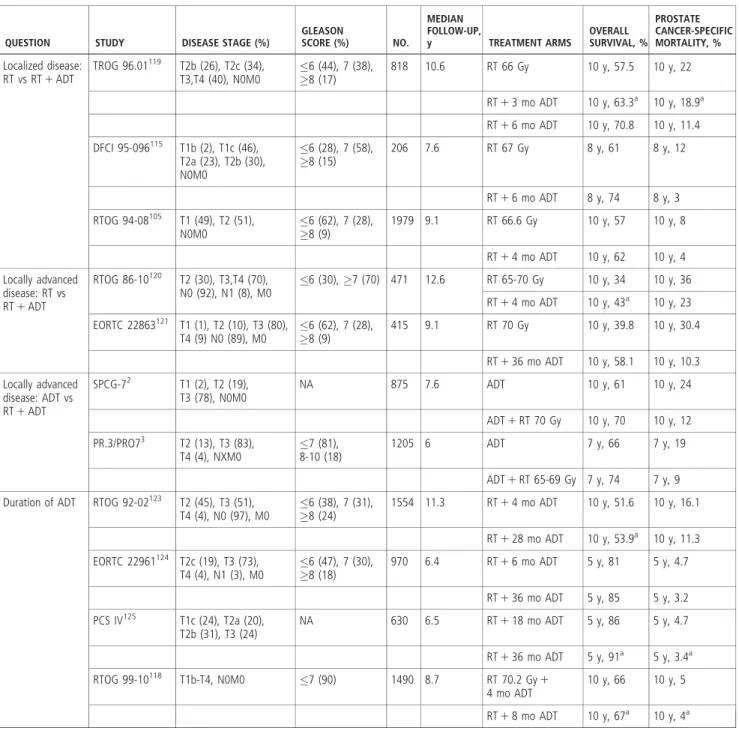
As shown in Table 4, several randomized studies have investigated the role of ADT with radiation for men with intermediate-risk to high-risk, localized prostate cancer rel-ative to standard dose radiation alone.105,115,118,119 These studies have used between 3 and 8 months of androgen deprivation and have demonstrated an advantage to the addition of hormonal therapy in both overall and prostate cancer-specific survival.
The Trans Tasman Radiation Oncology Group (TROG) performed a 3-arm randomized trial of radiation alone, radi-ation plus 3 months of androgen suppression, or radiradi-ation plus 6 months of androgen suppression for 818 men with T2b to T4 disease.119At a median follow-up of 10.6 years,
men on the 6-month arm had significant improvements in disease-free, metastasis-free, prostate-cancer specific, and overall survival relative to those treated with radiation alone. In a study from the Dana-Farber Cancer Institute, 206 men with clinical T1/T2 disease were randomized to radiation alone versus radiation and 6 months of combined androgen blockade.115The median PSA level in that trial was 11 ng/ mL, and 73% of men had Gleason score 7 or higher disease. With a median of 7.6 years of follow-up, men in the com-bined androgen blockade arm had longer overall and cancer-specific survival. In a post-hoc analysis, it appeared that the survival benefit was limited to only those men who had no or minimal comorbid illness. The RTOG 94-08 study randomized 1979 men with predominantly intermediate-risk disease to radiation alone versus radiation with 4 months of combined androgen blockade.105The 10-year overall sur-vival rate in that study was 62% in the androgen suppression arm compared with 57% in the radiation alone arm. In a post-hoc analysis, the 54% of men with intermediate-risk disease derived the greatest benefit, and the 35% in the low-risk group showed no significant benefit from the addition of hormonal therapy.
ADT and locally advanced disease
For men who have more extensive disease within the pros-tate, the role of hormonal therapy has been established over several decades. The RTOG 86-10 study randomized 456 men with large (25 cm3), stage T2b to T4, Nx tumors to either 4 months of total androgen suppression with radia-tion given after the first 2 months or radiaradia-tion alone.120 Those who received concurrent ADT had improved local control, disease-free survival, and freedom from metastases compared with patients who received 65 to 70 Gy of radia-tion alone. With a median follow-up of 12 years, the 10-year overall survival rates were no different between the 2 arms, but there was a significant improvement in disease-specific mortality in the concurrent ADT arm (36% vs 23%; P5.01). A randomized trial from the European Organiza-tion for Research in the Treatment of Cancer (EORTC) demonstrated an overall survival advantage for patients who received 3 years of goserelin starting with radiation versus radiation to 70 Gy alone.121Greater than 90% of the 415 patients had either T3 or T4 disease, and the remaining patients were eligible to participate because of high-grade tumors. With 9.1 years of follow-up, the addition of ADT was associated with significant improvements in both over-all survival and cause-specific survival.
Important to the question of whether local disease con-trol matters, 2 randomized trials investigated the question of whether the addition of radiation improved outcomes for men with locally advanced, localized disease receiving life-long androgen suppression.2,3In the study from Scandina-via, 875 men with a PSA level <70 ng/mL and N0, M0,
and mostly T3 disease were randomized to 3 months of combined androgen blockade followed by lifelong fluta-mide with or without 70 Gy of radiation.2With a median of 7.6 years of follow-up, the relative risk of cancer-specific death was 0.44 (95% CI, 0.30-0.66;P<.001) favoring the addition of radiation. Similar in design, the second study randomized 1205 men with mostly T3 and T4 disease to lifelong androgen suppression with or without 65 to 69 Gy
of radiation.3With a median of 6 years of follow -up, the hazard ratio was 0.54 (95% CI, 0.27-0.78) for cause-specific survival, and the 7-year overall survival rate was 74% for the combined arm versus 66% for the ADT alone arm.
Duration of ADT
There is continued debate regarding the optimal type, tim-ing, and duration of ADT in combination with radiation. TABLE 4. Randomized Trials of Androgen-Deprivation Therapy With Radiation
QUESTION STUDY DISEASE STAGE (%)
GLEASON SCORE (%) NO. MEDIAN FOLLOW-UP, y TREATMENT ARMS OVERALL SURVIVAL, % PROSTATE CANCER-SPECIFIC MORTALITY, % Localized disease: RT vs RT1ADT TROG 96.01119 T2b (26), T2c (34), T3,T4 (40), N0M0 6 (44), 7 (38), 8 (17) 818 10.6 RT 66 Gy 10 y, 57.5 10 y, 22 RT13 mo ADT 10 y, 63.3a 10 y, 18.9a RT16 mo ADT 10 y, 70.8 10 y, 11.4 DFCI 95-096115 T1b (2), T1c (46), T2a (23), T2b (30), N0M0 6 (28), 7 (58), 8 (15) 206 7.6 RT 67 Gy 8 y, 61 8 y, 12 RT16 mo ADT 8 y, 74 8 y, 3 RTOG 94-08105 T1 (49), T2 (51), N0M0 6 (62), 7 (28), 8 (9) 1979 9.1 RT 66.6 Gy 10 y, 57 10 y, 8 RT14 mo ADT 10 y, 62 10 y, 4 Locally advanced disease: RT vs RT1ADT RTOG 86-10120 T2 (30), T3,T4 (70), N0 (92), N1 (8), M0 6 (30),7 (70) 471 12.6 RT 65-70 Gy 10 y, 34 10 y, 36 RT14 mo ADT 10 y, 43a 10 y, 23 EORTC 22863121 T1 (1), T2 (10), T3 (80), T4 (9) N0 (89), M0 6 (62), 7 (28), 8 (9) 415 9.1 RT 70 Gy 10 y, 39.8 10 y, 30.4 RT136 mo ADT 10 y, 58.1 10 y, 10.3 Locally advanced disease: ADT vs RT1ADT SPCG-72 T1 (2), T2 (19), T3 (78), N0M0 NA 875 7.6 ADT 10 y, 61 10 y, 24 ADT1RT 70 Gy 10 y, 70 10 y, 12 PR.3/PRO73 T2 (13), T3 (83), T4 (4), NXM0 7 (81), 8-10 (18) 1205 6 ADT 7 y, 66 7 y, 19 ADT1RT 65-69 Gy 7 y, 74 7 y, 9 Duration of ADT RTOG 92-02123 T2 (45), T3 (51),
T4 (4), N0 (97), M0 6 (38), 7 (31), 8 (24) 1554 11.3 RT14 mo ADT 10 y, 51.6 10 y, 16.1 RT128 mo ADT 10 y, 53.9a 10 y, 11.3 EORTC 22961124 T2c (19), T3 (73), T4 (4), N1 (3), M0 6 (47), 7 (30), 8 (18) 970 6.4 RT16 mo ADT 5 y, 81 5 y, 4.7 RT136 mo ADT 5 y, 85 5 y, 3.2 PCS IV125 T1c (24), T2a (20), T2b (31), T3 (24) NA 630 6.5 RT118 mo ADT 5 y, 86 5 y, 4.7 RT136 mo ADT 5 y, 91a 5 y, 3.4a RTOG 99-10118 T1b-T4, N0M0 7 (90) 1490 8.7 RT 70.2 Gy1 4 mo ADT 10 y, 66 10 y, 5 RT18 mo ADT 10 y, 67a 10 y, 4a ADT indicates androgen-deprivation therapy; DFCI, Dana-Farber Cancer Institute; EORTC, European Organization for Research and Treatment of Cancer; Gy, grays; NA, not available; PCS, Prostate Cancer Study; RT, radiation therapy; RTOG, Radiation Therapy Oncology Group; SPCG, Scandinavian Prostate Cancer Group; TROG, Trans Tasman Radiation Oncology Group.aNot statistically significant.
The RTOG 99-10 study enrolled 1489 men with intermediate-risk disease to ADT with radiation after either 8 weeks or 28 weeks of neoadjuvant ADT.118With approximately 9 years of follow-up, the biochemical recurrence-free, disease-specific, and overall survival rates were no different between the 2 arms. These data suggest that, for intermediate-risk disease, 4 months of ADT are sufficient, although the study was not powered to show noninferiority for men with NCCN intermediate-risk dis-ease. Given retrospective data suggesting that men with favorable intermediate-risk disease may not benefit from ADT at all35,36and a meta-analysis122showing the poten-tial advantage of 6 months over 4 months for men with intermediate-risk disease, there is continued work to define subgroups in which more than 4 months of ADT will be necessary. Although we currently lack randomized data showing an advantage to anything more than 4 months of ADT for men with intermediate-risk disease, based on the level I evidence from randomized trials, both 4 months and 6 months of ADT should remain options for men with unfavorable intermediate–risk disease.105,115
Additional studies have investigated the optimal dura-tion of ADT among men with higher risk disease. In RTOG 92-02, 1521 patients with stage T2c to T4, N0 or N1, M0 disease were randomly assigned to either 4 months of total androgen suppression with radiation administered after 2 months or the same regimen followed by an addi-tional 2 years of goserelin.123 In patients who had tumors with Gleason scores from 8 to 10, there were significant improvements in overall survival and disease-specific sur-vival with the addition of the longer course ADT. In a sim-ilar study, the EORTC randomized 970 men with mostly T3 tumors to 6 months of combined androgen blockade and radiation to receive no further ADT versus 30 months of a luteinizing hormone-releasing hormone analog.124 That study was powered as a noninferiority study; but, at 6.4 years of follow- up, the 5-year cause specific mortality rate was 4.7% (95% CI, 2.7%-6.7%) versus 3.2% (95% CI, 1.6%-4.8%) favoring the 36 months of ADT.
A study from Quebec investigated radiation combined with 18 months of goserelin relative to radiation with 36 months of goserelin for men with high-risk disease.125 With 6.5 years of follow-up and only 147 deaths, the 5-year overall and cause-specific survival rates were not statistically different between the 2 arms. While the idea of using the shorter course is appealing given the morbidity of ADT and the upper end of the 95% CI of 1.59 on the point estimate for death (1.15), additional follow-up will be needed to determine whether 18 months are noninferior to 36 months.
While the addition of concurrent ADT with radiation has been associated with improved disease control and overall survival for men with intermediate-risk and high-risk disease,
numerous questions remain regarding the duration of ther-apy. With the advent of newer agents targeting the androgen axis,9,10 there are currently randomized trials investigating the integration of these therapies with radiation therapy for localized disease, including RTOG 11-15 (NCT01546987) and an International study (Australian New Zealand Clinical Trials Registry [ACTRN] 12614000126617). Ulti-mately, these studies may lead to optimized use of these agents, resulting in improved outcomes and decreased ADT-related morbidity.
Treatment of the Pelvic Lymph Nodes
Another area of debate in the current management of local-ized prostate cancer with external beam radiation is the question of whether at-risk regional lymph nodes should be treated. Several trials have investigated this question start-ing with RTOG 77-06, which randomized men with low-risk, lymph node-negative prostate cancer to radiation of the prostate and pelvis versus prostate alone.126 After a median follow-up of 7 years, that study failed to identify any advantage to the addition of the pelvic field for these lower risk men. RTOG 94-13 investigated this question again in a 4-arm study comparing whole-pelvis versus small-field radiation with 4 months of either neoadjuvant or adjuvant total androgen suppression. Eligible patients had adverse risk factors, such as high Gleason scores or ele-vated PSA levels, with an estimated risk of lymph node-positive disease greater than 15%. Initially, a difference was observed in progression-free survival at 4 years (54% vs 47% for whole pelvis vs prostate-only radiation, respec-tively;P5.022), but no difference was observed in overall survival.127An update of this study found that this benefit in progression-free survival did not persist based on the protocol definition or the more modern nadir plus 2 ng/mL definition.128A French study (European Urogenital Tumor Study Group [GETUG]-01) randomized 444 men to receive radiation either to the prostate or to the prostate and pelvis; and, with a median follow-up of 3.5 years, no significant improvement in disease control was reported with the addition of treatment to the pelvis.129In light of the results from these 3 randomized trials, the role of pelvic radiation in prostate cancer remains uncertain. Ongoing studies are addressing whether subgroups within unfavora-ble intermediate or high risk may benefit.
Postprostatectomy Radiation
An area of active investigation in prostate cancer manage-ment is the role of postprostatectomy radiation, including radiation to the prostate bed without evidence of PSA elevation, adjuvant radiation, and treatment only after evi-dence that the PSA is rising (“salvage” radiation). There are now 10-year results from 3 randomized trials investigating
the role of adjuvant radiation in men at high risk for recur-rence after radical prostatectomy.130-132 The inclusion cri-teria were similar for all 3 trials: men harboring 1 or more high-risk features of disease recurrence after prostatectomy (extracapsular extension, seminal vesicle invasion, or a positive margin) were randomized to either adjuvant radiation to the prostate bed or observation. The Southwest Oncology Group (SWOG) randomized 473 men,130 the EORTC 22911 trial randomized 1005 men,131 and the German ARO 96-02 trial randomized 385 men132; and, with at least 10 years of follow-up, all 3 showed a halving of the risk of biochemical failure with the addition of adju-vant radiation. At the most recent update, the SWOG study showed an improvement in overall survival of 48% versus 57% (P5.02) and a 29% reduced risk of developing metastatic disease. No metastasis or overall survival advant-age to the addition of adjuvant therapy was demonstrated by the other 2 trials. It is not immediately obvious why only 1 of the 3 found a survival and metastasis-free survival advantage. Differences in follow-up between the SWOG and EORTC studies may have led to ascertainment bias, although the median PSA at the time of salvage radiation in the SWOG study was lower than in the EORTC trial.131Another explanation may be that men in the obser-vation arm of the SWOG study, on average, were older and appeared to have higher rates of Gleason 8 to 10 disease.
In light of the observation of the potential benefit to initiating salvage radiation at a PSA <0.5 ng/mL from numerous retrospective studies,133 there are now several randomized trials investigating the role of adjuvant radia-tion versus early salvage, including the Radiaradia-tion Therapy and Androgen Deprivation Therapy in Treating Patients Who Have Undergone Surgery for Prostate Cancer (RAD-ICALS) (NCT00541047) trial and the Radiotherapy Adjuvant Versus Early Salvage (RAVES) (NCT00860652) trial. In these studies, early salvage radiation is defined as the initiation of treatment within 2 to 4 months of PSA failure. While we await the results from these studies, the American Society for Radiation Therapy and the American Urologic Association have issued a joint recommendation that adjuvant radiation should be offered to men with high-risk features after prostatectomy.134Before the issu-ance of these guidelines, the use of adjuvant radiation for men with higher risk features was very low.135Although it would need to be validated, a risk-adjusted approach could be considered in which radiation would be reserved for those at highest risk for a PSA recurrence.136
The role of ADT in the salvage setting is also under evalu-ation. The RTOG 96-01 study has reported preliminary results showing lower rates of metastatic disease among men with a rising PSA postprostatectomy who were random-ized to receive 2 years of bicalutamide 150 mg daily plus radiation relative to those who received radiation alone.137
The morbidity of bicalutamide was significant, with 89% of men developing grade 1 or 2 gynecomastia. Several of the adjuvant versus early salvage studies, including RADI-CALS, are investigating the role of ADT in the postpros-tatectomy stetting. Despite these advances, there is currently uncertainty regarding who to treat and whether to include ADT in both the salvage and adjuvant setting. The results of randomized studies, both now underway and completed, should provide additional guidance for this common situation.
Value-Based Health Care and Prostate Cancer While randomized trials have shown the advantage of treatment over observation for some men with prostate can-cer,138,139 there remains wide variation in how men with localized prostate cancer are treated.12 This variation appears to be as dependent on the treating center as it is on disease risk group and likely reflects potentially disparate treatment recommendations from experts140 and anxiety around strategies like active surveillance.141,142Patients are asked to decide between treatment options in the absence of randomized trials comparing treatment modalities or established measures of quality between treating centers.143
It is within this context that costly newer treatments can become popular.4,144 In the face of unsustainable increases in the costs of care, there is a recognition that aligning patients, providers, and payers around value can have a pro-found impact on how care is delivered and paid for.145 Defined as a patient’s outcomes divided by the cost to achieve those outcomes, value has been proposed as a unify-ing force to improve the quality of care.146 For prostate cancer, it is established that disease control, complications of treatment, and long-term quality of life are important in individual decision making.147 Relevant outcomes have been established for a variety of treatment approaches21; but, with the exception of selected institutional series,64,75 they are seldom collected or disseminated to patients. This problem is particularly significant in the area of patient-reported outcome measures, for which many validated instruments are available in the domains of urinary inconti-nence, urinary obstruction, bowel irritation, and sexual dys-function.21,148Because physicians frequently underestimate health-related quality of life in patients with prostate can-cer,149patient reported outcomes have been widely imple-mented.21,150Efforts to integrate both established disease control measures and other patient outcomes are taking place within registries,151,152but we do not yet have global standards on which measures should be included.
The systematic collection of outcome data has been shown to improve outcomes within an institution. For example, before initiating a study of MRI-guided brachy-therapy, 2 institutions collected patient-reported outcomes
for patients treated with standard ultrasound-guided brachytherapy.153 Patients from 1 of the centers reported worse urinary function after the procedure than patients from the other center. After implementation of best prac-tices, there was improvement in urinary symptoms after brachytherapy. Collecting standardized outcomes can also better inform which dosimetric parameters are associated with toxicity.62,63This approach will never replace random-ized trials, but it can rapidly disseminate improvements in practice, assist with shared decision making, and potentially drive improvements in value.
Conclusions and Future Research Directions Recent years have seen dramatic advances in the treatment of prostate cancer with radiation. Not only can we now match patients to appropriate treatments better than ever before, but we have also refined treatment options, in par-ticular around the use and duration of concurrent hormonal therapy. For men with low-risk and favorable intermediate-risk prostate cancer who favor treatment over active surveil-lance and wish to receive radiation, both brachytherapy and external beam radiation are options. Decisions between the 2 should be made based on patient factors and the antici-pated differences in short-term and long-term toxicities (ie, men with significant baseline urinary obstructive symptoms are likely to better tolerate external beam radiation). For men with unfavorable intermediate-risk prostate cancer who elect to have radiation, evidence suggests that 4 to 6 months of ADT should be added. For men with high-risk disease, the exact duration of ADT is not yet established but can range from 28 to 36 months and possibly can be as little as 18 months.
The technology for treatment delivery has also pro-gressed rapidly, resulting in dramatically more conformal treatments and increasingly shorter treatment times. In addition to evaluation of disease control and patient outcome measures, the implications of these advancements are increasingly assessed through the lens of cost. The com-ing years should brcom-ing results from hypofractionation trials to guide the widespread adoption of this potentially more convenient treatment approach. With the recently expanded availability of molecular and genetic tests of localized and resected prostate cancer, there should be fur-ther significant refinements in predicting the aggressiveness of disease in the near future. There is hope that these tests will more accurately identify men who can be safely fol-lowed with active surveillance as well as those best suited for more aggressive combined-modality therapy. Given the uncertainty regarding adjuvant and salvage radiation after prostatectomy, there is a pressing need to identify those men who will most benefit from radiation with or without hormonal therapy in these settings.
The future of prostate radiation may be foreshadowed by the manner in which breast cancer is approached today: increasingly guided by genetic predictors of recurrence and treated with shorter hypofractionated courses of radiation. That the treatment of these 2 common cancers is largely following similar paths is perhaps unsurprising, but prostate radiation continues to face significant questions regarding the optimal approach to deciding between a variety of effective options. Absent large, costly randomized trials, the routine collection and reporting of important patient outcomes through local or national quality registries should help our patients make informed decisions.䊏
References
1. Cuzick J, Swanson GP, Fisher G, et al. Prognostic value of an RNA expression sig-nature derived from cell cycle proliferation genes in patients with prostate cancer: a retrospective study. Lancet Oncol. 2011; 12:245-255.
2. Widmark A, Klepp O, Solberg A, et al. Endocrine treatment, with or without radiotherapy, in locally advanced prostate cancer (SPCG-7/SFUO-3): an open rando-mised phase III trial. Lancet. 2009;373: 301-308.
3. Warde P, Mason M, Ding K, et al. Com-bined androgen deprivation therapy and radiation therapy for locally advanced prostate cancer: a randomised, phase 3 trial.Lancet. 2011;378:2104-2111. 4. Nguyen PL, Gu X, Lipsitz SR, et al. Cost
implications of the rapid adoption of newer technologies for treating prostate cancer.J Clin Oncol. 2011;29:1517-1524. 5. Sheets NC, Goldin GH, Meyer AM, et al.
Intensity-modulated radiation therapy, proton therapy, or conformal radiation
therapy and morbidity and disease control in localized prostate cancer.JAMA. 2012; 307:1611-1620.
6. Alhassani A, Chandra A, Chernew ME. The sources of the SGR “hole.”N Engl J Med.2012;366:289-291.
7. Mitchell JM. Urologists’ use of intensity-modulated radiation therapy for prostate cancer.N Engl J Med. 2013;369:1629-1637. 8. Institute of Medicine (US). Committee on
Comparative Effectiveness Research Prioriti-zation. Initial National Priorities for Compar-ative Effectiveness Research. Washington, DC: National Academies Press; 2009. 9. Scher HI, Fizazi K, Saad F, et al. Increased
survival with enzalutamide in prostate cancer after chemotherapy.N Engl J Med. 2012;367:1187-1197.
10. de Bono JS, Logothetis CJ, Molina A, et al. Abiraterone and increased survival in met-astatic prostate cancer. N Engl J Med. 2011;364:1995-2005.
11. Tannock IF, de Wit R, Berry WR, et al. Docetaxel plus prednisone or mitoxan-trone plus prednisone for advanced
pros-tate cancer.N Engl J Med. 2004;351:1502-1512.
12. Cooperberg MR, Broering JM, Carroll PR. Time trends and local variation in primary treatment of localized prostate cancer.
J Clin Oncol. 2010;28:1117-1123. 13. Cooperberg MR, Vickers AJ, Broering JM,
Carroll PR. Comparative risk-adjusted mortality outcomes after primary surgery, radiotherapy, or androgen-deprivation therapy for localized prostate cancer. Can-cer. 2010;116:5226-5234.
14. Kibel AS, Ciezki JP, Klein EA, et al. Sur-vival among men with clinically localized prostate cancer treated with radical prosta-tectomy or radiation therapy in the pros-tate specific antigen era.J Urol. 2012;187: 1259-1265.
15. Zelefsky MJ, Eastham JA, Cronin AM, et al. Metastasis after radical prostatec-tomy or external beam radiotherapy for patients with clinically localized prostate cancer: a comparison of clinical cohorts adjusted for case mix.J Clin Oncol. 2010; 28:1508-1513.