Crystal structure of
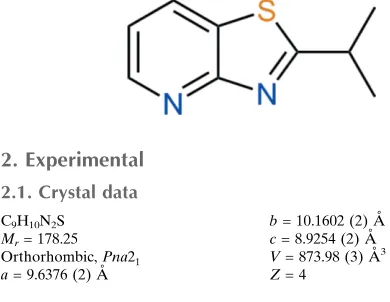
2-(1-methylethyl)-1,3-thiazolo[4,5-
b
]pyridine
Gamal A. El-Hiti,a* Keith Smith,bAmany S. Hegazy,b Saud A. Alanaziaand Benson M. Kariukib‡
aCornea Research Chair, Department of Optometry, College of Applied Medical,
Sciences, King Saud University, PO Box 10219, Riyadh 11433, Saudi Arabia, and
bSchool of Chemistry, Cardiff University, Main Building, Park Place, Cardiff
CF10 3AT, Wales. *Correspondence e-mail: [email protected]
Received 5 March 2015; accepted 25 March 2015
Edited by G. Smith, Queensland University of Technology, Australia
In the title molecule, C9H10N2S, one of the methyl groups is
almost co-planar with the thiazolopyridine rings with a deviation of 0.311 (3) A˚ from the least-squares plane of the thiazolopyridine group. In the crystal, weak C—H N hydrogen-bonding interactions lead to the formation of chains along [011].
Keywords:crystal structure; thiazolopyridine; hydrogen bonding.
CCDC reference:1056012
1. Related literature
For related compounds, see: Smithet al.(1994, 1995); El-Hiti (2003); Johnsonet al.(2006); Thomaeet al.(2008); Raoet al. (2009); Leeet al.(2010); Luoet al.(2015). For the X-ray crystal structures of related compounds, see: Yuet al.(2007); El-Hiti et al.(2014).
2. Experimental
2.1. Crystal data
C9H10N2S Mr= 178.25
Orthorhombic,Pna21 a= 9.6376 (2) A˚
b= 10.1602 (2) A˚
c= 8.9254 (2) A˚
V= 873.98 (3) A˚3
Z= 4
CuKradiation = 2.81 mm1
T= 150 K
0.230.200.14 mm
2.2. Data collection
Agilent SuperNova, Dual, Cu at zero, Atlas diffractometer Absorption correction: multi-scan
(CrysAlis PRO; Agilent, 2014)
Tmin= 0.897,Tmax= 0.940
2848 measured reflections 1366 independent reflections 1351 reflections withI> 2(I)
Rint= 0.012
2.3. Refinement
R[F2> 2(F2)] = 0.021 wR(F2) = 0.057 S= 1.08 1366 reflections 111 parameters
1 restraint
H-atom parameters constrained
max= 0.19 e A˚
3
min=0.20 e A˚
[image:1.610.45.240.580.731.2]3
Table 1
Hydrogen-bond geometry (A˚ ,).
D—H A D—H H A D A D—H A
C4—H4 N1i
0.95 2.51 3.391 (2) 153
Symmetry code: (i)xþ1 2;yþ
1 2;zþ
1 2.
Data collection: CrysAlis PRO (Agilent, 2014); cell refinement: CrysAlis PRO; data reduction: CrysAlis PRO; program(s) used to solve structure:SHELXS2013(Sheldrick, 2008); program(s) used to refine structure:SHELXL2013(Sheldrick, 2015); molecular graphics: ORTEP-3 for Windows(Farrugia, 2012); software used to prepare material for publication:WinGX(Farrugia, 2012) andCHEMDRAW Ultra (Cambridge Soft, 2001).
Acknowledgements
The authors extend their appreciation to the Cornea Research Chair, Department of Optometry, College of Applied Medical Sciences, King Saud University, for funding this research and to Cardiff University for continued support.
Supporting information for this paper is available from the IUCr electronic archives (Reference: ZS2329).
References
Agilent (2014).CrysAlis PRO. Agilent Technologies, Yarnton, England. Cambridge Soft (2001).CHEMDRAW Ultra. Cambridge Soft Corporation,
Cambridge, Massachusetts, USA.
El-Hiti, G. A. (2003).Monatsh. Chem.134, 837–841.
El-Hiti, G. A., Smith, K., Hegazy, A. S., Masmali, A. M. & Kariuki, B. M. (2014).Acta Cryst.E70, o932.
Farrugia, L. J. (2012).J. Appl. Cryst.45, 849–854.
Johnson, S. G., Connolly, P. J. & Murray, W. V. (2006).Tetrahedron Lett.47, 4853–4856.
Lee, T., Lee, D., Lee, I. Y. & Gong, Y.-D. (2010).J. Comb. Chem.12, 95–99. Luo, L., Meng, L., Peng, Y., Xing, Y., Sun, Q., Ge, Z. & Li, R. (2015).Eur. J.
Org. Chem.pp. 631–637.
Rao, A. U., Palani, A., Chen, X., Huang, Y., Aslanian, R. G., West, R. E. Jr, Williams, S. M., Wu, R.-L., Hwa, J., Sondey, C. & Lachowicz, J. (2009).
Bioorg. Med. Chem. Lett.19, 6176–6180. Sheldrick, G. M. (2008).Acta Cryst.A64, 112–122. Sheldrick, G. M. (2015).Acta Cryst.C71, 3–8.
Smith, K., Anderson, D. & Matthews, I. (1995).Sulfur Lett.18, 79–95.
data reports
o272
El-Hitiet al. doi:10.1107/S2056989015006039 Acta Cryst.(2015).E71, o272–o273ISSN 2056-9890
Smith, K., Lindsay, C. M., Morris, I. K., Matthews, I. & Pritchard, G. J. (1994).
Sulfur Lett.17, 197–216.
Thomae, D., Perspicace, E., Hesse, S., Kirsch, G. & Seck, P. (2008).
Tetrahedron,64, 9309–9314.
supporting information
sup-1 Acta Cryst. (2015). E71, o272–o273
supporting information
Acta Cryst. (2015). E71, o272–o273 [https://doi.org/10.1107/S2056989015006039]
Crystal structure of 2-(1-methylethyl)-1,3-thiazolo[4,5-
b
]pyridine
Gamal A. El-Hiti, Keith Smith, Amany S. Hegazy, Saud A. Alanazi and Benson M. Kariuki
S1. Chemical context
Thiazolopyridines have been efficiently synthesized and in high yield using different synthetic procedures (Smith et al.,
1994, 1995; El-Hiti, 2003; Johnson et al., 2006; Thomae et al., 2008; Rao et al., 2009; Lee et al., 2010; Luo et al., 2015).
During our continuing research towards the development of novel synthetic routes for the production of heterocyclic
compounds, we have synthesised the title compound 2-(methylethyl)-1,3-thiazolo[4,5-b]pyridine in high yield (Smith et
al., 1995). The X-ray structures for related compounds have been reported (Yu et al., 2007; El-Hiti et al., 2014).
S2. Structural commentary
The asymmetric unit of the title compound consists of a single molecule of C9H10N2S (Fig. 1). In the molecule, one of the
methyl groups is almost co-planar with the thiazolopyridine ring. The deviations from the least-squares plane of the
thia-zolopyridine group are 0.311 (3)Å and 1.269 (3)Å for C8 and C9 respectively, corresponding to torsion angles N1—C1—
C7—C8 and N1—C1—C7—C9 of 169.47 (19) and -65.9 (3)°, respectively.
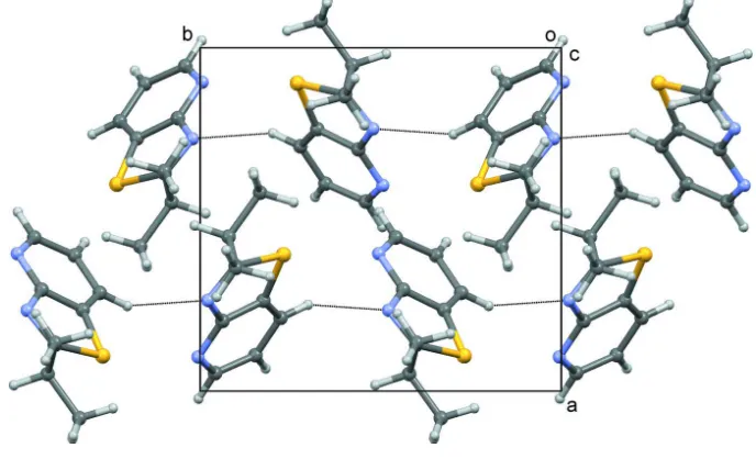
Weak C—H···N hydrogen-bonding interactions occur in the structure to form chains along [011] (Fig. 2, Table 1). No π–
π interactions are observed in the crystal structure.
S3. Synthesis and crystallization
2-(1-Methylethyl)-1,3-thiazolo[4,5-b]pyridine was obtained in 98% yield from acid hydrolysis (HCl, 5 M) of
3-(diiso-propylaminothiocarbonylthio)-2-(1-methylethylcarbonylamino)pyridine under reflux for 5 h (Smith et al., 1995).
Crystallization of the crude product from diethyl ether gave colourless crystals of the title compound. The spectroscopic
and analytical data for the title compound were consistent with those reported previously (Smith et al., 1995).
S4. Refinement details
H atoms were positioned geometrically and refined using a riding model with Uiso(H) constrained to be 1.2 times Ueq for
the atom it is bonded to except for methyl groups where it was 1.5 times with free rotation about the C—C bond.
Although not of relevance with this achiral compound, the absolute structure factor (Flack, 1983) was determined as
Figure 1
A molecule of C9H10N2S with atom labels and 50% probability displacement ellipsoids for non-hydrogen atoms.
Figure 2
Crystal structure packing viewed down the c axis with C—H···N interactions shown as dotted lines.
2-(1-Methylethyl)-1,3-thiazolo[4,5-b]pyridine
Crystal data
C9H10N2S
Mr = 178.25
Orthorhombic, Pna21
a = 9.6376 (2) Å
b = 10.1602 (2) Å
c = 8.9254 (2) Å
V = 873.98 (3) Å3
Z = 4
F(000) = 376
Dx = 1.355 Mg m−3
Cu Kα radiation, λ = 1.54184 Å
Cell parameters from 2276 reflections
θ = 6.3–73.8°
[image:4.610.132.476.341.550.2]supporting information
sup-3 Acta Cryst. (2015). E71, o272–o273
T = 150 K
Block, colourless
0.23 × 0.20 × 0.14 mm
Data collection
Agilent SuperNova, Dual, Cu at zero, Atlas diffractometer
Radiation source: sealed X-ray tube, SuperNova (Cu) X-ray Source
Mirror monochromator
Detector resolution: 10.5082 pixels mm-1
ω scans
Absorption correction: multi-scan (CrysAlis PRO; Agilent, 2014)
Tmin = 0.897, Tmax = 0.940
2848 measured reflections 1366 independent reflections 1351 reflections with I > 2σ(I)
Rint = 0.012
θmax = 74.0°, θmin = 6.3°
h = −11→10
k = −12→8
l = −10→10
Refinement
Refinement on F2
Least-squares matrix: full
R[F2 > 2σ(F2)] = 0.021
wR(F2) = 0.057
S = 1.08
1366 reflections 111 parameters 1 restraint
Hydrogen site location: inferred from neighbouring sites
H-atom parameters constrained
w = 1/[σ2(F
o2) + (0.0375P)2 + 0.1081P]
where P = (Fo2 + 2Fc2)/3
(Δ/σ)max = 0.001
Δρmax = 0.19 e Å−3
Δρmin = −0.20 e Å−3
Special details
Geometry. All e.s.d.'s (except the e.s.d. in the dihedral angle between two l.s. planes) are estimated using the full covariance matrix. The cell e.s.d.'s are taken into account individually in the estimation of e.s.d.'s in distances, angles and torsion angles; correlations between e.s.d.'s in cell parameters are only used when they are defined by crystal symmetry. An approximate (isotropic) treatment of cell e.s.d.'s is used for estimating e.s.d.'s involving l.s. planes.
Fractional atomic coordinates and isotropic or equivalent isotropic displacement parameters (Å2)
x y z Uiso*/Ueq
C1 0.13501 (18) 0.59856 (18) 0.8660 (2) 0.0227 (4)
C2 0.23657 (17) 0.67547 (17) 1.1015 (2) 0.0208 (3)
C3 0.29550 (18) 0.56187 (17) 1.0365 (2) 0.0203 (3)
C4 0.2865 (2) 0.72261 (17) 1.2365 (3) 0.0257 (4)
H4 0.2483 0.7985 1.2829 0.031*
C5 0.39534 (19) 0.6532 (2) 1.3007 (3) 0.0279 (4)
H5 0.4342 0.6813 1.3931 0.034*
C6 0.44760 (19) 0.54145 (19) 1.2283 (3) 0.0273 (4)
H6 0.5220 0.4959 1.2752 0.033*
C7 0.05003 (18) 0.57871 (19) 0.7259 (3) 0.0271 (4)
H7 0.0177 0.4852 0.7251 0.033*
C8 −0.0783 (2) 0.6663 (2) 0.7204 (3) 0.0365 (5)
H8A −0.1375 0.6475 0.8070 0.055*
H8B −0.1299 0.6488 0.6279 0.055*
H8C −0.0499 0.7589 0.7227 0.055*
C9 0.1411 (2) 0.5992 (3) 0.5875 (3) 0.0421 (6)
H9A 0.1666 0.6923 0.5796 0.063*
H9B 0.0897 0.5727 0.4977 0.063*
N1 0.23563 (16) 0.52156 (16) 0.9037 (2) 0.0241 (3)
N2 0.40055 (14) 0.49434 (16) 1.0985 (2) 0.0254 (4)
S1 0.10273 (4) 0.73079 (4) 0.98822 (7) 0.02515 (14)
Atomic displacement parameters (Å2)
U11 U22 U33 U12 U13 U23
C1 0.0205 (8) 0.0253 (8) 0.0224 (10) −0.0013 (7) 0.0032 (7) −0.0026 (8)
C2 0.0212 (8) 0.0210 (7) 0.0203 (9) −0.0011 (6) 0.0033 (6) −0.0004 (7)
C3 0.0199 (7) 0.0206 (7) 0.0204 (9) −0.0026 (6) 0.0032 (6) −0.0024 (7)
C4 0.0292 (9) 0.0258 (9) 0.0221 (9) −0.0028 (7) 0.0018 (8) −0.0061 (8)
C5 0.0302 (9) 0.0353 (11) 0.0184 (9) −0.0058 (7) −0.0024 (7) 0.0002 (9)
C6 0.0258 (8) 0.0303 (9) 0.0259 (9) 0.0005 (7) −0.0035 (8) 0.0042 (8)
C7 0.0266 (8) 0.0293 (9) 0.0254 (9) −0.0036 (7) −0.0032 (8) −0.0030 (8)
C8 0.0293 (9) 0.0428 (12) 0.0374 (13) 0.0037 (9) −0.0127 (9) −0.0071 (11)
C9 0.0351 (11) 0.0705 (16) 0.0207 (11) −0.0072 (11) −0.0024 (8) −0.0025 (11)
N1 0.0231 (7) 0.0264 (8) 0.0229 (8) 0.0012 (6) 0.0001 (6) −0.0067 (7)
N2 0.0238 (8) 0.0239 (7) 0.0284 (9) 0.0024 (6) −0.0016 (6) −0.0002 (7)
S1 0.0242 (2) 0.0266 (2) 0.0246 (2) 0.00703 (13) −0.0007 (2) −0.0048 (2)
Geometric parameters (Å, º)
C1—N1 1.291 (2) C6—N2 1.334 (3)
C1—C7 1.508 (3) C6—H6 0.9500
C1—S1 1.7585 (19) C7—C8 1.524 (3)
C2—C4 1.383 (3) C7—C9 1.530 (3)
C2—C3 1.411 (2) C7—H7 1.0000
C2—S1 1.7325 (19) C8—H8A 0.9800
C3—N2 1.342 (2) C8—H8B 0.9800
C3—N1 1.380 (2) C8—H8C 0.9800
C4—C5 1.388 (3) C9—H9A 0.9800
C4—H4 0.9500 C9—H9B 0.9800
C5—C6 1.400 (3) C9—H9C 0.9800
C5—H5 0.9500
N1—C1—C7 122.91 (17) C8—C7—C9 111.1 (2)
N1—C1—S1 115.73 (15) C1—C7—H7 107.6
C7—C1—S1 121.36 (14) C8—C7—H7 107.6
C4—C2—C3 120.08 (17) C9—C7—H7 107.6
C4—C2—S1 130.92 (14) C7—C8—H8A 109.5
C3—C2—S1 108.98 (14) C7—C8—H8B 109.5
N2—C3—N1 121.16 (16) H8A—C8—H8B 109.5
N2—C3—C2 123.53 (18) C7—C8—H8C 109.5
N1—C3—C2 115.30 (16) H8A—C8—H8C 109.5
C2—C4—C5 116.51 (17) H8B—C8—H8C 109.5
C2—C4—H4 121.7 C7—C9—H9A 109.5
C5—C4—H4 121.7 C7—C9—H9B 109.5
supporting information
sup-5 Acta Cryst. (2015). E71, o272–o273
C4—C5—H5 120.2 C7—C9—H9C 109.5
C6—C5—H5 120.2 H9A—C9—H9C 109.5
N2—C6—C5 124.73 (18) H9B—C9—H9C 109.5
N2—C6—H6 117.6 C1—N1—C3 110.99 (16)
C5—C6—H6 117.6 C6—N2—C3 115.54 (16)
C1—C7—C8 112.90 (18) C2—S1—C1 89.00 (9)
C1—C7—C9 109.85 (15)
C4—C2—C3—N2 −0.3 (3) C7—C1—N1—C3 −179.88 (16)
S1—C2—C3—N2 −179.06 (14) S1—C1—N1—C3 0.6 (2)
C4—C2—C3—N1 178.46 (16) N2—C3—N1—C1 178.62 (17)
S1—C2—C3—N1 −0.3 (2) C2—C3—N1—C1 −0.1 (2)
C3—C2—C4—C5 0.4 (3) C5—C6—N2—C3 0.0 (3)
S1—C2—C4—C5 178.89 (15) N1—C3—N2—C6 −178.61 (17)
C2—C4—C5—C6 −0.4 (3) C2—C3—N2—C6 0.1 (3)
C4—C5—C6—N2 0.2 (3) C4—C2—S1—C1 −178.11 (19)
N1—C1—C7—C8 169.47 (19) C3—C2—S1—C1 0.50 (14)
S1—C1—C7—C8 −11.0 (2) N1—C1—S1—C2 −0.64 (15)
N1—C1—C7—C9 −65.9 (3) C7—C1—S1—C2 179.79 (16)
S1—C1—C7—C9 113.64 (19)
Hydrogen-bond geometry (Å, º)
D—H···A D—H H···A D···A D—H···A
C4—H4···N1i 0.95 2.51 3.391 (2) 153