
Received 24 October 2018 Accepted 18 November 2018
Edited by D.-J. Xu, Zhejiang University (Yuquan Campus), China
Keywords:crystal structure; Schiff bases; 3-chloro-4-methylphenylimino; Hirshfeld surface.
CCDC reference:1873612
Supporting information:this article has supporting information at journals.iucr.org/e
Crystal structure and Hirshfeld surface analysis of
(
E
)-2,4-di-
tert
-butyl-6-[(3-chloro-4-methylphenyl-imino)methyl]phenol
Sevgi Kansiz,aMustafa Macit,b Necmi Degeaand Vadim A. Pavlenkoc*
a
Ondokuz Mayıs University, Faculty of Arts and Sciences, Department of Physics, 55139, Kurupelit, Samsun, Turkey,
b
Department of Chemistry, Faculty of Arts and Sciences, Ondokuz Mayıs, University, 55139, Samsun, Turkey, andcTaras Shevchenko National University of Kyiv, Department of Chemistry, Volodymyrska Str., 64, 01601 Kiev, Ukraine. *Correspondence e-mail: [email protected]
The title Schiff base compound, C22H28ClNO, shows mirror symmetry with all its
non-H atoms, except thetert-butyl groups, located on the mirror plane. There is an intramolecular O—H N hydrogen bond present forming anS(6) ring motif. In the crystal, the molecules are connected by C—H interactions, generating a three-dimensional supramolecular structure. Hirshfeld surface analysis and two dimensional fingerprint plots were used to analyse the intermolecular interactions present in the crystal, indicating that the most important contributions for the crystal packing are from H H (68.9%) and C H/ H C (11.7%) interactions.
1. Chemical context
In coordination chemistry, Schiff bases have found wide use as ligands (Calligariset al., 1972; Ho¨keleket al., 2004; Morozet al., 2012; Kansiz et al., 2018). Schiff bases are important for various areas of chemistry and biochemistry because of their biological activity (El-masry et al., 2000) and photochromic properties and have applications in various fields such as the measurement and control of radiation intensities in imaging systems and optical computers (Elmalı et al., 1999), electro-nics, optoelectronics and photonics (Iwan et al., 2007). They have been used as starting materials in the synthesis of many important medicinal substances. In the present study, a new Schiff base compound was synthesized and its crystal structure determined by X-ray diffraction. In addition, to understand the intermolecular interactions in the crystal structure, Hirshfeld surface analysis was performed.
2. Structural commentary
The molecular structure of the title compound is illustrated in Fig. 1. The title Schiff base compound shows mirror symmetry with all the non-H atoms, except thetert-butyl groups, located
on the mirror plane. The C14—O1 bond distance is 1.349 (3) A˚ , the C8 N1 and C5—N1 bond lengths are 1.278 (4) and 1.412 (4) A˚ , respectively, and the C7—Cl1 bond distance is 1.744 (3) A˚ . There is an intramolecular O—H N hydrogen bond present (Table 1), forming an S(6) ring motif.
3. Supramolecular features
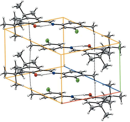
In the crystal, the molecules are connected by C1—H1B interactions, generating a three-dimensional supramolecular structure (Table 1 and Fig. 2).
4. Database survey
There are no previous reports of the title structure. However, several related structure have been reported (CSD, version 5.39, update May 2018; Groomet al., 2016), including bis{(E )- 1-[(3-chloro-4-methylphenylimino)methyl]naphthalen-2-ol-ate-N,O}copper(II) (SICXOU; Topraket al., 2018), 2-{(E )-[(3- chloro-4-methylphenyl)imino]methyl}-4-(trifluoromethoxy)-phenol (TERTUI; Atalay et al., 2017), {2,20
-[4-chloro-5-methyl-o -phenylenebis(nitrilomethylidyne)]diphenolato}nick-el(II) (WABDEK; Wang, 2010) and 4-[(E )-(3-chloro-4-methylphenyl)iminomethyl]-2-methoxy-3-nitrophenyl acetate (GAPPOE; Suet al., 2012). In all four compounds, the C—Cl bond lengths vary from 1.724 to 1.743 A˚ . In the title compound, the C7—Cl1 bond length is 1.744 (3) A˚ .
5. Hirshfeld surface analysis
The Hirshfeld surface analysis was performed using Crystal-Explorer(Turneret al., 2017). The Hirshfeld surfaces and their associated two-dimensional fingerprint plots were used to quantify the various intermolecular interactions in the synthesized complex. The Hirshfeld surfaces mapped over
dnorm,deanddiare illustrated in Figs. 3 and 4. The red spots on
1888
Kansizet al. C22H28ClNO Acta Cryst.(2018). E74, 1887–1890
[image:2.610.312.566.74.131.2]research communications
Table 1
Hydrogen-bond geometry (A˚ ,).
Cgis the centroid of the C9–C14 benzene ring.
D—H A D—H H A D A D—H A
O1—H1 N1 0.82 1.84 2.582 (3) 149 C1—H1B Cg1i 0.96 2.77 3.5072 (4) 134
[image:2.610.44.294.107.148.2]Symmetry code: (i)xþ1;yþ1 2;zþ1.
Figure 2
A view of the crystal packing of the title compound. Dashed lines denote the intramolecular and intermolecular hydrogen bonds.
Figure 3
[image:2.610.46.295.265.420.2]The Hirshfeld surface of the title compound mapped overdnorm,diandde.
Figure 1
The molecular structure of the title compound, showing the atom labelling. Displacement ellipsoids are drawn at the 10% probability level. Symmetry code: (i)x,y+1
[image:2.610.49.302.480.718.2]2,z.
Figure 4
Hirshfeld surface mapped overdnormfor visualizing the intermolecular
[image:2.610.313.563.567.712.2]the surfaces indicate the intermolecular contacts involved in strong hydrogen bonds and interatomic contacts (S¸en et al., 2017; Kansız & Dege, 2018; Sen et al., 2018; Gumus et al., 2018). The Hirshfeld surfaces were calculated using a standard (high) surface resolution with the three-dimensional dnorm
surfaces mapped over a fixed colour scale of0.031 (red) to
2.139 (blue) a.u. The red spots identified in Fig. 3 correspond to the near-type H contacts resulting from the C—H interactions (Table 1).
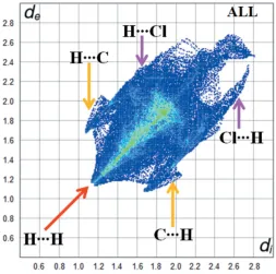
Fig. 5 shows the two-dimensional fingerprint of the sum of the contacts contributing to the Hirshfeld surface represented in normal mode. The graph shown in Fig. 6(a) (H H) shows the two-dimensional fingerprint of the (di, de) points
asso-ciated with hydrogen atoms. It is characterized by an end point that points to the origin and corresponds todi=de= 1.08 A˚ ,
which indicates the presence of the H H contacts in this study (68.9%). The graph shown in Fig. 6(b) shows the (C H/H C) contacts between the carbon atoms inside the surface and the hydrogen atoms outside the Hirshfeld surface and vice versa, which contribute 11.7%. There are two symmetrical wings on the left and right sides. Furthermore, there are also Cl H/H Cl (11%), C C (4.5%), C N/ N C (2.2%), O H/H O (1.3%) and N H/H N (0.4%) contacts.
A view of the three-dimensional Hirshfeld surface of the title compound plotted over electrostatic potential energy in the range0.030 to 0.044 a.u. using the STO-3G basis set at the Hartree–Fock level of theory is shown in Fig. 7 where the C—H donors and acceptors are shown as blue and red areas around the atoms related with positive (hydrogen-bond donors) and negative (hydrogen-bond acceptors) electrostatic potentials, respectively.
6. Synthesis and crystallization
[image:3.610.44.297.70.321.2]The title compound was prepared by refluxing a mixture of a solution containing 3,5-di-tert-butyl-2-hydroxybenzaldehyde (46.8 mg, 0.2 mmol) in ethanol (30 mL) and a solution containing 3-chloro-4-methylaniline (28.32 mg, 0.2 mmol) in ethanol (20 mL). The reaction mixture was stirred for 4 h under reflux. Crystals suitable for X-ray analysis were obtained by slow evaporation of an ethanol solution (m.p. 417–419 K; yield 84%).
Figure 7
A view of the three-dimensional Hirshfeld surface of the title compound plotted over electrostatic potential energy.
Figure 5
[image:3.610.45.298.450.695.2]Overall fingerprint plot for the title compound.
Figure 6
Two-dimensional fingerprint plots with a dnormview of the (a) H H
[image:3.610.319.563.543.712.2]7. Refinement
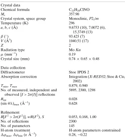
Crystal data, data collection and structure refinement details are summarized in Table 2. C-bound H atoms were positioned geometrically with C—H distances of 0.93–0.97 A˚ and refined as riding, withUiso(H) = 1.2Ueq(C).
Acknowledgements
The authors acknowledge the Faculty of Arts and Sciences, Ondokuz Mayıs University, Turkey, for the use of the Stoe
IPDS 2 diffractometer (purchased under grant F.279 of the University Research Fund).
References
Atalay, S¸., Gerc¸eker, S., Meral, S. & Bu¨lbu¨l, H. (2017).IUCrData,2, x171725.
Calligaris, M., Nardin, G. M. J. & Randaccio, C. (1972).Coord. Chem. Rev.7, 385–403.
Elmalı, A., Kabak, M., Kavlakog˘lu, E. & Elerman, Y. (1999).J. Mol. Struct.510, 207–214.
El-masry, A. H., Fahmy, H. H. & Ali Abdelwahed, S. (2000). Molecules,5, 1429–1438.
Farrugia, L. J. (2012).J. Appl. Cryst.45, 849–854.
Groom, C. R., Bruno, I. J., Lightfoot, M. P. & Ward, S. C. (2016).Acta Cryst.B72, 171–179.
Gumus, M. K., Kansiz, S., Dege, N. & Kalibabchuk, V. A. (2018).Acta Cryst.E74, 1211–1214.
Ho¨kelek, T., Bilge, S., Demiriz, S¸., O¨ zgu¨c¸, B. & Kılıc¸, Z. (2004).Acta Cryst.C60, o803–o805.
Iwan, A., Kaczmarczyk, B., Janeczek, H., Sek, D. & Ostrowski, S. (2007).Spectrochim. Acta A Mol. Biomol. Spectrosc.66, 1030–1041. Kansız, S. & Dege, N. (2018).J. Mol. Struct.1173, 42–51.
Kansiz, S., Macit, M., Dege, N. & Tsapyuk, G. G. (2018).Acta Cryst. E74, 1513–1516.
Moroz, Y. S., Demeshko, S., Haukka, M., Mokhir, A., Mitra, U., Stocker, M., Mu¨ller, P., Meyer, F. & Fritsky, I. O. (2012).Inorg. Chem.51, 7445–7447.
Sen, P., Kansiz, S., Golenya, I. A. & Dege, N. (2018).Acta Cryst.E74, 1147–1150.
S¸en, F., Kansiz, S. & Uc¸ar, I˙. (2017).Acta Cryst.C73, 517–524. Sheldrick, G. M. (2015a).Acta Cryst.A71, 3–8.
Sheldrick, G. M. (2015b).Acta Cryst.C71, 3–8. Spek, A. L. (2009).Acta Cryst.D65, 148–155.
Stoe & Cie (2002). X-AREAand X-RED32. Stoe & Cie GmbH, Darmstadt, Germany.
Su, D.-C., Wang, F.-T., Mao, C.-G. & Qian, S.-S. (2012).Acta Cryst. E68, o303.
Toprak, S¸., Tanak, H., Macit, M., Dege, N. & Orbay, M. (2018).J. Mol. Struct.1174, 184–191.
Turner, M. J., MacKinnon, J. J., Wolff, S. K., Grimwood, D. J., Spackman, P. R., Jayatilaka, D. & Spackman, M. A. (2017). CrystalExplorer17.5. University of Western Australia, Perth. Wang, H. (2010).Acta Cryst.E66, m1571.
1890
Kansizet al. C22H28ClNO Acta Cryst.(2018). E74, 1887–1890
[image:4.610.44.296.90.365.2]research communications
Table 2
Experimental details.
Crystal data
Chemical formula C22H28ClNO
Mr 357.90
Crystal system, space group Monoclinic,P21/m
Temperature (K) 296
a,b,c(A˚ ) 9.6753 (10), 7.0072 (6), 15.3749 (13) (
) 93.425 (7)
V(A˚3) 1040.51 (17)
Z 2
Radiation type MoK
(mm1) 0.19
Crystal size (mm) 0.740.650.48
Data collection
Diffractometer Stoe IPDS 2
Absorption correction Integration (X-RED32; Stoe & Cie, 2002)
Tmin,Tmax 0.879, 0.940
No. of measured, independent and observed [I> 2(I)] reflections
5895, 2300, 1298
Rint 0.028
(sin/)max(A˚
1
) 0.628
Refinement
R[F2> 2(F2)],wR(F2),S 0.053, 0.168, 1.00
No. of reflections 2300
No. of parameters 145
H-atom treatment H-atom parameters constrained max, min(e A˚
3
) 0.28,0.22
sup-1
Acta Cryst. (2018). E74, 1887-1890
supporting information
Acta Cryst. (2018). E74, 1887-1890 [https://doi.org/10.1107/S2056989018016377]
Crystal structure and Hirshfeld surface analysis of (
E
)-2,4-di-
tert
-butyl-6-[(3-chloro-4-methylphenylimino)methyl]phenol
Sevgi Kansiz, Mustafa Macit, Necmi Dege and Vadim A. Pavlenko
Computing details
Data collection: X-AREA (Stoe & Cie, 2002); cell refinement: X-AREA (Stoe & Cie, 2002); data reduction: X-RED (Stoe
& Cie, 2002); program(s) used to solve structure: SHELXLXT (Sheldrick, 2015a); program(s) used to refine structure:
SHELXL2017/1 (Sheldrick, 2015b); molecular graphics: ORTEP-3 for Windows (Farrugia, 2012); software used to
prepare material for publication: WinGX (Farrugia, 2012) and PLATON (Spek, 2009).
(E)-2,4-di-tert-butyl-6-[(3-chloro-4-methylphenylimino)methyl]phenol
Crystal data
C22H28ClNO
Mr = 357.90
Monoclinic, P21/m
a = 9.6753 (10) Å b = 7.0072 (6) Å c = 15.3749 (13) Å β = 93.425 (7)° V = 1040.51 (17) Å3 Z = 2
F(000) = 384
Dx = 1.142 Mg m−3
Mo Kα radiation, λ = 0.71073 Å Cell parameters from 5548 reflections θ = 2.1–30.7°
µ = 0.19 mm−1 T = 296 K Prism, orange
0.74 × 0.65 × 0.48 mm
Data collection
Stoe IPDS 2 diffractometer
Radiation source: sealed X-ray tube, 12 x 0.4 mm long-fine focus
Detector resolution: 6.67 pixels mm-1
rotation method scans
Absorption correction: integration (X-RED32; Stoe & Cie, 2002)
Tmin = 0.879, Tmax = 0.940
5895 measured reflections 2300 independent reflections 1298 reflections with I > 2σ(I)
Rint = 0.028
θmax = 26.5°, θmin = 2.1°
h = −12→12 k = −8→8 l = −18→19
Refinement
Refinement on F2
Least-squares matrix: full
R[F2 > 2σ(F2)] = 0.053
wR(F2) = 0.168
S = 1.00 2300 reflections 145 parameters 0 restraints
Hydrogen site location: mixed H-atom parameters constrained w = 1/[σ2(F
o2) + (0.0964P)2]
where P = (Fo2 + 2Fc2)/3
(Δ/σ)max < 0.001
Δρmax = 0.28 e Å−3
supporting information
sup-2
Acta Cryst. (2018). E74, 1887-1890 Special details
Geometry. All esds (except the esd in the dihedral angle between two l.s. planes) are estimated using the full covariance
matrix. The cell esds are taken into account individually in the estimation of esds in distances, angles and torsion angles; correlations between esds in cell parameters are only used when they are defined by crystal symmetry. An approximate (isotropic) treatment of cell esds is used for estimating esds involving l.s. planes.
Fractional atomic coordinates and isotropic or equivalent isotropic displacement parameters (Å2)
x y z Uiso*/Ueq
C1 0.2129 (4) 0.250000 0.2502 (2) 0.0799 (9)
H1A 0.122313 0.250000 0.272431 0.120*
H1B 0.223846 0.361862 0.215178 0.120*
C2 0.3181 (3) 0.250000 0.3253 (2) 0.0633 (8)
C3 0.2795 (3) 0.250000 0.4107 (2) 0.0703 (9)
H3 0.185649 0.250000 0.420637 0.084*
C4 0.3723 (3) 0.250000 0.4806 (2) 0.0700 (9)
H4 0.340566 0.250000 0.536544 0.084*
C5 0.5133 (3) 0.250000 0.46997 (18) 0.0556 (7)
C6 0.5560 (3) 0.250000 0.38600 (19) 0.0600 (7)
H6 0.649961 0.250000 0.376348 0.072*
C7 0.4596 (3) 0.250000 0.3165 (2) 0.0639 (8)
C8 0.5942 (3) 0.250000 0.61807 (19) 0.0568 (7)
H8 0.502444 0.250000 0.632881 0.068*
C9 0.7006 (3) 0.250000 0.68668 (18) 0.0521 (7)
C10 0.6637 (3) 0.250000 0.77307 (19) 0.0566 (7)
H10 0.570247 0.250000 0.784241 0.068*
C11 0.7595 (3) 0.250000 0.84146 (18) 0.0571 (7)
C12 0.8995 (3) 0.250000 0.82148 (19) 0.0620 (8)
H12 0.966318 0.250000 0.867572 0.074*
C13 0.9445 (3) 0.250000 0.7374 (2) 0.0590 (7)
C14 0.8423 (3) 0.250000 0.66939 (18) 0.0551 (7)
C15 1.0991 (3) 0.250000 0.7198 (2) 0.0722 (9)
C16 1.1330 (2) 0.0710 (4) 0.6677 (2) 0.0980 (10)
H16A 1.076649 0.068596 0.614163 0.147*
H16B 1.228999 0.073060 0.655013 0.147*
H16C 1.114737 −0.040681 0.701290 0.147*
C17 1.1899 (4) 0.250000 0.8041 (3) 0.1274 (18)
H17A 1.285592 0.250000 0.790824 0.191*
H17C 1.170713 0.138138 0.837395 0.191*
C18 0.7224 (3) 0.250000 0.9368 (2) 0.0753 (9)
C19 0.7730 (5) 0.4303 (8) 0.9793 (2) 0.176 (2)
H19A 0.734855 0.537623 0.947277 0.264*
H19B 0.744536 0.434730 1.037976 0.264*
H19C 0.872234 0.434730 0.979833 0.264*
C20 0.5646 (4) 0.250000 0.9437 (3) 0.1131 (15)
H20A 0.525969 0.136290 0.917256 0.170*
H20B 0.543785 0.250000 1.003968 0.170*
sup-3
Acta Cryst. (2018). E74, 1887-1890
N1 0.6191 (2) 0.250000 0.53735 (15) 0.0599 (6)
O1 0.8791 (2) 0.250000 0.58613 (13) 0.0748 (6)
H1 0.809294 0.250000 0.553116 0.112*
Atomic displacement parameters (Å2)
U11 U22 U33 U12 U13 U23
C1 0.088 (2) 0.066 (2) 0.083 (2) 0.000 −0.0157 (18) 0.000
C2 0.0734 (18) 0.0410 (17) 0.0749 (19) 0.000 −0.0011 (15) 0.000
C3 0.0559 (16) 0.077 (2) 0.077 (2) 0.000 0.0003 (16) 0.000
C4 0.0621 (17) 0.086 (2) 0.0633 (18) 0.000 0.0113 (15) 0.000
C5 0.0611 (16) 0.0480 (17) 0.0576 (16) 0.000 0.0032 (13) 0.000
C6 0.0630 (16) 0.0543 (18) 0.0631 (17) 0.000 0.0085 (14) 0.000
C7 0.0786 (19) 0.0547 (19) 0.0587 (16) 0.000 0.0068 (15) 0.000
C8 0.0531 (14) 0.0530 (18) 0.0649 (17) 0.000 0.0079 (13) 0.000
C9 0.0527 (14) 0.0458 (16) 0.0582 (15) 0.000 0.0074 (13) 0.000
C10 0.0546 (15) 0.0551 (18) 0.0616 (16) 0.000 0.0165 (14) 0.000
C11 0.0642 (16) 0.0530 (18) 0.0550 (15) 0.000 0.0112 (14) 0.000
C12 0.0595 (16) 0.065 (2) 0.0611 (17) 0.000 0.0016 (13) 0.000
C13 0.0554 (15) 0.0556 (18) 0.0669 (17) 0.000 0.0113 (14) 0.000
C14 0.0574 (15) 0.0524 (17) 0.0566 (16) 0.000 0.0128 (13) 0.000
C15 0.0519 (16) 0.082 (2) 0.083 (2) 0.000 0.0124 (15) 0.000
C16 0.0684 (14) 0.090 (2) 0.139 (2) 0.0156 (13) 0.0353 (16) −0.0004 (17)
C17 0.0520 (18) 0.219 (6) 0.110 (3) 0.000 −0.002 (2) 0.000
C18 0.079 (2) 0.091 (3) 0.0563 (17) 0.000 0.0149 (16) 0.000
C19 0.204 (4) 0.235 (5) 0.095 (2) −0.106 (4) 0.061 (3) −0.087 (3)
C20 0.102 (3) 0.168 (4) 0.073 (2) 0.000 0.036 (2) 0.000
Cl1 0.1117 (8) 0.1390 (10) 0.0595 (5) 0.000 0.0104 (5) 0.000
N1 0.0608 (13) 0.0617 (16) 0.0578 (14) 0.000 0.0075 (11) 0.000
O1 0.0613 (11) 0.1072 (18) 0.0574 (12) 0.000 0.0166 (10) 0.000
Geometric parameters (Å, º)
C1—C2 1.494 (5) C12—C13 1.389 (4)
C1—H1A 0.9600 C12—H12 0.9300
C1—H1B 0.9600 C13—C14 1.395 (4)
C1—H1Bi 0.9600 C13—C15 1.536 (4)
C2—C7 1.383 (4) C14—O1 1.349 (3)
C2—C3 1.387 (4) C15—C17 1.523 (5)
C3—C4 1.359 (4) C15—C16 1.534 (3)
C3—H3 0.9300 C15—C16i 1.534 (3)
C4—C5 1.383 (4) C16—H16A 0.9600
C4—H4 0.9300 C16—H16B 0.9600
C5—C6 1.379 (4) C16—H16C 0.9600
C5—N1 1.412 (4) C17—H17A 0.9600
C6—C7 1.376 (4) C17—H17C 0.9600
C6—H6 0.9300 C17—H17Ci 0.9600
supporting information
sup-4
Acta Cryst. (2018). E74, 1887-1890
C8—N1 1.278 (4) C18—C19 1.491 (4)
C8—C9 1.429 (4) C18—C20 1.537 (5)
C8—H8 0.9300 C19—H19A 0.9600
C9—C10 1.396 (4) C19—H19B 0.9600
C9—C14 1.412 (3) C19—H19C 0.9600
C10—C11 1.360 (4) C20—H20A 0.9600
C10—H10 0.9300 C20—H20B 0.9595
C11—C12 1.407 (4) C20—H20Ai 0.9600
C11—C18 1.530 (4) O1—H1 0.8200
C2—C1—H1A 108.6 O1—C14—C13 119.7 (2)
C2—C1—H1B 109.9 O1—C14—C9 119.5 (3)
H1A—C1—H1B 109.5 C13—C14—C9 120.8 (2)
C2—C1—H1Bi 109.91 (9) C17—C15—C16 108.31 (19)
H1A—C1—H1Bi 109.5 C17—C15—C16i 108.3 (2)
H1B—C1—H1Bi 109.5 C16—C15—C16i 109.7 (3)
C7—C2—C3 114.6 (3) C17—C15—C13 111.6 (3)
C7—C2—C1 123.8 (3) C16—C15—C13 109.45 (18)
C3—C2—C1 121.5 (3) C16i—C15—C13 109.45 (18)
C4—C3—C2 123.1 (3) C15—C16—H16A 109.5
C4—C3—H3 118.4 C15—C16—H16B 109.5
C2—C3—H3 118.4 H16A—C16—H16B 109.5
C3—C4—C5 121.1 (3) C15—C16—H16C 109.5
C3—C4—H4 119.5 H16A—C16—H16C 109.5
C5—C4—H4 119.5 H16B—C16—H16C 109.5
C6—C5—C4 117.6 (3) C15—C17—H17A 109.4
C6—C5—N1 116.2 (2) C15—C17—H17C 109.5
C4—C5—N1 126.1 (2) H17A—C17—H17C 109.5
C7—C6—C5 120.0 (3) C15—C17—H17Ci 109.48 (8)
C7—C6—H6 120.0 H17A—C17—H17Ci 109.5
C5—C6—H6 120.0 H17C—C17—H17Ci 109.5
C6—C7—C2 123.6 (3) C19i—C18—C19 115.8 (5)
C6—C7—Cl1 117.2 (2) C19i—C18—C11 109.23 (18)
C2—C7—Cl1 119.2 (3) C19—C18—C11 109.23 (18)
N1—C8—C9 123.2 (2) C19i—C18—C20 105.8 (2)
N1—C8—H8 118.4 C19—C18—C20 105.8 (2)
C9—C8—H8 118.4 C11—C18—C20 110.9 (3)
C10—C9—C14 119.1 (3) C18—C19—H19A 109.5
C10—C9—C8 119.2 (2) C18—C19—H19B 109.5
C14—C9—C8 121.7 (2) H19A—C19—H19B 109.5
C11—C10—C9 122.3 (2) C18—C19—H19C 109.5
C11—C10—H10 118.9 H19A—C19—H19C 109.5
C9—C10—H10 118.9 H19B—C19—H19C 109.5
C10—C11—C12 116.9 (2) C18—C20—H20A 109.5
C10—C11—C18 123.5 (2) C18—C20—H20B 109.4
C12—C11—C18 119.6 (3) H20A—C20—H20B 108.1
C13—C12—C11 124.2 (3) C18—C20—H20Ai 109.49 (9)
sup-5
Acta Cryst. (2018). E74, 1887-1890
C11—C12—H12 117.9 H20B—C20—H20Ai 108.1
C12—C13—C14 116.8 (2) C8—N1—C5 122.8 (2)
C12—C13—C15 121.8 (3) C14—O1—H1 109.5
C14—C13—C15 121.5 (3)
C7—C2—C3—C4 0.000 (1) C12—C13—C14—O1 180.000 (1)
C1—C2—C3—C4 180.000 (1) C15—C13—C14—O1 0.000 (1)
C2—C3—C4—C5 0.000 (1) C12—C13—C14—C9 0.000 (1)
C3—C4—C5—C6 0.000 (1) C15—C13—C14—C9 180.000 (1)
C3—C4—C5—N1 180.000 (1) C10—C9—C14—O1 180.000 (1)
C4—C5—C6—C7 0.000 (1) C8—C9—C14—O1 0.000 (1)
N1—C5—C6—C7 180.000 (1) C10—C9—C14—C13 0.000 (1)
C5—C6—C7—C2 0.000 (1) C8—C9—C14—C13 180.000 (1)
C5—C6—C7—Cl1 180.000 (1) C12—C13—C15—C17 0.000 (1)
C3—C2—C7—C6 0.000 (1) C14—C13—C15—C17 180.000 (1)
C1—C2—C7—C6 180.000 (1) C12—C13—C15—C16 119.9 (2)
C3—C2—C7—Cl1 180.000 (1) C14—C13—C15—C16 −60.1 (2)
C1—C2—C7—Cl1 0.000 (1) C12—C13—C15—C16i −119.9 (2)
N1—C8—C9—C10 180.000 (1) C14—C13—C15—C16i 60.1 (2)
N1—C8—C9—C14 0.000 (1) C10—C11—C18—C19i 116.2 (3)
C14—C9—C10—C11 0.000 (1) C12—C11—C18—C19i −63.8 (3)
C8—C9—C10—C11 180.000 (1) C10—C11—C18—C19 −116.2 (3)
C9—C10—C11—C12 0.000 (1) C12—C11—C18—C19 63.8 (3)
C9—C10—C11—C18 180.000 (1) C10—C11—C18—C20 0.000 (2)
C10—C11—C12—C13 0.000 (2) C12—C11—C18—C20 180.000 (1)
C18—C11—C12—C13 180.000 (1) C9—C8—N1—C5 180.000 (1)
C11—C12—C13—C14 0.000 (1) C6—C5—N1—C8 180.000 (1)
C11—C12—C13—C15 180.000 (1) C4—C5—N1—C8 0.000 (1)
Symmetry code: (i) x, −y+1/2, z.
Hydrogen-bond geometry (Å, º)
Cg is the centroid of the C9–C14 benzene ring.
D—H···A D—H H···A D···A D—H···A
O1—H1···N1 0.82 1.84 2.582 (3) 149
C1—H1B···Cg1ii 0.96 2.77 3.5072 (4) 134
![Crystal structure, Hirshfeld analysis and a molecular docking study of a new inhibitor of the Hepatitis B virus (HBV): ethyl 5 methyl 1,1 dioxo 2 {[5 (pentan 3 yl) 1,2,4 oxadiazol 3 yl]methyl} 2H 1,2,6 thiadiazine 4 carboxylate](data:image/gif;base64,R0lGODlhAQABAIAAAP///wAAACH5BAEAAAAALAAAAAABAAEAAAICRAEAOw==)