Diphenyl (isopropylamido)phosphate
Fahimeh Sabbaghi,a* Mehrdad Pourayoubi,bMarek Necˇascand Michal Babiakc
a
Department of Chemistry, Zanjan Branch, Islamic Azad University, Zanjan, Iran,
b
Department of Chemistry, Ferdowsi University of Mashhad, Mashhad, Iran, and
cDepartment of Chemistry, Faculty of Science, Masaryk University, Kotlarska 2, Brno
CZ-61137, Czech Republic
Correspondence e-mail: [email protected]
Received 14 November 2012; accepted 21 November 2012
Key indicators: single-crystal X-ray study;T= 120 K; mean(C–C) = 0.003 A˚; disorder in main residue;Rfactor = 0.027;wRfactor = 0.081; data-to-parameter ratio = 29.9.
The P atom in the title compound, C15H18NO3P, is in a
distorted tetrahedral P(O)(O)2N environment; the bond
angles at P are in the range 98.16 (6)–115.82 (6). In the
crystal, adjacent molecules are linked via N—H O P hydrogen bonds into a chain running parallel to the b axis. The methyl groups are disordered over two sets of sites in a 0.677 (14):0.323 (14) ratio. The crystal studied was a non-merohedral twin with a refined minor component of 22.31 (4)%.
Related literature
For bond lengths and angles in a related structure, see: Sabbaghiet al.(2011).
Experimental
Crystal data
C15H18NO3P Mr= 291.27 Monoclinic,Pn
a= 8.4432 (5) A˚
b= 5.3030 (4) A˚
c= 16.3443 (11) A˚
= 90.453 (6)
V= 731.78 (9) A˚3 Z= 2
MoKradiation
= 0.19 mm1 T= 120 K
0.450.420.40 mm
Data collection
Oxford Diffraction Xcalibur (Sapphire2) diffractometer Absorption correction: multi-scan
(CrysAlis RED; Oxford
Diffraction, 2009)
Tmin= 0.918,Tmax= 0.926
6226 measured reflections 6226 independent reflections 6040 reflections withI> 2(I)
Refinement
R[F2> 2(F2)] = 0.027 wR(F2) = 0.081 S= 1.06 6226 reflections 208 parameters 28 restraints
H atoms treated by a mixture of independent and constrained refinement
max= 0.17 e A˚
3
min=0.18 e A˚
3
Absolute structure: Flack (1983), 1229 Friedel pairs
Flack parameter: 0.05 (6)
Table 1
Hydrogen-bond geometry (A˚ ,).
D—H A D—H H A D A D—H A
N1—H1N O3i 0.81 (1) 2.23 (1) 3.0065 (17) 161 (2)
Symmetry code: (i)x;yþ1;z.
Data collection: CrysAlis CCD (Oxford Diffraction, 2009); cell refinement:CrysAlis CCD; data reduction:CrysAlis RED (Oxford Diffraction, 2009); program(s) used to solve structure:SHELXS97 (Sheldrick, 2008); program(s) used to refine structure:SHELXL97 (Sheldrick, 2008); molecular graphics:Mercury(Macraeet al., 2008); software used to prepare material for publication: SHELXTL (Sheldrick, 2008) andenCIFer(Allenet al., 2004).
Support of this investigation by Zanjan Branch, Islamic Azad University, is gratefully acknowledged.
Supplementary data and figures for this paper are available from the IUCr electronic archives (Reference: FF2090).
References
Allen, F. H., Johnson, O., Shields, G. P., Smith, B. R. & Towler, M. (2004).J. Appl. Cryst.37, 335–338.
Flack, H. D. (1983).Acta Cryst.A39, 876–881.
Macrae, C. F., Bruno, I. J., Chisholm, J. A., Edgington, P. R., McCabe, P., Pidcock, E., Rodriguez-Monge, L., Taylor, R., van de Streek, J. & Wood, P. A. (2008).J. Appl. Cryst.41, 466–470.
Oxford Diffraction (2009). CrysAlis CCD and CrysAlis RED. Oxford Diffraction Ltd, Yarnton, England.
Sabbaghi, F., Pourayoubi, M., Negari, M. & Necˇas, M. (2011).Acta Cryst.E67, o2512.
Sheldrick, G. M. (2008).Acta Cryst.A64, 112–122. Acta Crystallographica Section E
Structure Reports Online
supporting information
Acta Cryst. (2012). E68, o3459 [doi:10.1107/S1600536812047940]
Diphenyl (isopropylamido)phosphate
Fahimeh Sabbaghi, Mehrdad Pourayoubi, Marek Ne
č
as and Michal Babiak
S1. Comment
This work is a continuation of our studies of phosphoramidate compounds, during which the structures of various
di-phenyl amido phosphates, for example [C6H5O]2P(O)[NHCH(C2H5)(C6H5)] (Sabbaghi et al., 2011) were reported. Here,
we report the synthesis and crystal structure determination of the title compound, [C6H5O]2P(O)[NHCH(CH3)2].
The P═O (1.4602 (11) Å), P—O (1.5858 (11) and 1.5896 (11) Å) and P—N (1.6043 (14) Å) bond lengths are within the
expected values (Sabbaghi et al., 2011).
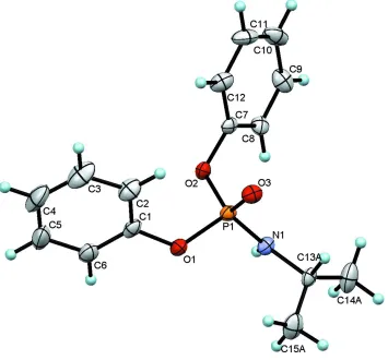
The P atom adopts a distorted tetrahedral configuration (Fig. 1). The bond angles at the P atom vary in the range 98.16
(6) [O1—P1—O2] to 115.82 (6)° [O3—P1—O2].
The C—O—P bond angles (124.07 (10) [C1—O1—P1] and 121.74 (10)° [C7—O2—P1]) and the C13A—N1—P1
(124.19 (11)°) bond angle are standard for this category of phosphoramidate compounds (Sabbaghi et al., 2011).
In the crystal structure, molecules are linked via N—H···O═P hydrogen bonds into extended chains running parallel to
the b axis (Table 1).
S2. Experimental
To a solution of [C6H5O]2P(O)Cl (2 mmol) in dry CH3CN (30 ml), a solution of isopropylamine (4 mmol) in the same
solvent (5 ml) was added at ice bath temperature under stirring. After 4 h, the solvent was removed and the product was
washed with distilled water and recrystallized from CH3CN/n-C6H14 (4:1) at room temperature. The single crystals
suitable for X-ray analysis were obtained from this solution after a few days at room temperature.
S3. Refinement
The crystal sample was non-merohedrally twinned. Using data reduction software, a HKLF 5 file was produced for a
two-component twin and used in the refinement. The fractional contribution of the minor twin component converged to
0.2231 (4). All carbon bound H atoms were placed at calculated positions and were refined as riding with their Uiso set to
either 1.2Ueq or 1.5Ueq (methyl) of the respective carrier atoms; in addition, the methyl H atoms were allowed to rotate
about the C—C bond. Nitrogen bound H atom was located in a difference Fourier map and its position was refined while
the N—H distance was fixed at 0.88 Å and the Uiso set to 1.2Ueq of N1. The disordered methyl groups were modeled over
two sites while restraining their anisotropic displacement parameters to be approximately isotropic (ISOR). To maintain a
correct hydrogen geometry, a dummy atom with zero occupancy was created and constrained to share the same site
Figure 1
The molecular structure of the title compound with ellipsoids shown at the 50% probability level and H atoms are drawn
as small spheres of arbitrary radii. The minor component of disordered part has been omitted for clarity and only one
orientation is shown for the disordered part.
Diphenyl (isopropylamido)phosphate
Crystal data
C15H18NO3P Mr = 291.27
Monoclinic, Pn a = 8.4432 (5) Å b = 5.3030 (4) Å c = 16.3443 (11) Å β = 90.453 (6)° V = 731.78 (9) Å3 Z = 2
F(000) = 308 Dx = 1.322 Mg m−3
Mo Kα radiation, λ = 0.71073 Å Cell parameters from 3821 reflections θ = 3.4–27.5°
µ = 0.19 mm−1 T = 120 K Block, colourless 0.45 × 0.42 × 0.40 mm
Data collection
Oxford Diffraction Xcalibur (Sapphire2) diffractometer
Radiation source: Enhance (Mo) X-ray Source Graphite monochromator
Detector resolution: 8.4353 pixels mm-1 ω scan
Absorption correction: multi-scan
(CrysAlis RED; Oxford Diffraction, 2009) Tmin = 0.918, Tmax = 0.926
Rint = 0.000
θmax = 25.0°, θmin = 3.8° h = −10→10
k = −6→6 l = −19→19
Refinement
Refinement on F2 Least-squares matrix: full R[F2 > 2σ(F2)] = 0.027 wR(F2) = 0.081 S = 1.06 6226 reflections 208 parameters 28 restraints
Primary atom site location: structure-invariant direct methods
Secondary atom site location: difference Fourier map
Hydrogen site location: inferred from neighbouring sites
H atoms treated by a mixture of independent and constrained refinement
w = 1/[σ2(Fo2) + (0.0626P)2 + 0.0183P] where P = (Fo2 + 2Fc2)/3
(Δ/σ)max < 0.001 Δρmax = 0.17 e Å−3 Δρmin = −0.18 e Å−3
Absolute structure: Flack (1983), 1229 Friedel pairs
Absolute structure parameter: 0.05 (6)
Special details
Geometry. All e.s.d.'s (except the e.s.d. in the dihedral angle between two l.s. planes) are estimated using the full covariance matrix. The cell e.s.d.'s are taken into account individually in the estimation of e.s.d.'s in distances, angles and torsion angles; correlations between e.s.d.'s in cell parameters are only used when they are defined by crystal symmetry. An approximate (isotropic) treatment of cell e.s.d.'s is used for estimating e.s.d.'s involving l.s. planes.
Refinement. Refinement of F2 against ALL reflections. The weighted R-factor wR and goodness of fit S are based on F2, conventional R-factors R are based on F, with F set to zero for negative F2. The threshold expression of F2 > σ(F2) is used only for calculating R-factors(gt) etc. and is not relevant to the choice of reflections for refinement. R-factors based on F2 are statistically about twice as large as those based on F, and R- factors based on ALL data will be even larger.
Fractional atomic coordinates and isotropic or equivalent isotropic displacement parameters (Å2)
x y z Uiso*/Ueq Occ. (<1)
P1 0.49359 (4) 0.20465 (6) 0.51025 (3) 0.01893 (9)
O1 0.46890 (12) 0.20756 (19) 0.41396 (6) 0.0229 (3)
O2 0.32869 (12) 0.3259 (2) 0.53519 (6) 0.0213 (2)
O3 0.52704 (12) −0.04455 (19) 0.54422 (6) 0.0247 (3)
N1 0.62616 (15) 0.4126 (2) 0.53057 (8) 0.0201 (3)
H1N 0.603 (2) 0.559 (3) 0.5224 (10) 0.024*
C1 0.33996 (19) 0.0959 (3) 0.37348 (10) 0.0241 (4)
C2 0.2687 (2) −0.1207 (3) 0.40086 (13) 0.0346 (4)
H2 0.3036 −0.2017 0.4496 0.042*
C3 0.1433 (2) −0.2176 (3) 0.35464 (15) 0.0454 (6)
H3 0.0915 −0.3663 0.3727 0.054*
C4 0.0931 (2) −0.1044 (4) 0.28402 (14) 0.0489 (6)
H4 0.0066 −0.1725 0.2537 0.059*
C5 0.1686 (2) 0.1079 (4) 0.25735 (13) 0.0440 (5)
H5 0.1359 0.1848 0.2075 0.053*
C6 0.2921 (2) 0.2121 (3) 0.30207 (11) 0.0307 (4)
H6 0.3430 0.3614 0.2838 0.037*
C7 0.27851 (18) 0.3331 (3) 0.61695 (10) 0.0197 (4)
C8 0.31935 (19) 0.5376 (3) 0.66473 (9) 0.0244 (4)
C9 0.2607 (2) 0.5528 (3) 0.74285 (10) 0.0319 (4)
H9 0.2874 0.6928 0.7765 0.038*
C10 0.1630 (2) 0.3656 (4) 0.77258 (11) 0.0376 (5)
H10 0.1232 0.3763 0.8267 0.045*
C11 0.1238 (2) 0.1643 (3) 0.72383 (12) 0.0360 (5)
H11 0.0562 0.0361 0.7443 0.043*
C12 0.1817 (2) 0.1460 (3) 0.64507 (12) 0.0306 (4)
H12 0.1547 0.0065 0.6113 0.037*
C13A 0.79551 (19) 0.3558 (3) 0.54259 (10) 0.0212 (4)
H13A 0.8017 0.1911 0.5722 0.025* 0.323 (14)
C14A 0.8579 (13) 0.557 (2) 0.6011 (9) 0.042 (3) 0.323 (14)
H14A 0.8401 0.7242 0.5773 0.064* 0.323 (14)
H14B 0.9716 0.5312 0.6104 0.064* 0.323 (14)
H14C 0.8021 0.5444 0.6533 0.064* 0.323 (14)
C15A 0.8791 (19) 0.324 (3) 0.4671 (9) 0.037 (3) 0.323 (14)
H15A 0.8307 0.1869 0.4355 0.056* 0.323 (14)
H15B 0.9902 0.2833 0.4788 0.056* 0.323 (14)
H15C 0.8736 0.4808 0.4354 0.056* 0.323 (14)
C13B 0.79551 (19) 0.3558 (3) 0.54259 (10) 0.0212 (4) 0.00
H13B 0.8085 0.2301 0.5878 0.025* 0.677 (14)
C14B 0.8824 (4) 0.5997 (8) 0.5652 (4) 0.0306 (11) 0.677 (14)
H14D 0.8633 0.7267 0.5227 0.046* 0.677 (14)
H14E 0.9963 0.5662 0.5696 0.046* 0.677 (14)
H14F 0.8434 0.6622 0.6177 0.046* 0.677 (14)
C15B 0.8680 (8) 0.2476 (13) 0.4620 (4) 0.0298 (11) 0.677 (14)
H15D 0.8160 0.0877 0.4482 0.045* 0.677 (14)
H15E 0.9818 0.2188 0.4701 0.045* 0.677 (14)
H15F 0.8517 0.3683 0.4174 0.045* 0.677 (14)
Atomic displacement parameters (Å2)
U11 U22 U33 U12 U13 U23
P1 0.01606 (19) 0.01911 (19) 0.02163 (19) 0.00093 (19) 0.00011 (14) 0.0017 (2)
O1 0.0195 (6) 0.0253 (6) 0.0238 (6) −0.0048 (5) −0.0003 (5) −0.0003 (5)
O2 0.0173 (6) 0.0247 (6) 0.0218 (6) 0.0019 (5) −0.0019 (4) 0.0017 (5)
O3 0.0239 (6) 0.0191 (6) 0.0313 (6) 0.0014 (4) 0.0029 (5) 0.0024 (5)
N1 0.0184 (7) 0.0154 (7) 0.0265 (8) 0.0024 (5) −0.0008 (5) 0.0027 (6)
C1 0.0155 (8) 0.0276 (9) 0.0291 (10) −0.0002 (7) 0.0017 (7) −0.0118 (7)
C2 0.0318 (11) 0.0258 (9) 0.0462 (12) −0.0045 (8) −0.0004 (8) −0.0038 (9)
C3 0.0349 (12) 0.0286 (11) 0.0727 (17) −0.0092 (9) 0.0004 (11) −0.0148 (11)
C4 0.0245 (11) 0.0615 (14) 0.0604 (15) −0.0062 (10) −0.0054 (10) −0.0292 (11)
C5 0.0239 (11) 0.0722 (16) 0.0359 (12) 0.0046 (10) −0.0064 (8) −0.0101 (11)
C6 0.0207 (10) 0.0402 (11) 0.0313 (10) 0.0021 (8) 0.0023 (8) −0.0036 (9)
C7 0.0137 (9) 0.0204 (8) 0.0251 (9) 0.0058 (6) 0.0003 (7) 0.0059 (7)
C8 0.0262 (10) 0.0223 (8) 0.0248 (9) −0.0025 (7) −0.0027 (7) 0.0031 (7)
C9 0.0431 (12) 0.0242 (10) 0.0284 (10) 0.0084 (9) −0.0014 (8) 0.0006 (8)
C10 0.0368 (12) 0.0450 (12) 0.0312 (10) 0.0186 (9) 0.0138 (9) 0.0091 (9)
C12 0.0237 (10) 0.0255 (9) 0.0427 (11) 0.0001 (7) 0.0052 (8) 0.0003 (8)
C13A 0.0166 (9) 0.0217 (8) 0.0253 (9) 0.0034 (6) −0.0049 (7) 0.0023 (7)
C14A 0.033 (4) 0.043 (4) 0.051 (4) 0.008 (3) −0.019 (3) −0.007 (3)
C15A 0.030 (4) 0.038 (5) 0.043 (4) 0.010 (4) −0.006 (3) −0.011 (4)
C13B 0.0166 (9) 0.0217 (8) 0.0253 (9) 0.0034 (6) −0.0049 (7) 0.0023 (7)
C14B 0.0200 (15) 0.0309 (18) 0.041 (2) 0.0000 (12) −0.0056 (15) −0.0077 (16)
C15B 0.0230 (18) 0.035 (3) 0.0317 (19) −0.0010 (19) 0.0069 (14) −0.007 (2)
Geometric parameters (Å, º)
P1—O3 1.4602 (11) C9—C10 1.381 (3)
P1—O1 1.5858 (11) C9—H9 0.9500
P1—O2 1.5896 (11) C10—C11 1.371 (3)
P1—N1 1.6043 (14) C10—H10 0.9500
O1—C1 1.4008 (19) C11—C12 1.384 (2)
O2—C7 1.4057 (17) C11—H11 0.9500
N1—C13A 1.4728 (19) C12—H12 0.9500
N1—H1N 0.811 (13) C13A—C15A 1.436 (15)
C1—C2 1.373 (2) C13A—C14A 1.524 (8)
C1—C6 1.378 (2) C13A—H13A 1.0000
C2—C3 1.393 (3) C14A—H14A 0.9800
C2—H2 0.9500 C14A—H14B 0.9800
C3—C4 1.366 (3) C14A—H14C 0.9800
C3—H3 0.9500 C15A—H15A 0.9800
C4—C5 1.367 (3) C15A—H15B 0.9800
C4—H4 0.9500 C15A—H15C 0.9800
C5—C6 1.383 (3) C14B—H14D 0.9800
C5—H5 0.9500 C14B—H14E 0.9800
C6—H6 0.9500 C14B—H14F 0.9800
C7—C12 1.367 (2) C15B—H15D 0.9800
C7—C8 1.379 (2) C15B—H15E 0.9800
C8—C9 1.376 (2) C15B—H15F 0.9800
C8—H8 0.9500
O3—P1—O1 114.19 (6) C12—C7—O2 119.04 (14)
O3—P1—O2 115.82 (6) C8—C7—O2 118.95 (13)
O1—P1—O2 98.16 (6) C9—C8—C7 118.79 (15)
O3—P1—N1 114.26 (7) C9—C8—H8 120.6
O1—P1—N1 106.55 (6) C7—C8—H8 120.6
O2—P1—N1 106.26 (6) C8—C9—C10 120.31 (17)
C1—O1—P1 124.07 (10) C8—C9—H9 119.8
C7—O2—P1 121.74 (10) C10—C9—H9 119.8
C13A—N1—P1 124.19 (11) C11—C10—C9 119.82 (16)
C13A—N1—H1N 116.9 (13) C11—C10—H10 120.1
P1—N1—H1N 117.1 (13) C9—C10—H10 120.1
C2—C1—C6 121.57 (16) C10—C11—C12 120.63 (15)
C2—C1—O1 122.74 (15) C10—C11—H11 119.7
C1—C2—C3 117.70 (19) C7—C12—C11 118.59 (17)
C1—C2—H2 121.2 C7—C12—H12 120.7
C3—C2—H2 121.2 C11—C12—H12 120.7
C4—C3—C2 121.69 (19) C15A—C13A—N1 113.1 (7)
C4—C3—H3 119.2 C15A—C13A—C14A 116.8 (6)
C2—C3—H3 119.2 N1—C13A—C14A 105.7 (4)
C3—C4—C5 119.3 (2) C15A—C13A—H13A 106.9
C3—C4—H4 120.4 N1—C13A—H13A 106.9
C5—C4—H4 120.4 C14A—C13A—H13A 106.9
C4—C5—C6 120.8 (2) H14D—C14B—H14E 109.5
C4—C5—H5 119.6 H14D—C14B—H14F 109.5
C6—C5—H5 119.6 H14E—C14B—H14F 109.5
C1—C6—C5 118.93 (17) H15D—C15B—H15E 109.5
C1—C6—H6 120.5 H15D—C15B—H15F 109.5
C5—C6—H6 120.5 H15E—C15B—H15F 109.5
C12—C7—C8 121.86 (15)
O3—P1—O1—C1 67.82 (12) C2—C1—C6—C5 0.3 (3)
O2—P1—O1—C1 −55.32 (12) O1—C1—C6—C5 177.98 (15)
N1—P1—O1—C1 −165.06 (11) C4—C5—C6—C1 1.1 (3)
O3—P1—O2—C7 48.50 (13) P1—O2—C7—C12 −95.01 (17)
O1—P1—O2—C7 170.46 (11) P1—O2—C7—C8 89.47 (14)
N1—P1—O2—C7 −79.57 (12) C12—C7—C8—C9 0.0 (2)
O3—P1—N1—C13A 30.26 (15) O2—C7—C8—C9 175.40 (15)
O1—P1—N1—C13A −96.81 (13) C7—C8—C9—C10 0.2 (3)
O2—P1—N1—C13A 159.24 (12) C8—C9—C10—C11 −0.4 (3)
P1—O1—C1—C2 −33.7 (2) C9—C10—C11—C12 0.3 (3)
P1—O1—C1—C6 148.68 (12) C8—C7—C12—C11 −0.1 (3)
C6—C1—C2—C3 −1.2 (3) O2—C7—C12—C11 −175.45 (15)
O1—C1—C2—C3 −178.68 (16) C10—C11—C12—C7 −0.1 (3)
C1—C2—C3—C4 0.7 (3) P1—N1—C13A—C15A 80.0 (6)
C2—C3—C4—C5 0.7 (3) P1—N1—C13A—C14A −150.9 (7)
C3—C4—C5—C6 −1.7 (3)
Hydrogen-bond geometry (Å, º)
D—H···A D—H H···A D···A D—H···A
N1—H1N···O3i 0.81 (1) 2.23 (1) 3.0065 (17) 161 (2)