Printed in Great Britain © The Company ofBiobgists Limited 1985
POSTSYNAPTIC POTENTIALS OF LIMITED
DURATION IN VISUAL NEURONES OF A LOCUST
BY PETER J. SIMMONS
Department of Zoology, University of Newcastle upon Tyne, Newcastle upon Tyne, NE1 7RU
Accepted 27 November 1984
SUMMARY
Large, second-order neurones of locust ocelli ('L-neurones') make both excitatory and inhibitory connections amongst each other. A single L-neurone can be presynaptic at both types of connection. At the excitatory connections, transmission can be maintained for long periods without decre-ment. In contrast, inhibitory postsynaptic potentials (IPSPs) never last for more than 15—35 ms. This paper examines mechanisms which could limit the duration of these IPSPs.
1. An IPSP begins 4-5 ms after a presynaptic neurone starts to depolarize from its resting potential, and the time-to-peak is 7 ms.
2. The amplitude of an IPSP depends both upon the amplitude of the peak presynaptic potential and upon the potential at which a presynaptic neurone is held before it is depolarized.
3. The rate at which a postsynaptic neurone hyperpolarizes to produce an IPSP is proportional to the rate at which the presynaptic neurone depolarizes, independent of the potential from which the presynaptic depolarization starts. A maximum rate of postsynaptic hyperpolarization is reached when the presynaptic neurone depolarizes at lOmVms"1.
4. Once an IPSP has occurred, both the amplitudes and the rates of hyperpolarization of subsequent IPSPs are depressed. The connection recovers its full ability to transmit over a period of 1-5 s. Larger IPSPs are followed by initially greater depression than smaller IPSPs.
5. A connection can begin to recover from depression while the presynap-tic neurone is held depolarized from resting.
6. Transmission fails when a presynaptic neurone is depolarized by pulses shorter than 2 ms.
7. The most likely reason why the duration of the IPSPs is limited is that calcium channels in the presynaptic terminal inactivate within 7 ms of first opening.
INTRODUCTION
Large, second-order neurones of locust ocelli ('L-neurones') provide useful sub-jects for investigations on the transmission of signals both within a single neurone, and across synapses between neurones. Pairs of microelectrodes can be inserted into single
study the operating characteristics of output connections from an L-neurone (Sim-mons, 1981, 1982a). Signals decrement very little throughout the length of an L-neurone (Wilson, 19786; Simmons, 1982a), and L-L-neurones have very simple geometries (C. S. Goodman, 1976; L. J. Goodman, Patterson & Mobbs, 1975; Simmons, 1982a). This means that an electrode placed in the axon of an L-neurone will record fairly accurately the amplitudes and waveforms of potential changes which occur at pre- or postsynaptic sites.
The output connections which L-neurones make are of particular interest because two types of them, which have contrasting properties, have been found. A single L-neurone can make both types of output connection. L-L-neurones make excitatory connections with some large brain neurones which descend in the nerve cord (Sim-mons, 1981), and make both excitatory and inhibitory connections laterally amongst each other (Simmons, 1982a). At the excitatory connections, small, slowly changing variations in presynaptic potential are transmitted to the postsynaptic neurone, as long as the presynaptic neurone is depolarized from a threshold potential. Because the normal resting potential of an L-neurone is depolarized above the threshold for release of transmitter at the excitatory connections, small hyperpolarizations, such as those that occur when ocellar illumination is increased, are communicated across these connections (Simmons, 1981, 1982a). Transmission at the excitatory connections can be sustained for long periods without decrement. In contrast, at the inhibitory con-nections which L-neurones make amongst each other, the postsynaptic potential (PSP) has a duration limited to less than 35 ms, no matter for how long the presynaptic terminal is held depolarized (Simmons, 1982a). Two of the possible mechanisms which would limit the duration of these PSPs are that presynaptic transmitter becomes depleted, or that postsynaptic receptors desensitize. If either of these processes occurred rapidly, then slow changes in presynaptic potential would not be transmitted across the inhibitory connections, and fast depolarizing potentials, such as spikes, would be required to effect transmission. L-neurones produce single spikes, or, rarely, short bursts of spikes when illumination is reduced (Chappell & Dowling, 1972; Patterson & Goodman, 1974; Wilson, 1978a) or when a pulse of hyperpolariz-ing current injected into an L-neurone ends (Wilson, 19786). The amplitude of a spike in an L-neurone depends on both the amplitude and duration of the preceding hyperpolarization (Simmons, 1982a) and L-neurones do not produce trains of spikes when depolarized. In this paper it is shown that neither transmitter depletion, nor desensitization of postsynaptic receptors is likely to occur at the inhibitory connec-tions among L-neurones. Instead, transmission may be limited in duration by proper-ties of calcium channels in the presynaptic membrane. The importance of rapid depolarization, such as a spike provides, in effective transmission at these connections is demonstrated.
METHODS
sealed with wax, and the locust was fixed upright on a plasticene block. The lateral ocellar nerves and the dorsal surface of the brain were exposed. The head was stabilized against movement by inserting pins through the genae into the plasticene. A constant supply of saline (Eibl, 1978) bathed the brain. The tracheae of the brain were not disturbed, and ventilation was provided by the animal's own continual pumping movements. The laboratory was darkened as much as possible during an experiment. The temperature of the preparation was 18-22°C.
In most preparations, to ease the passage of electrodes through the connective tissue sheath of the ocellar nerves and brain, a 1 % solution of protease in saline was applied for 2min. Routinely, up to three independent glass microelectrodes were employed. They were filled with 2 m o i r1 potassium acetate and had d.c. resistances of 40-80 MQ. Each electrode was connected to a d.c. amplifier which incorporated a bridge circuit, allowing current to be injected through the electrode. Current was measured by using a virtual ground amplifier, which measured current flowing along the chloridized silver wire which was employed as the indifferent electrode. This electrode lay along the inside of the thorax, with its free end placed as close as possible to the brain. Results were recorded with an FM tape recorder and were analysed later with the aid of a North Star Horizon microcomputer, fitted with a Comart 7A + D analogue/digital converting card. Pre-triggered displays on the oscilloscope screen and signal averaging were achieved by use of programmes written in Pascal M / T (Simmons, 1985). The figures in this paper were drawn by an X-Y plotter, driven by the microcomputer.
e
1
2
mv,
post- 20
RESULTS
The time course of the inhibitory postsynaptic potential
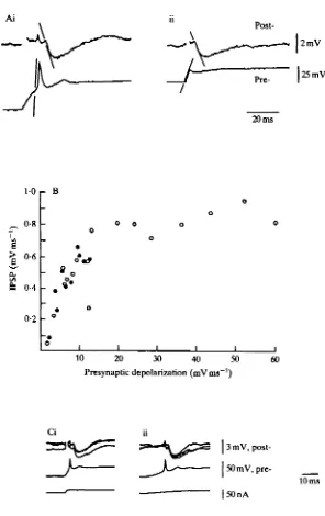
The inhibitory postsynaptic potential (IPSP) at a connection between two L-neurones was examined by producing in the presynaptic neurone either a spike (Fig. 1A) or a more long-lasting depolarization (Fig. IB). A spike was produced by switch-ing off a hyperpolarizswitch-ing current and lasted 4 ms, measured between departure from and return to nesting potential (Fig. 1A). Resting potential was — 30 to — 35 mV (Simmons, 1981). To produce an IPSP by long depolarization, the presynaptic neurone was first held hyperpolarized by 4 mV, before a depolarization lasting 200 ms (Fig. IB)
Latency was measured from the time the presynaptic neurone depolarized from resting potential, and was always 4-5 ms. Time-to-peak was 7 ms. Duration was 10-35 ms, varying with the amplitude of the IPSP, probably as a function of the membrane time constant.
Pulses of current injected to depolarize an L-neurone from resting were never observed to produce discrete spikes. However, such pulses did appear to activate regenerative channels in L-neurones, as the rate of potential change of an L-neurone was greater when the neurone was depolarized from resting than when it was repolarized back to resting potential. In addition, following the initial peak depolarization, there was usually a slight hyperpolarization in the membrane potential of an L-neurone, before it climbed again to a more sustained plateau depolarization, e.g. Fig. IB. In 20 experiments, the potential was recorded at two different locations of an L-neurone while current was injected through a third electrode. The response recorded in the ocellar neuropile had a very similar waveform to that recorded in the axon in the brain. Measurements from five of these experiments gave values for the space constant of an neurone of 2-5 to 3-5 times the length of one of the lateral L-neurones (Goodman, 1976; Simmons, 1982a). In contrast to the statement in the summary of Wilson's (19786) paper, spikes were conducted with little attenuation along L-neurone axons (some of Wilson's paired recordings may have been from two L-neurones linked by an excitatory connection rather than from a single axon; see also Simmons, 1982a). All recordings in this paper were made from L-neurone axons within 200 fim of the junction of the ocellar nerve with the brain. Because of the simple geometry (C.S. Goodman, 1976; L. J. Goodmanet al. 1975; Simmons, 19826) and the large space constants of L-neurones, the potentials recorded from this location were probably at least 70% of the value of the potentials occurring at any other location in an L-neurone, except for the cell body and its neurite. In ultrastructural studies, synapses have been found between large axons in ocellar nerves near to the brain (Goodman, Mobbs & Guy, 1977; Koontz & Edwards, 1984), although a detailed description of the distribution of synapses made by an L-neurone has not been made.
•^20 mV -35 mV
Ci D
Average
3 mV,
post-50 mV,
pre-r - 5 m V
20 ms
2-0
1-5
1-0
0-5 E
0
o a
-o
o
a
•
i
0 O O
10 20 30 Peak of presynaptic potential (mV)
Fig. 2
the increase in PSP amplitude and to an increase in membrane resistance (Wilson, 1978A). The decay of an IPSP had a similar waveform and duration to the decay in potential at the end of a small pulse of hyperpolarizing current injected through an electrode into an L-neurone. In six experiments, the conductance of a postsynaptic neurone was measured both before and 40 ms following the start of an IPSP by injecting a 5 ms current pulse into the neurone. No conductance change following an IPSP was found.
The relationship between pre- and postsynaptic potentials
The amplitude of the IPSP was influenced by the potential at which the presynaptic neurone was held before depolarizing current was injected into it as well as by the peak of the presynaptic depolarizing potential (Fig. 2). In Fig. 2A, spikes were initiated in the presynaptic neurone by terminating pulses of hyperpolarizing current. These pulses hyperpolarized the neurone by 20 mV from its resting potential, and spikes of different amplitudes were produced by varying the duration of the hyperpolarizing pulses (Simmons, 1982a). For spikes reaching between 5 and 10 mV above resting potential, each 1-mV increase in spike amplitude caused an increment of 0*2 mV in IPSP amplitude (open circles, Fig. 2E). For larger spikes, the proportionate increase in IPSP amplitude fell off until, for spikes of 25 mV and greater, the IPSP reached its maximum amplitude of 2mV. In Fig. 2C depolarizing pulses of current were injected to depolarize the presynaptic neurone from its resting potential. These presynaptic potentials (filled circles, Fig. 2E) produced smaller IPSPs than did presynaptic spikes reaching equivalent potentials from resting (open circles, Fig. 2E). For the depolarizations from resting, each 1-mV increase in peak depolarization of the presynaptic neurone caused an increment of 0-1 mV in IPSP amplitude, between presynaptic potentials of 10 and 20 mV. Fig. 2D and the open squares in Fig. 2E show IPSPs produced when the presynaptic neurone was hyperpolarized 5 mV from resting potential between depolarizing pulses. These IPSPs were intermediate in amplitude between those produced by spikes and those produced when the presynaptic potential returned to resting between pulses.
There was considerable variation in the maximum amplitudes of IPSPs which could be elicited in different preparations, although a detailed statistical analysis was not made. A few IPSPs reached 10 mV in amplitude, but most studies were made of IPSPs between 2 and 3 mV high. Often smaller IPSPs were recorded,
Post-2mV
25 mV
20 ms
7
Q
1-0
0-8
0-6
0-4
0-2
r
B--
.**
o
o
o
o
0 0
o
1 1
o o
0 0
10 20 30 40 50 Presynaptic depolarization (mVms"')
3 mV, post-|50mV,
[image:8.451.90.386.79.549.2]pre-10 ms |50nA
but measurements on these were difficult as they are about the same amplitude as fluctuations in potential caused by the random arrival of photons at photoreceptors (Wilson, 1978c).
The relationship between rates of change in pre-and postsynaptic potentials The reason why the starting potential of the presynaptic neurone influences IPSP amplitude became clear when the rates of change of pre- and postsynaptic potentials were measured. For the experiment shown in Fig. 2, the rates of potential change in the two neurones were plotted (Fig. 3A), and the rates of presynaptic depolarization were plotted against rates of postsynaptic hyperpolarization both for presynaptic spikes and for more long lasting depolarizations (Fig. 3B). The rate of postsynaptic hyperpolarization, causing an IPSP, depended on the rate of presynaptic depolariza-tion, and was independent of the initial potential of the presynaptic neurone. When a pulse of depolarizing current was injected into an L-neurone, the neurone depolarized more rapidly if it was initially hyperpolarized than if it was initially at resting potential. In the plot of Fig. 3B, for each 1 mVms"1 increase in the rate of presynaptic depolarization up to a rate of 10 mV ms"1, the rate of postsynaptic hyper-polarization increased by 0-08 mV ms"1. For more rapid presynaptic depolarizations, which were achieved only by spikes, there was little or no increase in the rate of postsynaptic hyperpolarization beyond a maximum of 0'8mVms"'. The maximum recorded rate of hyperpolarization during an IPSP was 3 mV ms"1, and the amplitude of an IPSP was linearly proportional to its rate of rise. In a total of five experiments of this type, similar results were obtained. The rate of presynaptic depolarization at which the maximum rate of postsynaptic hyperpolarization was achieved was always within 2 mV ms"1 of 10 mV ms"1. The rate of postsynaptic hyperpolarization, as well as the maximum amplitude of an IPSP, varied from experiment to experiment.
When gradual ramps of current were injected into a presynaptic neurone to depolarize it from an initially hyperpolarized potential, the neurone usually spiked and the spike mediated an IPSP (Fig. 3C). Judged from the inflection in the rate of depolarization, spikes were always initiated as the membrane potential was depolarized to resting, no matter how rapid the depolarization. Attempts to determine the minimum rate of presynaptic depolarization that would mediate an IPSP were thwarted by the tendency of L-neurones to spike. Ramps of current which produced depolarizations at less than 1 mV ms"1 produced neither spikes nor detectable IPSPs.
Recovery of the ability of inhibitory connections to transmit
2nd: 120ms 2nd: 220ms 2nd: 500ms
2nd: 120 ms 2nd: 220 ms 2nd: 500 ms
10 mV, post-1 50 mV,
pre-96 nA I
20 ms
100 90 80 70 60 50 40 30 20 10
- C
- o * o •
O 0 • •
- o • - •
-1 -1 -1 -1 -1 -1 -1 -1 -1
1 2 3 4 5 6 7 8 9 10
Interval between spikes (x 100 ms) Fig. 4
Both the latency of an IPSP, and the time taken for it to reach its peak hyper-polarization, were the same for first or second IPSPs of a pair, or for IPSPs following large or small spikes. This is illustrated in Fig. 4D, where the traces are aligned with each other according to the times of the presynaptic spikes.
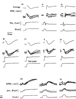
current pulses to the presynaptic neurone. The first spike of each pair produced an IPSP 2-2mV in amplitude, and the second spike produced an IPSP of 0-8 mV, or 36 % of the amplitude of the first IPSP. In Fig. 5B, pairs of depolarizing pulses were injected into the presynaptic neurone. In each pair, the first pulse lasted for 48 ms, and the start of the second pulse occurred 100 ms after the start of the first pulse. The IPSP caused by each first pulse was l-9mV in amplitude, and the amplitude of the IPSP caused by each second pulse was 0-7 mV, or 37 % of the amplitude of the first IPSP. Therefore, the connection was recovering some of its ability to transmit before the presynaptic potential returned to resting at the end of the first pulse. The depolarizing pulses caused no more long-lasting depression than did single spikes. Similar results were obtained with much longer lasting pulses of depolarizing current injected into the presynaptic neurone (not shown). However, in three experiments, there was a relatively short interval between the end of the first pulse and the start of the second, and the second IPSP was usually reduced or absent. The reduction in IPSP amplitude was accompanied by a change in the shape of the initial depolarizing potential in the presynaptic neurone, which indicates that the short interval between pulses probably provided insufficient time for the conductance of the presynaptic membrane to return to its resting value. For example, in Fig. 5C, the interval between the start of the first and second pulses of each pair was 450 ms. The first pulse of each pair (Fig. 5Ci) produced an IPSP l-4mV in amplitude. IPSPs produced by the second pulses, when the duration of the first pulse was 390 ms, had amplitudes of 11 mV, or 79% of the amplitudes of the first IPSPs (Fig. 5Cii). When the first pulse was lengthened to 410 ms, no IPSP was detected following each second pulse (Fig. 5Ciii). Here, the rate of depolarization of the presynaptic neurone at the start of the second pulse was reduced, as was the amplitude of the hyperpolarization which followed the initial peak depolarizing potential.
PSPs following short depolarizing pulses in presynaptic neurones
As pulses of current injected presynaptically were reduced to a length of 4 ms and less, both the IPSPs and EPSPs they mediated became smaller (Fig. 6). The pulse was 50 ms long in Fig. 6Ai, raising the presynaptic potential by 4mV, and mediated and IPSP of 2-5mV. In Fig. 6Aii, the presynaptic pulse reached an amplitude of 4 mV
-L «L J
) 1
J
J
J
~1
!
r
e
a.
E
BO
E
for 3 ms, and mediated an IPSP of 1-5 mV. With a shorter pulse (Fig. 6Aiii) the presynaptic neurone was depolarized to a maximum of 2mV for 0-7 ms. There was no detectable IPSP following this depolarization.
Further evidence that presynaptic depolarizations shorter than 4 ms are less effective than longer pulses at mediating transmission was provided by examining the recovery of connections from depression. When two presynaptic stimuli, each longer than 4 ms, were separated by 100 ms, the amplitude of the second IPSP was reduced to 20-35% of its value in a non-depressed connection (Fig. 4C). The reduction could be expected to be less if transmission during the first IPSP were to be reduced. This was examined by following each of the presynaptic pulses in Fig. 6A, after 100 ms, by a 50-ms test pulse (Fig. 6C). The amplitudes of the IPSPs elicited by these test pulses were compared with those of IPSPs elicited by single presynaptic pulses of the same amplitude and duration (Fig. 6B). The presynaptic pulses in Fig. 6B reached an amplitude of 12 mV, and elicited IPSPs of amplitude 4-5 mV. The ability of the electrode to depolarize the neurone declined during the last part of the experiment (Fig. 6Ciii), but this did not affect the results adversely. Following a first pulse 50 ms long (Fig. 6Ai), the test IPSP (Fig. 6Ci) had an amplitude of 2-5 mV, or 55 % of the amplitude of the IPSP in Fig. 6B. As the length of the first pulse decreased (Fig. 6Aii, iii) the amplitudes of the test IPSPs (Fig. 6Cii, iii) increased to 90% and 100% of the amplitude of the control IPSP in Fig. 6B. The IPSPs which followed presynaptic pulses 5 ms long had the same amplitude as those which followed longer presynaptic pulses, and 5-ms presynaptic pulses caused the same decrement in test IPSPs as did longer pulses.
Reduction in efficiency of transmission as presynaptic pulses were reduced in length was also observed at excitatory connections between L-neurones. In all three recordings of Fig. 6D current pulses depolarized the presynaptic neurone to 15 mV from resting. In Fig. 6D the pulse lasted for 50 ms, and the postsynaptic neurone depolarized to 2mV above resting. The peak presynaptic depolarization of 15 mV lasted for 2-5 ms in Fig. 6D, and the postsynaptic potential reached 1 mV above resting. For a shorter depolarizing pulse, where the presynaptic membrane returned towards resting as soon as the peak depolarization was arrived at, the postsynaptic potential had an amplitude of 0-4 mV (Fig. 6Diii).
Ai
[image:14.451.89.392.73.469.2]Average
IPSP, 3 mV |
Pre, 15 mV
20 nA
10 ms
I
Test pulse
_r
_r
UI
DISCUSSION
Possible mechanisms for limiting the duration of a PSP are: (1) that stores of transmitter in the presynaptic neurone become depleted; (2) that postsynaptic recep-tors for the transmitter desensitize; (3) that the PSP is followed by a second PSP of opposite sign, curtailing it; and (4) that prolonged entry of calcium ions into the presynaptic sites is impossible, for reasons discussed below. At the inhibitory connec-tions between L-neurones the first three mechanisms can be discounted. It is unlikely that stores of transmitter become depleted at presynaptic sites of L-neurones because if a presynaptic L-neurone remains depolarized for some time following an IPSP, the connection starts to recover its ability to transmit during this depolarization (Fig. 5). If transmitter stores did become rapidly depleted, the connection would not start to recover its ability to transmit until the end of the depolarizing pulse. Transmitter depletion has been demonstrated at a synapse between large neurones in the hatchet-fish (Highstein & Bennett, 1975), when fairly intense trains of spikes are elicited presynaptically, and depletion may occur in some circumstances at synapses between giant neurones in the squid (Charlton, Smith & Zucker, 1982). However, there is no instance where depletion has been demonstrated to occur when naturally-occurring bursts of spikes invade presynaptic sites, and it has not been shown to occur at any synapse where the presynaptic neurone operates without producing trains of spikes. In some synapses, a spike is thought to cause the release of a single vesicle of trans-mitter from a presynaptic bouton (Korn, Mallet, Triller & Faber, 1982; Neale et al. 1983), and when such small stores are held ready for release at presynaptic terminals, depletion could play a role in shaping PSPs. The experiments to study the recovery from depression following long presynaptic depolarizing pulses (Fig. 5) also show that desensitization of postsynaptic receptors does not play an important role in limiting the duration of these IPSPs. If desensitization were important, transmitter would have to be released throughout the duration of a presynaptic depolarization, and the receptors would not begin to resensitize until the pulse ended. The possibility that the postsynaptic receptors would desensitize if exposed to transmitter for some time cannot be excluded, since the presynaptic neurone may not be able to release trans-mitter continually. A way to test for densitization would be to apply the transtrans-mitter directly to the postsynaptic membrane, as has been done at a neuromuscular junction in a frog (Katz & Thesleff, 1957), and with some neurones in Aplysia (Gardner & Kandel, 1977). The time courses both of desensitization and of resensitization in these two examples are considerably slower than the time courses of decrement and recovery of transmission at the inhibitory connections between L-neurones.
of PSP is followed by a second, both PSPs mediated by the same presynaptic neurone, have been reported for nudibranch molluscs (e.g. Wachtel & Kandel, 1967; Getting, 1981).
One mechanism which could reduce or eliminate prolonged entry of calcium ions into a presynaptic neurone is closure of the calcium channels by hyperpolarization of the membrane. This hyperpolarization could be caused by an increase in potassium conductance triggered by a depolarizing potential, or by synaptic feedback onto the L-neurone. Following a rapid depolarization, an L-neurone does produce a hyper-polarizing potential (Fig. 1A,B). However, this hyperpolarization is relatively small throughout the length of an L-neurone, and it is much less prolonged than the depression in the ability of an inhibitory connection to transmit following the produc-tion of an IPSP (Fig. 4). After a spike, changes in L-neurone conductance can persist for several tens of milliseconds, and for this reason care was taken to ensure that the two spikes were of equal amplitude, when pairs of spikes were produced in studying the recovery of the ability of a connection to transmit (Fig. 4). No difference in the duration of two spikes of such pairs was found, as would be expected if a long-lasting increase in potassium conductance was reducing transmitter release caused by the second spike. Because a good correlation between rates of pre- and postsynaptic potential change during transmission has been observed (Fig. 3B), and because changes in the shape of a presynaptic depolarization can be correlated with changes in IPSP amplitude (Fig. 5C), it is likely that any important hyperpolarizing potential that did occur at the presynaptic sites would have been recorded. Synaptic feedback onto the presynaptic neurone is unlikely because, if it occurred, a connection would not start to recover its ability to transmit until the end of a long depolarizing pulse applied to the presynaptic neurone. These inhibitory connections do start to recover their ability to transmit while the presynaptic neurone is depolarized from resting (Fig. 5).
inactivation of voltage-gated sodium channels in the membrane of squid giant axons (Hodgkin & Huxley, 1952a,6), and at other presynaptic sites calcium channels do not inactivate, behaving more like potassium channels which cause the hyperpolarizing phase of the action potential in the squid giant axon (Hodgkin & Huxley, \952a,b). L-neurones possibly have at least two types of calcium channel: inactivating cal-cium channels, which mediate transmitter release at inhibitory connections to other L-neurones; and non-inactivating calcium channels, which mediate the release of transmitter at excitatory connections to other L-neurones (Simmons, 1982a) or to large, descending brain neurones (Simmons, 1981). In the leech, different output synapses of a single neurone have been found to facilitate at different rates, and the most likely explanation for this is that the neurone contains two different types of calcium channel (Muller & Nicholls, 1974). A number of non-spiking neurones make connections at which transmission can apparently be maintained for long periods without decrement (for example - vertebrate retina: Dowling & Ripps, 1973; Toyoda & Kujiraoka, 1982; arthropod central ganglia: Burrows & Siegler, 1978; Siegler, 1981; Blight & Llinds, 1980) and the presynaptic membrane of these neurones prob-ably contains calcium channels which do not inactivate. Vertebrate photoreceptors have been shown to possess a non-inactivating calcium conductance (Bader, Bertrand & Schwartz, 1982), in contrast to horizontal cells maintained in culture, which produce a calcium current that inactivates after about 2 s (Tachibana, 1983). At some other synapses where the presynaptic neurone normally does not spike, a long pulse of depolarizing current injected presynaptically produces a PSP which has an initial peak amplitude, followed by a more sustained, lower plateau level of potential (spiny lobster stomatogastric ganglion, Graubard, 1978; dragonfly ocellar photoreceptors to L-neurones, Simmons, 19826). At these synapses, the presynaptic neurones may contain an intermediate type of calcium channel, or else a mixture of calcium chan-nels, some of which do and some of which do not inactivate.
when isolated from the vertebrate retina, can release a neurotransmitter when depolarized in the absence of the entry of calcium ions (Schwartz, 1982). The second line of experiments is to measure the flow of ionic currents across the membrane of an L-neurone. The eventual aim would be to measure the entry of calcium into a presynaptic neurone, and correlate this with the production of PSPs. The following predictions about this calcium current can be made. First, its rate will be determined by the rate at which the presynaptic membrane depolarizes, and the current will reach a maximum value when the membrane depolarizes at lOmVms"1. Second, it will inactivate within 7 ms, and recover from this over about 1-5 s. It will recover even if the presynaptic neurone is held depolarized.
(Fuchs, Henderson & Nicholls, 1982; Henderson, Kuffler, Nicholls & Zhang, 1983), synapses in the stomatogastric ganglion of the spiny lobster (Graubard, 1978), synapses in Tritonia (Getting, 1981) and synapses between photoreceptors and L-neurones in dragonfly ocelli (Chappell & Dowling, 1972; Dowling & Chappell, 1972; Simmons, 19826). In their patch clamp study on single calcium channels in the cell body of a snail neurone, Lux & Brown (19846) found that there could be a waiting time of 4-5 ms between the onset of a depolarization applied across the membrane and the opening of a calcium channel. Calcium channels in presynaptic membranes may well vary in the time they take to open, as well as in whether or not they inactivate. One function of the spike in L-neurones is to ensure that these neurones can produce the rapid depolarizations that are required for transmission at the inhibitory connections between L-neurones. The ionic basis of this spike remains to be inves-tigated. Similar spikes in second-order neurones of barnacle ocelli are mediated by calcium ions (Stuart & Oertel, 1978). In locust L-neurones, the channels which produce the rising phase of a spike are probably not the same channels through which calcium ions enter the presynaptic membrane to mediate transmitter release. The reasons for this suggestion are: first, there is a relatively long latency, of 4-5ms, between the start of a spike and the start of a PSP; and second, when a pair of spikes, separated by a few 100 ms, are produced in the presynaptic neurone, the rate of hyperpolarization during the second IPSP is less than the rate during the first IPSP, although the rates of depolarization during the two spikes are the same (Fig. 4). In a crab, the flow of currents across the membrane of a non-spiking stretch receptor have been studied (Mirolli, 1983). Here, there is a fast initial transient to depolarizing potentials, which is mediated by sodium ions and is sensitive to tetrodotoxin, in segments of the neurone that are isolated from presynaptic membrane. In presynaptic regions of these neurones, fast transient depolarizing potentials may also be produced by the entry of calcium ions (Blight & Lima's, 1980; Mirolli, 1983). Fast depolarizing transients may well be necessary for transmission at other synapses, besides that described in this paper, which produce PSPs of limited duration. The reason why such PSPs have been found between at connections between L-neurones is that it is feasible to record pre- and postsynaptic potentials simultaneously. Possibly PSPs of limited duration are quite common in nervous systems, but the experiments needed to reveal them might rarely be feasible.
This work was supported by a grant from The Science and Engineering Research Council. I would like to thank Claire Rind for help and encouragement.
R E F E R E N C E S
BADER, C. R., BERTRAND, D. & SCHWARTZ, E. A. (1982). Voltage-activated and calcium-activated currents studied in solitary rod inner segments from the salamander retina. J. PhysioL, Land. 331, 253-284. BERRY, M. S. & PENTREATH, V. W. (1976). Criteria for distinguishing between monosynaptic transmission.
Brain Res. 105, 1-20.
BLIGHT, A. R. & LUNAS, R. (1980). The non-impulsive stretch receptor complex of the crab: a study of depolarisation-release coupling at a tonic sensorimotor synapse. Phil. Trans. R. Soc. B. 290, 219—276. BURROWS, M. & SIECLER, M. V. S. (1978). Graded synaptic transmission between local interneurones and
motor neurones in the metathoracic ganglion of the locust. J. PhysioL, Land. 285, 231-255.
Synaptic structure. J'. gen. Physiol. 60, 148—165.
DOWUNG, J. E. & RIPPS, H. (1973). Effect of magnesium on horizontal cell activity in the skate retina. Nature, Land. 242, 101-103.
EIBL, E. (1978). Morphology of the sense organs in the proximal parts of the tibiae of Gryllus campestris and Cryllus bimaculatus de Geer (Insecta, Ensifera). Zoomorphologie 89, 185-205.
FUCHS, P. A., HENDERSON, L. P. & NICHOLLS, J. G. (1982). Chemical transmission between individual Retzius and sensory neurones of the leech in culture. J. Physiol., Land. 323, 195-210.
GARDNER, D . & KANDEL, E. R. (1977). Physiological and kinetic properties of cholinergic receptors activated by multiaction interneurons in buccal ganglia of Aplysia.J. Neuwphysiol. 40, 333-348.
GETTING, P. A. (1981). Mechanisms of pattern generation underlying swimming in Tritonia. I. Neuronal network formed by monosynaptic connexions. J. Neurophysiol. 46, 65—79.
GOODMAN, C. S. (1976). Anatomy of the ocellar neurones of Acridid grasshoppers. I. The large interneurones. Cell Tissue Res. 175, 203-225.
GOODMAN, L. J., MOBBS, P. G. & GUY, R. G. (1977). Information processing along the course of a visual interneuron. Experientia 33, 748-750.
GOODMAN, L. J., MOBBS, P. G. & KIRKHAM, J. B. (1979). The fine structure of the ocelli of Schistocerca gregaria. The neural organisation of the synaptic plexus. Cell Tissue Res. 196, 487-510.
GOODMAN, L. J., PATTERSON, J. A. & MOBBS, P. G. (1975). The projections of ocellar neurons within the brain of the locust. Cell Tissue Res. 157, 467-492.
GRAUBARD, K. (1978). Synaptic transmission without action potentials: input-output properties of a non-spiking presynaptic neuron. J . Neurophysiol. 41, 1014-1025.
HENDERSON, L. P., KUFFLER, D . P., NICHOLLS, J. & ZHANG, R.-J. (1983). Structural and functional analysis
of synaptic transmission between identified leech neurones in culture. .7. Physiol., Land. 340, 347—358. HIGHSTEIN, S. M. & BENNETT, M. V. L. (1975). Fatigue and recovery of transmission at the Mauthner-giant
fiber synapse of the hatchetfish. Brain Res. 98, 229-242.
HODGKIN, A. L. & HUXLEY, A. F. (1952a). Currents carried by sodium and potassium ions through the membrane of the giant axon olLoligo.J. Physiol., Land. 116, 449—472.
HODGKIN, A. L. & HUXLEY, A. F. (19526). A quantitative description of membrane and current and its application to conduction and excitation in nerve. J'. Physiol., Land. 117, 500—544.
KATZ, B. & THESLEFF, S. (1957). A study of the 'desensitization' produced by acetylcholine at the motor end-plate. J. Physiol., Land. 138, 63-80.
RLEIN, M. & KANDEL, E. R. (1980). Mechanism of calcium current modulation underlying presynaptic facilitation and behavioral sensitization inAplysia. Proc. natn. Acad. Sd. U.SA. 71, 6912-6916.
KOONTZ, M. A. & EDWARDS, J. S. (1984). Central projections of first-order interneurons in two orthopteroid insects. Cell Tissue Res. 236, 133-146.
KORN, H., MALLET, A., TRILLER, A. & FABER, D. S. (1982). Transmission at a central inhibitory synapse. I I . Quanta] description of release, with a physical correlate for binomial n.J. Neurophysiol. 48, 679—707. LLINXS, R., STEINBERG, I. Z. & WALTON, K. (1981). Relationship between presynaptic calcium current and
postsynaptic potential in squid giant synapse. Biophys.J. 33, 323-351.
LLJNAS, R., SUGIMOJII, M. & SIMON, S. M. (1982). Transmission by presynaptic spike-like depolarisation in the squid giant synapse. Proc. natn. Acad. Sd. U.SA. 79, 2415-2419.
Lux, H. D. & BROWN, A. M. (1984a). Patch and whole cell calcium currents recorded simultaneously in snail neurons. J. gen. Physiol. 83, 727-750.
Lux, H. D. & BROWN, A. M. (19846). Activation and inactivation of single calcium channels in snail neurons. J. gen. Physiol. 83, 751-769.
Lux, H. D . & NAGY, K. (1981). Single channel Ca currents in Helixpomatia neurons. Pflugers Arch, ges. Physiol. 391, 252-254.
MIROLLI, M. (1983). Inward and outward currents in isolated dendrites of crustacean coxal receptors. Cell. molec. Neurobiol. 3, 355-370.
MUI.T.F.R, K. J. & NICHOLLS, J. G. (1974). Different properties of synapses between a single sensory neurone and two different motor cells in the leech C.N.S. J. Physiol., Land. 238, 357-369.
NEALE, E. A., NELSON, P. G., MACDONALD, R. L., CHRISTIAN, C. N. & BOWERS, L. M. (1983). Synaptic interactions between mammalian central neurons in cell culture. I I I . Morphological correlates of quantal synaptic transmission. J . Neurophysiol. 49, 1459-1468.
NICHOLLS, J. & WALLACE, B. G. (1978). Modulation of transmission at an inhibitory synapse in the central nervous system of the leech. J. Physiol., Land. 282, 157-170.
PATTERSON, J. A. & GOODMAN, L. J. (1974). Intracellular recordings from receptor cells and second-order cells in the ocelli of the desert locust, Schistocerca gregaria. J. comp. Physiol. 95, 237-250.
SIEGLER, M. V. S. (1981). Posture and history of movement determine membrane potential and synaptic events in nonapiking intemeurons and motor neurons of the locust.,7. Neurophysiol. 46, 296—309.
SIMMONS, P. J. (1981). Synaptic transmission between second- and third-order neurones of a locust ocellus. J. comp. Physiol. 145, 26S-276.
SIMMONS, P. J. (1982a). Transmission mediated with and without spikes at connexions between large second-order neurones of locust ocelli. J. comp. Pkysiol. 147, 401-414.
SIMMONS, P. J. (19826). The operation of connexions between photoreceptors and large second-order neurones in dragonfly ocelli. 7. comp. Pkysiol. 149, 389-398.
SIMMONS, P. J. (198S). Signal averaging by microcomputer using a program written in a high level language.
J. Neuwsci. Methods 12, 235-240.
STUART, A. E. & OERTEL, D. (1978). Neuronal properties underlying processing of visual information in the barnacle. Nature, land. 275, 287-290.
TACHIBANA, M. (1983). Solitary horizontal cells in culture. I. Their electrical properties. Vision Res. 23, 1209-1216.
TOYODA, J.-I. & KUJIRAOKA, T . (1982). Analyses of bipolar cell responses elicited by polarization of horizontal cells. J. gen. Physiol. 79, 131-6145.
WACHTEL, H. & KANDEL, E. R. (1967). A direct aynaptic connection mediating both excitation and inhibition. Science, N.Y. 158, 1206-1208.
WILSON, M. (1978a). The functional organisation of locust ocelli. J . comp. Physiol. 124, 297-316.
WILSON, M. (19786). Generation of graded potential signals in the second order cells of locust ocellus. J . comp. Physiol. 124, 317-331.