STRUCTURAL AND FUNCTIONAL STUDIES OF BACTERIAL OUTER MEMBRANE PROTEINS
Hubing Lou
A Thesis Submitted for the Degree of PhD at the
University of St Andrews
2010
Full metadata for this item is available in Research@StAndrews:FullText
at:
http://research-repository.st-andrews.ac.uk/
Structural and functional studies of bacterial outer
membrane proteins
Hubing Lou
A thesis presented for the degree of Doctor of Philosophy
School of Chemistry
University of St Andrews
Supervisor: Prof. James H. Naismith
Declarations
I, Hubing Lou, hereby certify that this thesis, which is approximately 25,000 words in
length, has been written by me, that it is the record of work carried out by me and that it
has not been submitted in any previous application for a higher degree.
Date ………Signature of candidate ………
I was admitted as a research student in April, 2006 and as a candidate for the degree of
Doctor of Philosophy in April, 2007; the higher study for which this is a record was
carried out in the University of St Andrews between 2006 and 2009.
Date ………Signature of candidate ………
I hereby certify that the candidate has fulfilled the conditions of the Resolution and
Regulations appropriate for the degree of PhD in the University of St Andrews and that
the candidate is qualified to submit this thesis in application for that degree.
Date ……….Signature of supervisor ………
In submitting this thesis to the University of St Andrews we understand that we are
the University Library for the time being in force, subject to any copyright vested in the
work not being affected thereby. We also understand that the title and the abstract will be
published, and that a copy of the work may be made and supplied to any bona fide library
or research worker, that my thesis will be electronically accessible for personal or
research use unless exempt by award of an embargo as requested below, and that the
library has the right to migrate my thesis into new electronic forms as required to ensure
continued access to the thesis. We have obtained any third-party copyright permissions
that may be required in order to allow such access and migration, or have requested the
appropriate embargo below.
The following is an agreed request by candidate and supervisor regarding the electronic
publication of this thesis:
Embargo on both of printed copy and electronic copy for the same fixed period of one
year on the following ground:
publication would preclude future publication.
Date …… …………signature of candidate …… ………
Acknowledgements
With no doubt, this thesis would never have been possible without Prof. James Naismith
(Jim). It is his tremendous enthusiasm for scientific research, his firm support, complete
trust, and freedom he has given that has inspired me throughout my PhD research.
Prof. Hagan Bayley at Oxford University also deserves my appreciation. During 4
months of working at his lab, Hagan was unconditionally supportive and he generously
showed me a way of doing good research. It was a pleasant time working at Oxford, his
postdocs Min Chen, Dvir Rotem, and Orit Braha were very kind and helpful.
Mention should also be made to the whole JHN group with a few people in particular.
Kostas Beis, Jim’s former student, shared with me his expertise on membrane protein
crystallography. I also admired Changjiang Dong, Huanting Liu and Muse Oke, not only
because of their knowledge and wisdom, but also because of their very gentle souls
enlightened me during my dark time.
Last but not least, I thank my parents and younger sister who stand behind me whenever
Abstract
This thesis studies two particular bacterial outer membrane proteins called OmpC and
Wzi, focusing on their expression, purification, crystallization and X-ray structure
determination.
A series of four naturally occurring OmpC mutants were isolated from a single patient
with anE. coliinfection of liver cysts. The isolatedE. colistrains progressively exhibited
increasing breadth of antibiotic resistance in which OmpC was predicted to take a partial
role. We carried out an assay in which a strain of E. coli lacking OmpC was used to
express the first (antibiotic sensitive) and the last (antibiotic resistant) of the clinical
OmpC mutants and drug permeation assessed. Single channel conductance measurements
were carried out and the X-ray structures for all the isolates were determined. Protein
stability was assessed. With these data we propose that changes in the transverse electric
field, not the pore size, underlie the clinically observed resistance to the antibiotics. This
is the first demonstration of this strategy for antibiotic resistance.
Wzi is a novel outer membrane protein involved in the biosynthesis and translocation
mechanism of the K30 antigen fromE. coli. The mechanism is a complicated process that
requires several proteins including outer and inner membrane proteins. The protein Wzi
was expressed, purified and crystallized. Initial crystals were tested and diffracted to 15
List of abbreviations
Amino acids
Ala A Alanine
Arg R Arginine
Asn N Asparagine
Asp D Aspartic acid
Cys C Cysteine
Gln Q Glutamine
Glu E Glutamic acid
Gly G Glycine
His H Histidine
Ile I Isoleucine
Leu L Leucine
Lys K Lysine
Met M Methionine
Phe F Phenylalanine
Pro P Proline
Ser S Serine
Thr T Threonine
Trp W Tryptophan
Tyr Y Tyrosine
Val V Valine
Others
βOG n-octyl-β-D-glucopyranoside
CCP4 Collaborative Computational Project Number 4
C8E4 octyl-tetraethylene glycol
EPPS 3-[4-(2-hydroxyethyl)-1-piperazinyl] propanesulfonic acid
HEPES 4-(2-hydroxyethyl)-1-piperazineethanesulfonic acid
IM inner membrane
LB lysogeny broth
LDAO Lauryldimethylamine-oxide
LPS lipopolysaccharide
Mass-spec Mass spectrometry
MIC minimum inhibition concentration
MTT (3-(4,5-Dimethylthiazol-2-yl)-2,5-diphenyltetrazolium bromide
NAD+ Nicotinamide adenine dinucleotide (oxidized form)
NADH Nicotinamide adenine dinucleotide (reduced form )
NADPH Nicotinamide adenine dinucleotide phosphate (reduced form)
NCS non-crystallographic symmetry
OD optical density
OM outer membrane
OMP outer membrane protein
OPOE n-octylpolyoxyethylene
PDB protein data bank
PDC protein-detergent complex
PEG polyethylene glycol
PEG 2000 MME polyethylene glycol 20000 monomethyl ethane
SDS-PAGE sodium dodecyl sulfate polyacrylamide gel electrophoresis
Contents
Chapter 1
Introduction 1
Overview 2
1.1 The Gram-negative bacteria cell envelope 3
1.2 The importance of membrane proteins and challenges of crystallization 5
1.3 Structures of bacterial outer membrane proteins 8
1.3.1 General and substrate-specific porins 8
1.3.2 Dimeric β-barrel proteins 12
1.3.3 TonB-dependent active transporters 15
1.3.4 Monomeric β-barrel proteins 19
1.3.5 α-helix outer membrane protein 28
Chapter 2
The role of OmpC porin mutants in clinical drug resistance 31
Summary 32
2.1 Introduction 33
2.1.1 β-lactam antibiotics 33
2.1.2 Antibiotic resistance 36
2.1.3 MTT assay 40
2.1.4 Project background 42
2.2 Materials and methods 47
2.2.1 Expression of OmpC 47
2.2.3 MTT assay with antibiotics 50
2.3 Results 52
2.3.1 OmpC expression 52
2.3.2 MTT assay without antibiotics 54
2.3.3 MTT assay with antibiotics 66
2.4 Discussion and future work 74
Chapter 3
Crystal structures of clinical OmpC porin 79
Summary 80
3.1 Materials and methods 81
3.1.1 Expression and purification of OmpC 81
3.1.2 Crystallization of OmpC and optimization 83
3.1.3 Data collection and Structure determination 86
3.2 Results and discussion 98
3.2.1 Overall structure of OmpC 98
3.2.2 The mutation sites 102
3.3 Discussion 108
Chapter 4
Channel properties of the clinical OmpC mutantsSummary 111
Summary 112
4.1 Introduction 113
4.2 Materials and methods 119
4.2.2 Thermal stability test 120
4.3 Results and discussion 121
4.3.1 Single channel measurements 121
4.3.2 Antibiotic (gentamicin) interaction with OmpC20 and OmpC33 127
4.3.3 Thermal stability 131
4.4. Discussion and Conclusion 132
Chapter 5
Crystallization of a new outer membrane protein Wzi fromE. coliSummary 135
Summary 136
5.1 Introduction 137
5.1.1E. coliCapsules 137
5.1.2 Wzy-dependent pathway 137
5.1.3 Wzi and aim of this project 139
5.2 Material and Methods 140
5.2.1 Expression, extraction and purification of Wzi 140
5.2.2 Initial crystallization of Wzi 146
5.2.3 X-ray characterisation and optimization of wzi crystals 147
5.2.4 X-ray data collection 154
5.2.5 Is Wzi a monomer? 155
5.2.6 Structure prediction by online server 157
5.3 Discussions and prospectives 158
Chapter 1
Overview
Structure determination for membrane proteins is considered the biggest challenge among
crystallography community today. Indeed, among more than 60000 PDB entries only 554
are membrane protein structures (October 2009); in other words, less than 1% of known
structures are membrane proteins. However, it is predicted that 20-30% of the genomes of
eubacterial, archaean and eukaryotic organisms encodes for membrane proteins and 70%
of today’s drugs act via membrane proteins. Therefore, it is crucial to determine the
membrane proteins structures which will provide insights and basis for rational drug
design.
One may ask why it is so difficult to obtain membrane protein crystal structures. In this
chapter, the environment of membrane proteins is discussed and their difficult nature for
crystallization explained. All the currently known structures of bacterial outer membrane
1.1 The Gram-negative bacteria cell envelope
The cell envelope comprises three layers in Gram-negative bacteria such as Escherichia
coli. These layers include the outer membrane (OM), inner membrane (IM) and a
periplasmic layer in between (Nikaido and Nakae, 1979). Both membranes are bilayers
composed by two leaflets, while the periplasmic layer is a gel-like fluidic band comprises
proteins and peptidoglycan (Nikaido and Nakae, 1979). The inner (cytoplasmic) membrane
is a symmetrical bilayer which is exclusively composed of phospholipids. Because of the
hydrophobic nature of phospholipids, the IM represents a real diffusion barrier for
hydrophilic solutes. Polar molecules such as sugars, nucleotides and ions move across the
IM via specific integral membrane protein transporters (Nikaido, 2003). In contrast to the
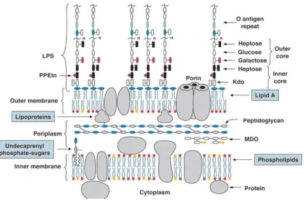
IM, the OM is a highly asymmetric bilayer which is composed by phospholipids in the
inner leaflet and lipopolysaccharide (LPS) in the outer leaflet. The LPS comprises three
domains: lipid A, core oligosaccharide, and O polysaccharides (O antigen repeats) (Figure
1.1.1) (Raetz and Whitfield, 2002). Many saccharides in the LPS are acidic and negatively
charged. The charged nature of the outside LPS layer confers bacteria with high resistance
to hydrophobic compounds; the lipid portion is almost impermeable to all charged
molecules. Cells must import and export molecules to and from the environment; therefore,
the OM contains channel-forming proteins, mainly a class of channels called porins that
Figure 1.1.1 Schematic view of the inner and outer membranes of E. coli K-12. In the
figure MDO is membrane-derived oligosaccharides; Kdo stands for
3-deoxy-D-manno-oct-2-ulosonic acid; PPEtn is short for pyrophosphorylethanolamine
1.2 The importance of membrane proteins and challenges of
crystallization
Although the lipid bilayer comprises the backbone of Gram-negative cell membranes,
membrane proteins perform the most of the specific functions, including respiration, signal
transduction and molecular transport (Byrne and Iwata, 2002). Genome analysis of
eubacterial, archaean, and eukaryotic organisms predicted that 20–30% of the open reading
frames encode integral membrane proteins (Wallin and von Heijne, 1998). Moreover,
many membrane proteins are critical components of genetic disorders and it is estimated
that membrane proteins are targeted by more than 70% of today’s drugs (Byrne and Iwata,
2002). It is therefore important to obtain high resolution membrane protein structures to
understand their functions, which will provide insights and basis for new drugs and new
therapies. The sequence and function of many membrane proteins are known, but only a
small number of structures are elucidated to date. According to the “Membrane Proteins of
Known 3D Structure” website of S. H. White
(http://blanco.biomol.uci.edu/Membr-ane_Proteins_xtal.html), there are currently (October 2009) 554 membrane protein
structures of which 204 are unique, in comparison to the more than 60000 structures of
water-soluble proteins deposited in PDB (Protein Data Bank, October 2009,
As nowadays, structure biology is becoming routine: expression, purification,
crystallization and X-ray data analysis. Why it is still so hard to solve membrane protein
structures? The barriers impact upon every step of structural determination. The
recombinant expression and the following purification of sufficient amounts of membrane
protein are often difficult. Moreover, numerous crystallization trials are necessary to get
crystals that diffract to high resolution. Because of these reasons, membrane proteins of
known structure in the database in early days are mainly naturally abundant. However,
most membrane proteins are present in the cell at low concentrations (Hunte and Michel,
2002).
All membrane proteins that are transported to the outer membrane contain an N-terminal
signal peptide, usually ~20 amino acids long, which directs them through the translocon in
the IM to the periplasm. The signal peptide is removed during translocation by signal
peptidases prior to protein folding and insertion into the membrane. For the recombinant
production of outer membrane proteins it is important to take into account the signal
peptide.E. coli is usually used as the host organism for the overexpression of either native
or heterologous recombinant outer membrane proteins. In some cases the E. coli signal
peptidase cannot process the signal peptide of heterologous proteins, resulting in the
formation of inclusion bodies or no expression (Baneyx, 1999). Manipulation of the signal
peptide of the target protein can resolve this problem (Hearn et al., 2008). Absence of the
signal peptide from the recombinant protein results in the production of the target protein
the absence of a signal peptide can be extracted and purified in the presence of urea in a
relatively straightforward manner. A successful example of this method for protein
production is the crystal structure of OmpG fromE. coli(Yildiz et al., 2006).
Extraction and solubilisation of membrane proteins is challenging because of the extensive
hydrophobic/amphiphilic surfaces. When the native membrane is removed during
extraction and purification of the protein, suitable detergents have to replace the lipid that
was in the native environment. Once extracted and purified, the membrane protein is in a
form of protein-detergent complexes, or PDCs, which is the real starting material for
membrane protein crystallization.
Membrane proteins do not crystallize readily. PDCs form crystals at the polar surfaces of
the protein protruding from the detergent micelle, and most PDCs have only limited
accessible hydrophilic surfaces (Hunte and Michel, 2002). The use of additives, such as
heptane-triol, has played a significant role in membrane protein crystallization (Wiener,
2004). A goal has been to enlarge the polar surface of the protein, most often by attaching
polar domains with specifically binding antibody fragments, to enhance crystallization
(Ostermeier et al., 1995). The use of recombinant antibody fragments, in a form of either
Fv (variable fragment, ~28 kDa) or Fab (fragment antibody binding, ~56 kDa), has been
shown to improve the crystallization of several membrane proteins (Hunte et al., 2000;
1.3 Structures of bacterial outer membrane proteins
1.3.1 General and substrate-specific porins
Porins were the first outer membrane proteins (Omps) to be studied. The first x-ray
structure of a porin from Rhodobacter capsulatus was determined in 1990 (Weiss et al.,
1990) quickly followed by OmpF and PhoE fromEscherichia coli (Cowan et al., 1992), a
porin from Rhodopseudomonas blastica (Kreusch and Schulz, 1994), OmpK36 from
Klebsiella pnueumoniae (Dutzler et al., 1999), Omp32 from Comamonas acidovorans
(Zeth et al., 2000) and Delftia acidovorans (Zachariae et al., 2006), and more recently
OmpC fromE. coli K12 (Basle et al., 2006). These porins have the same basic architecture
consisting of 16-stranded hollow β-barrels. They have all crystallized as trimers, with a
three fold axis normal the membrane plane. The crystal structure of OmpC (Basle et al.,
2006) is shown in Figure 1.3.1.1. The hydrophobic interactions between monomeric barrel
surfaces stabilize the trimer. The barrels all show eight tight turns on the periplasmic side
and eight large, irregular loops (L1 to L8) on the extracellular side. L2 interacts with the
neighboring monomer to stabilize the trimer (Phale et al., 1998). L3 folds into the barrel
forming a “constriction zone” at about midway into the barrel. As a result when viewed via
a space-filling model, the central pore has an hourglass shape. In the constriction zone,
there are acidic residues in L3 with a cluster of basic residues at the opposite barrel. These
opposite charged residues created a transverse electrostatic field which is thought to make
Figure 1.3.1.1 Structure of OmpC porin fromE. coli (PDB code: 2J1N). (A) View of the
trimer from the outside of membrane, loop L2 is colored in orange, L3 is in purple. (B)
Side view of the monomer. (C) View of the constriction zone of the channel, featuring the
opposite-charged residues that form the electrostatic field.
These porins are known as “general porins” and are water-filled channels with no
particular substrate specificity. Their biological role is to control the passage of hydrophilic
solutes based on the substrate molecular size (Nikaido, 2003). In addition to general porins,
bacteria have substrate-specific porins which are also trimeric β-barrel proteins. The
substrate-specific porins include the maltose-specific channel LamB from E. coli
(Schirmer et al., 1995) and its homolog fromSalmonella typhimurium (Phale et al., 1997),
the sucrose-specific channel ScrY from S. typhimurium (Forst et al., 1998), and from
Pseudomonas aeruginosa the phosphate-specific transporters OprP (Moraes et al., 2007),
OprD (Biswas et al., 2007) and OpdK (Biswas et al., 2008).
Monomers of LamB and ScrY have 18 β-stranded structures instead of the 16 observed in
general porins and also form a homotrimer, with the three-fold parallel to the membrane
normal. Once again L3 folds back into the β-barrel (Figure 1.3.1.2). X-ray structure
analysis of sugar-soaked LamB crystal identified a substrate translocation pathway
(Schirmer et al., 1995) with of a number of aromatic amino acids as the “greasy slide”
(Figure 1.3.1.2B) lined by polar residues the “ionic track” (Schirmer et al., 1995). The
hydrophobic face of sugar makes van der Waals’ contacts with the greasy slide while the
sugar hydroxyl groups make hydrogen bonds with the ionic track. It has been proposed that
substrates move through the channel via a sequence of hydrogen bond making and
breaking steps (Schirmer et al., 1995). The channel-lining residues are mostly conserved
between LamB and ScrY, but the differences confer the specificity.
Figure 1.3.1.2 Structure of LamB (PDB code: 1MAL). (A) View of the trimer from
extracellular side. (B) Aromatic residues that contribute to the greasy slide are shown in
purple.
The P. aeruginosa OprP is a 16-stranded antiparallel β-barrel but unusually OprP has an
extended periplasmic N terminus (Figure 1.3.1.3A) involved in stabilizing the trimer
(Moraes et al., 2007). Inside the channel, there is a nine-residue arginine “ladder” spanning
from the extracellular surface down to the constriction zone (Figure 1.3.1.3B). This ladder
is proposed to control the transit of the phosphate anion (Moraes et al., 2007). The inner
periplasmic surface is coated by lysine residues, creating an ‘electropositive sink’ that
attracts the phosphates through the channel.
Figure 1.3.1.3 Structure of OprP (PDB code: 2O4V). (A) Side view of OprP trimer, the
tricorn N terminal strands are colored red. (B) The arginine ladder located on the upper
portion of the monomer of OprP is shown in stick representation
OprD and OpdK are 18 β-strand substrate-specific porins from P. aeruginosa;OprD takes
up basic amino acids such as lysine and arginine (Trias and Nikaido, 1990) and OpdK is
responsible for taking up vanillate and related small aromatic acids (Biswas et al., 2008).
They both crystallized as monomers as shown by their crystal packing. However,
biochemical and biophysical studies showed that they form trimers in the bacterial outer
membrane. Similar to OprP, OprD contains a ladder of arginine and lysine residues. In
OpdK, only some of the ladder residues are present. Both OprD and OprK have similar
charge distribution of the residues lining the pore. However, in OprD, the periplasmic
funnel surface is negatively charged whilst in OpdK it is positively charged. This
difference seems to underlie the differing substrate specificities.
1.3.2 Dimeric β-barrel proteins
Besides trimeric 16 to 18 β-strand porins, bacteria also have dimeric β-barrel proteins. The
first dimeric structure was the outer membrane phospholipase A (OMPLA). OMPLA is an
integral membrane enzyme which catalyzes the hydrolysis of acyl ester bonds in
phospholipids in a Ca2+-dependent manner (Ubarretxena-Belandia et al., 1998).
Dimerization is essential for its function (Ubarretxena-Belandia et al., 1998).The monomer
of OMPLA is a 12-stranded antiparallel β-barrel and like the porins, has long extracellular
loops and short periplasmic turns. The interior OMPLA is polar with a hydrogen-bonding
network, however the central pore of OMPLA does not apparently function as a channel
(Snijder et al., 1999). The dimer interface is formed by one side of the barrel (Figure
1.3.2.1) and two Ca2+ ions are adjacent to the interface. The active sites lie at the outer
PapC is the translocation pore responsible for the assembly of adhesive pili on the surface
of gram-negative pathogenic bacteria acting as P pilus usher (Remaut et al., 2008). P pili
are complex extended fibers that are produced by pyelonephritic strains ofE. coli. PapC is
considered to the prototype for bacterial usher proteins and is found as a dimer arranged
similarly to that seen for OMPLA. Each PapC monomer is a 24 β-stranded monomer (the
largest barrel observed to date). A long sequence connects the two β-strands β6 and β7 and
is located in the centre of the pore forming a “plug” domain. The plug domain is a
six-stranded β-sandwich, the plane of the strands is parallel to the assumed membrane
plane. The plug completely occludes the translocation pore and is held in place by a
β-hairpin (connecting strands β5 and β6) that folds into the channel lumen. The only helix
in the structure sits above (on the extracellular side) the β-hairpin (Figure 1.3.2.2). The
inward curvature of the β5-6 hairpin creates a gap in the side of the β-barrel that is
expected to extend into the outer membrane bilayer (Figure 1.3.2.2). This partly “missing
stave” is thus far a unique feature.
Figure 1.3.2.1 Structure of dimeric OMPLA
(PDB code: 1QD6) viewed from the
extracellular side. The two black spheres
represent two calcium ions, the substrate
analogue, hexadecanesulphonyl-fluoride, at
Figure 1.3.2.2 Structure of PapC translocation domain (PDB code: 2VQI). (A) The PapC
translocation channel viewed from the extracellular side. The plug domain, the β5-6
hairpin, and the α-helix are colored purple, orange and blue, respectively. (B) β-barrel
viewed from side. Structural elements are colored as in (A), the abnormal β5-6 is labeled.
β5-6
1.3.3 TonB-dependent active transporters
In addition to trimers and dimers, monomeric β-barrel protein structures have been
determined and they include structural proteins, enzymes and transporters. The
TonB-dependent active transporters are 22 β-stranded barrel and use the proton motive
force to transport specific substrates. The proton motive force acts from the cytoplasmic
membrane through the TonB-ExbB-ExbD energy transducing complex across the bacterial
outer membrane (Moeck and Coulton, 1998). Despite low sequence similarities (~15%) the
iron-siderophore transporters FhuA, FepA, FecA from E. coli, FptA and FpvA from P.
aeruginosa, the cobalamins (e.g., cyanocobalamin, vitamin B12) transporter BtuB from E.
coli, and the colicin I receptor Cir from E. coli all belong to this class (Cobessi et al.,
2005). The strands of the β-barrel are connected by extracellular long loops and short
periplasmic turns as already seen in porins. The N-terminal domain, often referred to as
either the “plug” or “cork” domain, contains mixed four stranded β-sheets connected by a
series of short β-strands, α-helices and irregular secondary structure elements. This domain
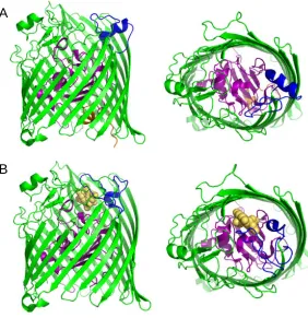
Figure 1.3.3.1 Crystal structure of (A) FecA (PDB 1KMP) and (B) FecA in complex with
iron-free dicitrate (PDB 1PO0). The two extracellular loops L7 and L8, which undergo
major conformational changes upon ligand binding, are shown in blue. The plug domain is
colored purple. The switch helix colored orange, located in the periplasmic pocket of
FecA, only observed in the unliganded conformation. The substrate iron-free dicitrate is
shown as yellow spheres.
Siderophores are secreted by bacteria to acquire iron (Ferguson and Deisenhofer, 2002)
and the iron siderophore complex is then transported through the outer membrane. All the
iron-siderophore transporters characterized to date have essentially the same structure. The
A
barrels are 60-70Å in height and have an elliptical cross section. The transporters can exist
in three states, (I) apo, (II) bound to siderophore and (III) bound to the iron-siderophore
complex. All three binding states have been characterized for FecA (Ferguson et al., 2002;
Yue et al., 2003). Comparison between the unloaded FecA and FecA bound to siderophore
(citrate) reveals only minor differences (~1Å) in the extracellular loops L7 and L8. Major
conformational changes occur when FecA binds the ferric citrate complex, including 10Å
movements in the two extracellular loops L7 and L8. Conformational changes also occur in
the plug domain and an N-terminal segment inside the periplasmic pocket, termed the
‘switch helix’. The helix is thought to unwind and become disordered (Figure 1.3.3.1B).
Structures of FhuA and FhuA - iron-ferrichrome complex have been reported by two
independent groups (Ferguson et al., 1998; Locher et al., 1998). The barrel domain and the
extracellular loops undergo only minor changes and the key differences lie in the plug
domain and the ‘switch helix’. Upon ligand binding, the plug domain is translated upward
towards the ligand and the switch helix is completely unwound and rotated ~180° in the
opposite direction of the former helix axis (Ferguson et al., 1998).
Compared to the structures of iron-siderophore transporters, BtuB and Cir have shorter
transmembrane barrels with BtuB around 55Å and Cir around 40Å. In general these
structures possess shorter extracellular loops, except that in Cir loops L7 and L8 are very
long (Buchanan et al., 2007). Upon binding its substrate colicin Ia, Cir undergoes some
conformation became more open compared with the uncomplexed Cir structure (Figure
[image:29.595.206.334.179.354.2]1.3.3.2) (Buchanan et al., 2007).
Figure 1.3.3.2 Conformational changes in Cir upon ligand binding. A superposition of
uncomplexed Cir (PDB code: 2HDF) and colicin-bound Cir (PDB code: 2HDI) shows that
the most significant change occurs in the extracellular loops L7 and L8. These loops are
colored green in the apo Cir and orange in the Cir-colicin complex.
When BtuB binds the cyanocobalamin, small conformational changes occur in several
extracellular loops and once again large changes can be observed in the plug domain. The
Ton box, a highly conserved stretch of seven amino acid residues near the N-terminus of
TonB-dependent transporters (Chimento et al., 2003) also undergoes change. Interestingly
the conformational change in FhuA upon binding TonB is relatively minor (Pawelek et al.,
2006) whilst the conformation of Ton box in BtuB-TonB complex is significant (Shultis et
conformational change of the luminal domain is required for to create a path for substrate.
The nature of this change is not known.
1.3.4 Monomeric β-barrel proteins
OmpA, OmpX, OmpW, PagP from E. coli, and NspA from Neisseria meningitides all
belong to the same 8 β-stranded barrel family (Figure 1.3.4.1). The basic architecture
comprising of long extracellular loops and short periplasmic turns is again found in these
proteins. However, there are important differences: PagP has a periplasmic N-terminal
amphipathic α-helix (Figure 1.3.4.1d) and the interior of the OmpW barrel is a
hydrophobic channel possibly involved in the transport of small hydrophobic molecules
(Hong et al., 2006). Inside OmpA, OmpX and NspA, there is an extensive
hydrogen-bonding network but no channel through which ions or other molecules could be
transported (Pautsch and Schulz, 1998; Vandeputte-Rutten et al., 2003; Vogt and Schulz,
1999). PagP has an unusual interior: the hydrophobic upper half devoid of waters and the
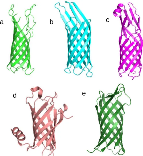
Figure 1.3.4.1 A gallery of 8-stranded β-barrel proteins. (a) OmpA (PDB code: 1BXW)
(b) OmpX (PDB code: 1QJ8), (c) OmpW (PDB code: 2F1V), (d) PagP (PDB code:
1THQ), (e) NspA (PDB code: 1P4T).
Currently, there are only two outer membrane proteins with 10-strands have been
structurally characterized. One is the protease OmpT from E. coli (Vandeputte-Rutten et
al., 2001) and the other is adhesin OpcA fromNeisseria meningitides(Prince et al., 2002).
A comparison of these two structures showed that the overall fold was similar: a long
transmembrane β-barrel that protrudes far from the lipid bilayer into the extracellular space
(Figure 1.3.4.2). The top of OmpT barrel is circular whereas in the central part of the
molecule is elliptical. In contrast, the OpcA barrel has an elliptical cross section along its
a b c
barrel axis. Both barrels are apparently accessible to water from the periplasmic side, but
both are blocked on the extracellular face.
Twelve-stranded b-barrel membrane proteins have also been reported. These include theE.
colinucleoside transporter Tsx (Basle et al., 2004) and theN. meningitidesautotransporter
NalP β-domain (Oomen et al., 2004), which has an additional α-helical peptide (Figure
1.3.4.3A) in the middle. The autotransporter EspP from E. coli has a similar structure as
NalP (Barnard et al., 2007) but unlike NalP, the β-domain of EspP begins with a short
A
[image:32.595.90.248.204.505.2]B
Figure 1.3.4.2 Structures of OmpT (PDB
code: 1I78) and OpcA (PDB code: 1K24). (A)
Side view of OmpT with 90 degree turn with
respect to each other. (B) Side view of OpcA
α-helix. However, it is predicted that full length EspP contains an amphipathic α-helix
spanning the length of the barrel pore as seen in NalP (Barnard et al., 2007). The
autotransporter Hia from Haemophilus influenzae is a 12-stranded β-barrel domain
superficially similar to that in NalP (Meng et al., 2006). Strikingly in Hia the barrel is
assembled by three subunits with each contributing four β-strands. Three α-helices, one
from each subunit, fill the central pore (Figure 1.3.4.3B). This is radically different from
other bacterial outer membrane proteins discussed thus far whose barrels are typically
formed from a single monomer.
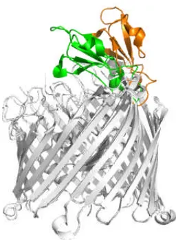
Figure 1.3.4.3 Structure of the β-domain from NalP (PDB code: 1UYN) and Hia (PDB
code: 2GRB). (A) Side view of β-domain of NalP, the N-terminal α-helix is colored red.
(B) Side view of Hia β-domain, each subunit is colored in a different color.
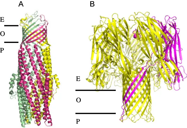
This multi subunit single barrel is also found in TolC (Koronakis et al., 2000), VceC
(Federici et al., 2005), OprM (Akama et al., 2004), MspA (Faller et al., 2004) and
α-haemolysin (Song et al., 1996). Although very different in sequence, E. coli TolC, V.
choleraeVceC andP. aeruginosa OprM share a high degree of structural similarity. They
have a 12 stranded β-barrel that is anchored to the outer membrane and is attached to a
long α-helical periplasmic barrel. Both barrels are formed from three protomers (Figure
1.3.4.4). The β-barrel is completely open in TolC, while in both OprM and VceC three
extracellular loops form a constriction. The α-helical barrel is closed at the periplasmic end
in all three proteins and is assumed to represent a “resting state”. As these three proteins
are responsible for the export of drugs and other toxic compounds from the cytoplasm
(Federici et al., 2005) the periplasmic domain must open during function.
α-haemolysin is assembled from seven subunits to form a 14 β-stranded barrel with a large
extracellular domain shaped like a mushroom with a large central hydrophilic channel
(Figure 1.3.4.4B). MspA fromMycobaterium smegmatis has a 16-stranded β-barrel formed
Figure 1.3.4.4 Structure of TolC (PDB code: 1EK9) and α-haemolysin (PDB code: 7AHL).
(A)the three TolC subunits are colored differently.. (B) In the structure of α-haemolysin,
one subunit is colored purple. The approximate position of the outer membrane (O), the
extracellular side (E) and the periplasmic side (P) are indicated.
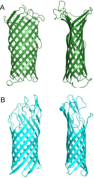
OmpG is a single protomer and is 14-stranded β-barrel functioning as a porin. OmpG has a
large channel and lacks the constriction zone seen in other porins (Subbarao and van den
Berg, 2006; Yildiz et al., 2006). Instead, OmpG has flexible extracellular loops which
undergo conformational changes under different pH conditions. At neutral pH the pore is
open (Figure 1.3.4.5A) but at pH 5.6 (or lower) the pore is blocked by loop L6 which folds
across and into the channel (Figure 1.3.4.5B).
A B
E
E O
P
O
P
A B
E
E O
P
O
Figure 1.3.4.5 Structure of OmpG in two conformation states viewed from the extracellular
side of the membrane. Loop L6 which undergoes largest conformational change is labeled.
(A) Open conformation of OmpG (PDB code: 2IWV). (B) Closed conformation of OmpG
(PDB code: 2IWW).
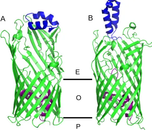
The fatty acid transporter FadL from E. coli (van den Berg et al., 2004), the aromatic
hydrocarbon transporter TodX from Pseudomonas putida and TbuX from Ralstonia
pickettii (Hearn et al., 2008) have a similar 14 β-stranded barrel. In each structure the
lumen occluded by an N-terminal “hatch domain” consisting of three short helices. The L3
consists of two antiparallel α-helices which forms a hydrophobic cleft that is thought to
bind substrate. In TodX and TbuX, the loop lies flat on the top of the barrel while in FadL
it protrudes into the extracellular environment (Figure 1.3.4.6). Based on structural data, a
model has emerged in which substrate is bound by the extracellular loop L3 before transit
into the central hydrophobic channel. A conformational change of the hatch domain allows
substrate to diffuse into the periplasm.
L6 L6
Figure 1.3.4.6 Structure features of TodX (A, PDB code: 3BSO) and FadL (B, PDB code:
1T16). The N-terminal hatch domain is colored in purple, the extracellular loop L3 is in
blue. The position of outer membrane (O), the extracellular side (E) and the periplasm (P)
are indicated.
As has been seen despite the variability, the dominant feature of outer membrane proteins
is the β-barrel architecture. β-barrel proteins are transported and assembled by a specialised
protein transport machinery involving multiple proteins (Kim et al., 2007). The outer
membrane protein 85-two-partner secretion B (Omp85-TpsB) superfamily is at the heart of
this system. Omp85-TpsB is thought to contain a C-terminal β-barrel region and an
N-terminal region with putative polypeptide-transport-associated (PORTA) domains
(Clantin et al., 2007). FhaC, an outer membrane protein from the Omp85-TpsB transporter
family, plays a role in the secretion of filamentous hemagglutinin (FHA) from Bordetella
pertussis (Clantin et al., 2007). FhaC is a monomer comprising of a 16 stranded β-barrel
A B
E
O
with the loop L6 forming a hairpin inserted into the barrel. The extracellular N-terminus of
the protein is a long α-helix (H1), and this helix spans through the transmembrane β-barrel.
The helix connects to a periplasmic module that precedes the β-barrel. The periplasmic
module consists of two structurally related PORTA domains. The domains, both 75
residues with the same topology of strand-helix-strand-strand, consist of a three-stranded
β-sheets and one α-helix (Figure 1.3.4.7). The two domains are thought to recognize the
N-terminus of FHA, providing a template for assembly and export. Translocation is
proposed to start with FHA which adopts an extended β-hairpin structure during transit
which refolds at the cell surface. After the C-terminus of FHA has reached the cell surface,
the N-terminus of FHA dissociates from PORTA1 and completes translocation (Clantin et
al., 2007). This templating function of the POTRA domains is predicted to be the basis by
which β-barrels themselves are formed. A stretch of amino acids in the unfolded protein
binds to the POTRA domain creating the first β-strand against which the other strands of
the protein assemble. This process once started is presumably spontaneous. The β-barrel
structure is extremely strong, being held together by extensive main chain hydrogen bonds.
In fact many β-barrels can be removed from the membrane by very harsh detergents
without unfolding, they are relatively insensitive (at least in the membrane portion) to
Figure 1.3.4.7 Structure of FhaC (PDB code: 2QDZ). The α-helix H1 is colored red,
PORTA 1 yellow, PORTA 2 blue, loop L6 in olive.
1.3.5 α-helix outer membrane protein
Until recently all outer membrane proteins were presumed to follow the β-barrel paradigm
and POTRA templating system provided a simple rationale for this. The structure of Wza
showed this not to be the case, it has an α-helical barrel which spans the outer membrane
(Dong et al., 2006). Wza is assembled from eight protomers with a very large central
periplasmic cavity (reminiscent of TolC) (Figure 1.3.5.1). Although the α-helical barrel is
[image:39.595.97.355.109.380.2]central cavity would seem essential for function but the trigger for any such opening is
unknown.
Figure 1.3.5.1 Structure of Wza (PDB code: 2J58). (A) Side view of Wza. The outer
membrane (OM) position is marked. (B) The channel of Wza, viewed from outside of the
cell.
The structure of Cytolysin A (ClyA) fromE. coliK12 at its membrane state (Mueller et al.,
2009) is the second example of α-helical outer membrane proteins. ClyA is a pore forming
toxin (PFT). PFTs are produced as soluble proteins by their host organisms and can
transform into the membrane integrated state (Parker and Feil, 2005). PFTs are of two
kinds: α-PFTs or β-PFTs, depending on their types of pores integrated into the membrane
(Gouaux, 1997). The structure of ClyA is of cylinder shape comprised by 12 α-helical
protomers (Mueller et al., 2009) (Figure 1.3.5.2). The crystal structure of ClyA in the
membrane state is different from its soluble state, indicating an extensive conformational
change have to take place (Mueller et al., 2009).
Figure 1.3.5.2 Structure of ClyA (PDB code: 2WCD). View from top (left). View from
side (middle). The outer membrane (OM) position is marked. View from bottom (right).
One protomer is colored magenta and the rest are green.
O
M
Chapter 2
The role of OmpC porin mutants in clinical
Summary
Clinical (as opposed to laboratory) antibiotic resistance is a complex process in which
numerous factors contribute. The clinical strains referred to in this study, were isolated
from a patient with anE. coli infection of liver cysts and suffering from Caroli Syndrome
(Low et al., 2001). The patient was treated by a variety of antibiotics over a period of four
years from December 1994. The clinical strains, isolated from the last two years of the
patient’s life, progressively exhibited greater antibiotic resistance in which the last isolate
showed resistance to a broad spectrum of antibiotic (Low et al., 2001). The later isolates all
evolved from the first single focal infection with an E. coli strain which was deficient in
OmpF expression and carried a unique ompC gene (OmpC20). While deciphering each
factor that contributed to antibiotic resistance would be beyond the scope of our study, we
wished to determine the role of OmpC mutants in the antibiotic resistance. We carried out
the MTT assay in which a control strain of E. coli lacking OmpC was transformed with
OmpC20 (isolated from the antibiotic sensitive strain) and OmpC33 (isolated from the
antibiotic resistant strain) and their ability to uptake antibiotics was compared. Results
2.1 Introduction
2.1.1 β-lactam antibiotics
Antimicrobial compounds are considered one of the most significant discoveries of the 20th
century. They have been in wide application since the 1940’s after Alexander Fleming
discovered penicillin in 1920’s. Ever since then, different antibiotics have been discovered
and developed and they fall into several different classes, the most common of which are
described below.
The β-lactam class are the most frequently used antibiotics in clinical practice and account
for about 50% of antibiotic use world-wide (Livermore, 1998). β-lactam antibiotics can be
divided into groups according to their chemical structure. They share the common
structural feature of the four-membered β-lactam (azetidin-2-one) ring. The β-lactam class
includes the penicillins and cephalosporins. Penicillins are N-acylated derivatives of
6-β-amino penicillanic acid as shown in figure 2.1.1.1. The basic structure of penicillins is
a tripeptide intermediate which can be synthesed by two amino acids: valine and cysteine.
The third amino acid (R in the diagram) of this tripeptide confers the diversity of
Figure 2.1.1.1 Molecular structures of some penicillin antibiotics (Diagram adapted from
website http://helios.bto.ed.ac.uk/bto/microbes/penicill.htm).
Cephalosporins are N-acylated derivatives of 7-β-aminocephalosporanic acid. These
compounds have the β-lactam ring fused to a 6-membered dihydrothiazine ring containing
sulphur and a double bond (Figure 2.1.1.2). Two sites (marked as R and X in the diagram)
can be modified, giving rise to a huge number of semi-synthetic derivatives.
Cephalosporins are grouped into a series of generations (Babic et al., 2006). Early
compounds such as cephaloridine are defined as the first generation; compounds resistant
to β-lactamases such as cefuroxime are the second generation and compounds not only
resistant to β-lactamases but also with enhanced antibacterial activity, such as cefotaxime,
cephalosporins have been recognized and described (Babic et al., 2006). The newer
cephalosporins generations have more enhanced Gram-negative antimicrobial abilities.
Figure 2.1.1.2 Structure of cephalosporin antibiotics. (a) cephalosporin backbone.
(b) cephaloridine. (c) cefuroxime. (d) cefotaxime.
(a) (b)
(c)
2.1.2 Antibiotic resistance
At the early days of antibiotic application, due to the effectiveness of the antibiotics,
various bacterial diseases were considered to be defeated (Kumar and Schweizer, 2005).
However, the widespread use and misuse of antibiotics has selected antibiotic-resistant
pathogens, including the multi-drug resistant strains (Levy, 1998; Neu, 1992). These
resistant bacteria are of two kinds: opportunistic and professional (Neu, 1992). These
bacteria make use of a variety of ways to combat with antibiotics. They can degrade the
drug, inactivate the drug by enzymatic modification, or alter the drug target (Davies,
1994). The resistance of Gram-negative bacteria to the β-lactam is a combination of
β-lactamases, outer membrane permeability and efflux systems (Babic et al., 2006; Kumar
and Schweizer, 2005; Lambert, 2005).
In Gram-negative bacteria, one important mechanism of resistance is the inactivation of
β-lactam antibiotics by periplasmic enzymes called β-lactamases (Massova and
Mobashery, 1998). Beta-lactamases are proposed to be descended from cell wall
biosynthetic enzymes, the so-called penicillin-binding proteins (PBPs) (Medeiros et al.,
1987). PBPs are present on the surface of the cytoplasmic membrane in almost all bacteria
and they are essential enzymes that catalyze the peptidoglycan synthesis process. The
structures of β-lactams are similar to one of the subunits that comprise peptidoglycan;
therefore when PBPs use penicillin as the substrate mistakenly for cell wall synthesis, the
following steps in cell wall synthesis are hindered and eventually bacteria killed (Babic et
it reaches the PBP target. The structural closeness between β-lactamases and PBPs allows
β-lactamases to hydrolyze and inactivate the β-lactam (Massova and Mobashery, 1998).
The permeation of antibiotics through the outer membrane of Gram-negative bacteria is
largely governed by porins (Nikaido, 2003) and altered porin properties have been
observed in many drug resistant bacteria (reviewed in (Pages et al., 2008). It has been
shown that loss of porin in K. pnuemoniae confers resistance to cephalothin and
cefotaxime (Chevalier et al., 2000). Several studies reported the alteration of porin patterns
contribute to the exclusion of antibiotics. In K. pnuemoniae strains highly resistant to a
number of cephalosporins, there is an expression shift: the narrower-channel porin,
OmpK36 (an OmpC homolog) is expressed in favor of the larger-channel porin, OmpK35
(an OmpF homolog) (Hernandez-Alles et al., 1999). It has been shown thatE. coliresistant
to certain β-lactam antibiotics have increased OmpC levels at the expense of OmpF, even
at low osmolarity (Harder et al., 1981). There are also reports on mutations in the
constriction zones in porins from pathogens to confer antibiotic resistance, such as
Enterobacter aetogenes (Thiolas et al., 2004) and Neisseria gonorrhoeae (Olesky et al.,
2002).
Active drug efflux systems in bacteria are believed to be another important mechanism of
resistance. They can be divided into four families on the basis of supermolecular assembly,
mechanism, and sequence homology (Nikaido, 1994a). These are the major facilitator
et al., 1992), resistance-nodulation-division family (RND) (Saier et al., 1994), and ABC
[adenosine triphosphate (ATP)-binding cassette] transporters (Nikaido and Saier, 1992).
To pump out drugs, the first three families use a proton-motive force as a source of energy,
whereas the ABC transporters utilize ATP. Some efflux pumps are selective and only
pump out specific antibiotics. However, there are also multi-drug resistance (MDR) pumps
that have the ability to remove a large number of unrelated and structurally diverse drugs
from the cell (Lewis, 1994). It is well known that in Gram-negative bacteria, there are
three-component export systems that contain a transporter which is a cytoplasmic
membrane protein, an outer membrane channel and a periplasmic protein. Such systems
have been identified and include: AcrAB-TolC (Figure 2.1.2.1) (Koronakis, 2003;
Figure 2.1.2.1 Structure of TolC and model of drug efflux. (Left) The structure of the outer
membrane protein TolC. (Right) The drug efflux is achieved by TolC interacting with
AcrA, a membrane fusion protein, and AcrB, an inner membrane efflux transporter (Okusu
2.1.3 MTT assay
MTT assay is one of the colorimetric assays used for measuring the activities of enzymes
reacting with MTT. The key component, MTT, in this system is
3-[4,5-dimethylthiazol-2-yl]-2,5-diphenyl tetrazolium bromide (Figure 2.1.3.1 left). MTT
is one of the tetrazolium salts that have various applications in cell biology (Berridge et al.,
2005). At the core of the tetrazolium structure, four nitrogen atoms form a positively
charged quaternary tetrazole ring (see the structure of MTT in figure 2.1.3.1), and this ring
is usually surrounded by three aromatic groups (Berridge et al., 2005). Tetrazolium salts
are water-soluble, after reduction, their colorless or weakly colored solutions can be
transformed into intensively colored formazan precipitate. The dissolved formazan can be
measured spectrophotometrically and the reaction has become the basis of biochemical
application of tetrazolium salts.
Aqueous solutions of MTT are yellowish in color. Due to the presence of phenyl rings that
provide the hydrophobic properties of the molecule, MTT is able to enter cells readily. It is
believed that MTT is reduced by NAD(P)H-dependent oxidoreductases and
dehydrogenases of metabolically active cells (Berridge et al., 2005). The reduction results
in intracellular insoluble blue-magenta colored formazan crystals (Figure 2.1.3.1 right)
which however can be dissolved in acidified isopropanol resulting in a purple solution
(Denizot and Lang, 1986; Mosmann, 1983). The assay first described by Mosmann was
and its modified version have been used extensively to characterize the cell metabolic
activity. The reduction takes place only when the cells are metabolically active; dead cells
do not react with MTT (Berridge et al., 2005; Ulukaya et al., 2008) . Cytotoxic compounds
damage or kill the cells, which result in the reduced ability for the reduction of MTT.
Therefore this reaction is often used as a measure of living cells as higher number of live
cells results in higher amount of MTT formazan hence higher spectrophotometerical
absorbance. When the live cells are treated with an agent, say antibiotics, the resulting
purple formazan can be compared with that produced by the control cells; the effectiveness
of the antibiotics can be deduced from this difference.
Figure 2.1.3.1 Yellow MTT (left) (3-(4,5-Dimethylthiazol-2-yl)-2,5-diphenyltetrazolium
2.1.4 Project background
This project has been in collaboration with Professor Ian Booth, University of Aberdeen.
We have chosen to study naturally occurring mutants of the OmpC porin from a clinicalE.
coliinfection. The OmpC mutants were isolated from a patient with anE. coli infection of
liver cysts and suffering from Caroli Syndrome. The patient was treated by a variety of
antibiotics over a period of four years from December 1994.E. coliisolates were collected,
either from blood samples or directly from liver abscesses over a two year time period
(Low et al., 2001). Measurement of the minimum inhibitory concentrations (MIC) for each
of the antibiotics used during treatment of the patient had shown that the E. coli isolates
had, in general, progressively greater antibiotic resistance (Figure 2.1.4.1). The last isolate
shows broad spectrum antibiotic resistance (Low et al., 2001). The later isolates all evolved
from the first single focal infection with an E. coli strain which was deficient in OmpF
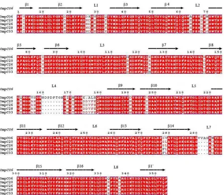
expression and carried a uniqueompCgene (OmpC20) (Low et al., 2001). OmpC20 differs
from the “standard” uropathogenic OmpCO6 (Welch et al., 2002) strain at 27 amino acids,
and with an 8 residue deletion in loop L4 and 4 residue deletion in L7 (Figure 2.1.4.2).
There is no data on when or where the changes between OmpCO6 and OmpC20 took place
(may not have been in patient), our study is focused on the change from the first isolate
(antibiotic sensitive containing OmpC20) to the final isolate (antibiotic resistant containing
OmpC33). During the infection, the OmpC gene mutated to OmpC26 (mutation D18E),
then to OmpC28 (additional mutation S271F), and finally to OmpC33 (additional mutation
R124H) (Figure 2.1.4.2). These changes correlate with increase in clinical antibiotic
Figure 2.1.4.1 Antibiotic resistance in the seven isolates (Figure from Low et al., 2001).
The OmpC protein from clinical E. coli strain No. 1, 2 and 3 are denoted as OmpC20,
isolate 4 as OmpC26, isolate 5 as OmpC28 while OmpC33 refers to isolate No.6 and 7.
The antibiotic therapy received by the patient is indicated on a time line below the MIC
table. Filled arrows indicate dates on which the antibiotic combination was changed.
β-lactam antibiotics are indicated in bold. Open arrows indicate when each isolate was
Figure 2.1.4.2 Sequence Alignment of OmpC from clinical isolates stated in Low et al
2001, and OmpCO6 from uropathogenic E. coli O6:H1 (accession no. Q8CVW1). The
alignment was carried out in Multalin (Corpet, 1988). Above the alignment, the β-strands
forming the barrel and the extracellular loops connecting the β-strands are numbered. The
β-strands are labeled β1-β16 and β1’, strand β1 from the N-terminal and strand β1’ from
the C-terminal are two parts of the same strand. The residue numbering is according to that
Clinical antibiotic resistance is often multifactorial and no single change in any protein can
be said to “cause” its occurrence. In order to investigate the contribution of the OmpC
porins to antibiotic resistance, the wild typeompCgene fromE. coliK12 (OmpCK12) was
expressed from the plasmid pOmpCWT in the first (containing OmpC20) and the last
(containing OmpC33) clinical isolates, respectively. Results showed that in the first
clinical isolate there were no significant differences in MIC in the presence or absence of
OmpCK12. However, the last transformed clinical isolate recovered susceptibility to
antibiotics, particularly for the two carbapenem antibiotics, meropenem and imipenem
(Table 2.1.4.1, courtesy Professor Ian Booth) suggesting that mutations in OmpC33 are in
[image:56.595.66.531.430.650.2]part responsible for clinical antibiotic resistance.
Table 2.1.4.1. Minimum Inhibitory Concentrations of Antibiotics (µg / ml)
Strain Meropenem Imipenem Ciprofloxacin Cefotaxime Ceftazidime
First clinical isolate (OmpC20)
0.062 0.25 1.0 0.5 0.5
First clinical isolate (OmpC20),
pOmpCWT
0.031 0.25 1.0 0.125 0.5
Last clinical isolate (OmpC33)
2.0 4.0 8.0 32 64
Last clinical isolate (OmpC33),
pOmpCWT
As the difference between OmpC20 and OmpC33 is only three residues, we wished to
isolate the factor of OmpC porin and determine the role of mutation in the clinically
2.2 Materials and methods
2.2.1 Expression of OmpC
The OmpC expression constructs were made by Professor Ian Booth, our collaborator, at
the University of Aberdeen and Dr. Vicki Bamford who worked as a postdoc in our
laboratory. The plasmid containing the variantOmpC genes were transformed into OmpC
andOmpFminusE. coliHN705 cells (Sugawara and Nikaido, 1992).
The transformed HN705 cell cultures were stored in 10% glycerol at -80ºC. A small
amount of cells were used to inoculate 10mL LB medium containing 30 µg / ml
chloramphenicol and 0.5 % (w/v) glucose for inhibiting the LamB expression. For the
growth of HN705 cells, no antibiotics were added during fermentation. Cells were grown
at 37 °C and were harvested by centrifugation. To check the expression level of OmpC,
10-15µl intact cells were incubated with SDS at 100ºC for 15min and centrifuged to
remove the unsolubilised cells before running the gel. Gels were run at 200V for 35mins in
MES running buffer, pH 7.4, following the manufacturer’s protocols (Invitrogen). Proteins
were stained by Coomassie blue staining. Molecular weight values were estimated from
the migration rates of Mark-12 markers (Invitrogen).
The intensity of the bands corresponding to OmpC protein was analyzed by the software
ImageGauge V4.21. The corresponding bands were sent to Mass-Spec for the identity
2.2.2 MTT assay without antibiotics
2.2.2.1 Influence of cell numbers and the growth stage
MTT assay were carried out using a modified procedure for the Cell Growth
Determination Kit (Sigma-Aldrich). MTT solution at concentration of 5mg/ml was directly
from the manufacturer Sigma-Aldrich. Other solutions were from the manufacturer or
made fresh in the lab and sterilised using 0.22μm membrane filter. To define the suitable
concentration of cell culture reacting with MTT, cells at exponential phase (OD600
~1.0)and stationary phase (OD600 ~2.0) were harvested, respectively. Cells were
resuspended in sterilized phosphate buffered saline (PBS, pH 7.4) and were washed three
times with PBS. A series of dilutions were made to give an OD600value range of 0.1 to 1.0.
The reaction took place at a cuvette (10mm wide) by adding 90 µl of resuspended cells
along with 10 µl 5mg/ml MTT. After incubation for 4 hours at 20 ºC, 900 µl of 0.1N HCl
(~36%, Fisher Scientific) dissolved in absolute isopropanol (99.9%, VWR International)
was added to the cuvette to solubilise the formazans. Pipetting up and down was necessary
to fully solubilise the formazans. The absorbance of each sample was measured at
wavelength of 570 nm with the background absorbance at 690 nm subtracted. The solution
for zeroing the background was 900µl 0.1N HCl dissolved in absolute isopropanol plus
100µl PBS solution. Each cell concentration was measured at least three times within an
2.2.2.2 Influence of added MTT amount
To measure the influence of the amount of MTT added to the cell culture, 5μl, 10μl, 15μl
5mg/ml MTT solution was added to 90μl cell culture, corresponding to ~5%, 10%, ~15%
of the total reaction volume, respectively. Experiments were carried out triplicate at room
temperature for 4 hours and following the protocol as described above.
2.2.2.3 Influence of cell viability
Cells at an OD600 0.4 were killed by heating at 100 ºC for 1h. 10µl MTT solution was
added to 90µl dead cell solutions and left at room temperature for reaction. MTT activity
was checked by eye-observation after 24 hours.
2.2.2.4 Influence of incubation time with MTT
Cells growing to late exponential stage (OD600 ~1.0) were harvested and washed three
times by PBS. Cells were diluted to give an OD600 0.4. 10µl MTT solution was added to
90µl cell solutions and left at room temperature for reaction for a time period of 1h, 2h, 3h,
2.2.3 MTT assay with antibiotics
2.2.3.1 The influence of antibiotic itself on MTT activity
Three antibiotics, gentamicin sulfate (Melford, UK), carbenicillin disodium (Melford, UK),
cefotaxime sodium (Sigma, Germany) were tested for their reaction with MTT. Antibiotic
solutions were made in PBS as 1M concentration as stock. The solutions were made fresh
on the day of experiment. 10µl 5mg/ml MTT solution was added to 10µl antibiotic
solutions along with 90µl PBS. The final concentration of antibiotics is approximately
100mM. After incubation for 4 hours at 20ºC, 890µl 0.1N HCl dissolved in isopropanol
was added to the solution. Measurement was carried out using the spectrophotometer
which used to measure the MTT activity with cells. The zeroing solution was 890µl 0.1N
HCl dissolved in isopropanol plus 110µl PBS.
2.2.3.2 The control strainE. coliHN705
The metabolic activity ofE. coliHN705 (OmpCandOmpFknocked out), was investigated
by the MTT assay. Cells growing to the exponential stage and stationary stage were
harvested, washed and diluted with PBS to give a series of OD value ranging from 0.1 to
1.0. The measurement was taken place as described in section 2.2.2.1.
2.2.3.3 MTT assay with antibiotics
Three antibiotics, gentamicin sulfate (Melford, UK), carbenicillin disodium (Melford, UK),
HN705 transformed with OmpC20 (HN705-OmpC20) and cells of HN705 with OmpC33
(HN705-OmpC33), respectively. Cells were all grown to OD ~1.0 and harvested by
centrifugation, washed with PBS three times and diluted into OD 0.4. Antibiotics dissolved
in PBS at 1M stock were made fresh on the day of experiment or stored at -20ºC for future
use.
We first tested a big range of antibiotic concentrations then zoom into a smaller scale for
further tests. A series of 10µl antibiotic solutions along with 10µl 5mg/ml MTT were
added to 90µl of resuspended cells. After 4 hours incubation at 20ºC the cells containing
the resulting purple crystals were resuspended by addition of 890 µl of 0.1N HCl dissolved
in isopropanol. The absorbance of each assay was measured at wavelength of 570 nm with
the background absorbance at 690 nm subtracted. Each assay was performed at least with
two independent experiments with each sample triplicate. The metabolic activity of cells
without the addition of antibiotics was defined as 100%. After incubation with antibiotics,
the metabolic activity of cells decreased. The metabolic inhibition rate is defined as the
ratio of the amount of cell death to the total number of live cells. It is calculated from the
570nm absorbance mean value. For example, the mean value of 570nm absorbance of cells
without any antibiotics is 0.8, the 570nm absorbance value of cells incubated with 25mM
carbenicillin is 0.3, then by definition, the metabolic inhibition is (0.8-0.3)/0.8 *100%=
2.3 Results
2.3.1 OmpC expression
HN705 cells, the strain with ompF and ompC knocked out, was grown at the same LB
medium as that for OmpC20 and OmpC33 cells but without antibiotics and glucose.
HN705 cells were used as a control for the OmpC expression. Figure 2.3.1.1 shows the
result of whole cell SDS-PAGE. It shows OmpC20 and OmpC33 are both expressed
(indicated by the pointing arrow) with no significant difference on the expression level.
Analysis by ImageGauge confirmed this observation with corresponding gel bands the
same UV absorbance (indicated by the numbers listed). It is noticed that HN705 cells have
a different expression pattern except for the OmpC protein (figure 2.3.1.1). The OmpC
Figure 2.3.1.1 Comparison of expression level of OmpC20 and OmpC33 in HN705. Whole
cells in two different batches were solubilized and analysed in separate gels (left, right).
The lanes are marked as shown above the gel. The arrow indicates the location of OmpC
which is absent in HN705 cells. The intensity of the corresponding bands were analysed by
the software ImageGauge and the UV absorbance value was indicated.
1714279 1712703
2.3.2 MTT assay without antibiotics
2.3.2.1 Influence of cell numbers and growth stage
With a defined concentration, MTT must have a maximum ability to act on the cells. To
find out the suitable cell concentration, a series of dilution from the concentrated cell
culture were made and tested. Table 2.3.2.1a shows the MTT formazan absorbance from
OmpC20 and OmpC33 exponential stage cells. Each reaction carried out at individual
cuvette, the measured value at 570nm subtracting the measured value at 690nm is to
exclude the difference factor of cuvette absorbance. It was noticed that at 690nm the
absorbance value were negative, this was because the zeroing solution had a higher
absorbance at 690nm. We define real 570nm absorbance value by the following equation:
Real 570nm absorbance = Measured 570nm absorbance – measured 690nm absorbance
We plotted the real 570nm absorbance against the cell OD600values. Figure 2.3.2.1a shows
the effect of cell numbers on the 570nm absorbance corresponding to the MTT reduction
activity. It suggests before OD600 0.4, the cell number dominates while after OD600 0.4,
MTT reaches its saturation level as a higher number of cells do not result in relatively
higher activity.
In a similar manner, Table 2.3.2.1b shows the absorbance measured for OmpC20 and
OmpC33 cells harvested from the stationary phase. Figure 2.3.2.1b showed a similar trend
of 570nm absorbance values verses the cell numbers. The two independent experiments