The tactile afferents that innervate the inside of the hand
signal the transformation of soft tissues that occurs when the hand interacts with objects and thus provide infor‑ mation about the physical properties of the object and the contact between the object and the hand. People with impaired tactile sensibility have difficulties with many everyday activities because the brain lacks the infor‑ mation about mechanical contact states that is needed to plan and control object manipulations. Vision pro‑ vides only indirect information about such mechani‑ cal interactions, and proprioceptive afferents exhibit low
sensitivity to mechanical fingertip events1–4.
In this Review, we address emerging concepts regard‑ ing the use of tactile information by the brain in manipu‑ lation tasks. In doing so, we discuss the notion that the planning and control of manipulation tasks is centred on mechanical events that mark transitions between consecutive action phases and that represent subgoals of the overall task. We highlight recent findings that help explain the speed with which the brain detects and classi‑ fies tactile fingertip events in object manipulation. Finally, we discuss multisensory representation of action goals in object manipulation. Our account differs from a recent review of tactile signals in manipulation5 by emphasizing the use of these signals in the control of manipulatory tasks, by considering how other sensory signals contrib‑ ute to this control and by discussing the central neural mechanisms involved in manipulation tasks.
Tactile sensors encoding fingertip transformations When humans manipulate objects, the brain uses tac‑ tile afferent information related to the time course, magnitude, direction and spatial distribution of contact forces, the shapes of contacted surfaces, and the friction between contacted surfaces and the digits. The inside of
the human hand is equipped with four functionally dis‑ tinct types of tactile afferents (TABLE 1; reviewed in more
detail in REfS 5,6). FA‑I (fast‑adapting type I) and SA‑I
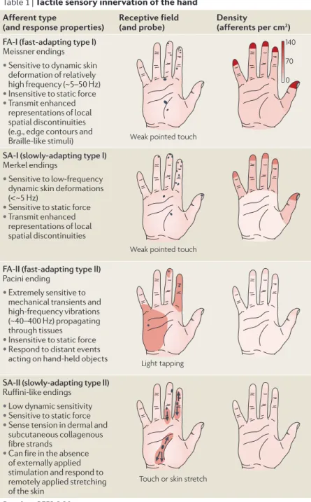
(slow‑adapting type I) afferents terminate superficially in the skin, with a particularly high density in the fingertips. FA‑Is exhibit sensitivity to dynamic skin deformations of relatively high frequency7,8, whereas SA‑Is are most easily excited by lower‑frequency skin deformations7,8 and can respond to sustained deformation. There are more FA‑I afferents than SA‑I afferents in the fingertips (TABLE 1),
reflecting the importance of extracting spatial features of dynamic mechanical events, such as the skin forming and breaking contact with objects or scanning across a textured surface.
FA‑II and SA‑II afferents innervate the hand with a lower and roughly uniform density and terminate deeper in dermal and subdermal fibrous tissues. FA‑II afferents are optimized for detecting transient mechani‑ cal events7–10. Hundreds of FA‑II afferents, distributed throughout the hand, can be excited when hand‑held objects contact or break contact with other objects11. SA‑II afferents can respond to remotely applied lateral stretching of the skin12,13 and can be sensitive to the tan‑ gential shear strain to the skin that occurs during object manipulation2,11. SA‑II‑like afferents are found in most fibrous tissues (such as muscle fascias and joint capsules and ligaments)14 and there is evidence that they can act as proprioceptors (BOX 1).
Traditional studies on tactile sensing that examine correlations between afferent signals and perceptual (declarative) phenomena evoked by gently touching pas‑ sive digits (for reviews see REfS 6,14–20) provide little
information about the encoding and use of tactile infor‑ mation in object manipulation for several reasons: the control processes that are active in manipulation operate
*Physiology Section, Department of Integrative Medical Biology, Umeå University, SE‑901 87 Umeå, Sweden.
‡Department of Psychology and Centre for Neuroscience Studies, Queen’s University, Kingston, Ontario, K7L 3N6, Canada.
Correspondence to R.S.J. e‑mail: roland.s.johansson@ physiol.umu.se
doi:10.1038/nrn2621 Published online 8 April 2009 Tactile afferents
fast-conducting myelinated afferent neurons that convey signals to the brain from low-threshold
mechanoreceptors in body areas that actively contact objects — that is, the inside of the hand, the sole of the foot, the lips, the tongue and the oral mucosa.
Proprioceptive afferents fast-conducting myelinated afferents that provide information about joint configurations and muscle states. These include mechanoreceptive afferents from the hairy skin, muscles, joints and connective tissues.
Coding and use of tactile signals
from the fingertips in object
manipulation tasks
Roland S. Johansson* and J. Randall Flanagan
‡Abstract | During object manipulation tasks, the brain selects and implements action-phase
controllers that use sensory predictions and afferent signals to tailor motor output to the
physical properties of the objects involved. Analysis of signals in tactile afferent neurons and
central processes in humans reveals how contact events are encoded and used to monitor
and update task performance.
Table 1 | Tactile sensory innervation of the hand Afferent type
(and response properties) receptive field (and probe) Density (afferents per cm2)
FA-I (fast-adapting type I)
Meissner endings • Sensitive to dynamic skin
deformation of relatively high frequency (~5–50 Hz) • Insensitive to static force • Transmit enhanced
representations of local spatial discontinuities (e.g., edge contours and Braille-like stimuli)
SA-I (slowly-adapting type I)
Merkel endings
• Sensitive to low-frequency dynamic skin deformations (<~5 Hz)
• Sensitive to static force • Transmit enhanced
representations of local spatial discontinuities
FA-II (fast-adapting type II)
Pacini ending
• Extremely sensitive to mechanical transients and high-frequency vibrations (~40–400 Hz) propagating through tissues
• Insensitive to static force • Respond to distant events
acting on hand-held objects
SA-II (slowly-adapting type II)
Ruffini-like endings • Low dynamic sensitivity • Sensitive to static force • Sense tension in dermal and
subcutaneous collagenous fibre strands
• Can fire in the absence of externally applied stimulation and respond to remotely applied stretching of the skin
Data from REfS 6,20.
Nature Reviews | Neuroscience
Weak pointed touch
Weak pointed touch
Light tapping
Touch or skin stretch
140 70 0
Action-phase controller A learned sensorimotor ‘control policy’ that uses specific sensory information and sensory predictions to generate motor commands to attain a sensory goal. Sensorimotor control point A planned contact event in which predicted and actual sensory signals are compared to assess the outcome of an executed action-phase controller.
largely subconsciously and very rapidly, the use of tac‑ tile signals differs across tasks and task phases, and the forces that are involved in manipulation typically differ from the forces that are present during gentle touch.
The information that a mechanoreceptive affer‑ ent conveys depends on several factors, including the branching of the nerve terminal, the mechanical proper‑ ties of the end organs of the nerve endings, the anchor‑ ing of the end organs in the surrounding tissues and, not least, the overall mechanical deformational properties of these tissues. Thus, the distributed patterns of stresses and strains that develop in the skin and the underlying tissues when a fingertip interacts with an object affect both afferents that terminate in contact areas and affer‑ ents that terminate remotely21–23. This implies that the
actual receptive field of an afferent can be considerably larger than the classical cutaneous receptive field deline‑ ated by lightly touching the hand with a pointed object
(TABLE 1). Consequently, models of neural encoding of
tactile stimuli that visualize the receptor mosaic as a two‑dimensional pixel‑like array of densely localized sensors distributed over a flat skin surface15–17 are not viable for predicting tactile signalling in manipulation tasks. Importantly, the functional overlap of large recep‑ tive fields can enhance rather than degrade the encoding of spatiotemporal information24,25.
Owing to the mechanical properties of the fingertip, the mapping between fingertip events and afferent responses is highly complex16,22,23. Simply looking at how the pattern of stress develops in the contact area when the fingertip contacts a flat surface demonstrates this complexity (BOX 2). Researchers have attempted to model
the mechanics of the fingertip while incorporating its composite material properties, with the goal of predict‑ ing the responses of populations of tactile afferents to various fingertip stimuli26–34. However, no model yet possesses the level of realism that satisfies this goal. Contact events and action goals in manipulation Dexterous manipulation tasks can be broken down into a series of action phases, usually delimited by the mechanical events that represent subgoals of the task (see REfS 5,35 for details). For example, when picking
up a hammer to strike a nail, contact between the digits and the handle marks the end of the reach phase; the braking of contact between the hammer and the support surface marks the end of the load phase; and contact between the hammer head and the nail marks the end of the swing phase.mechanical events involved in manipu‑ lation generate specific patterns of activity in the tactile afferents and often also in auditory and visual afferents. Thus, manipulation tasks can be specified as a sequence of specific sensory events linked to subgoals.
To achieve these subgoals the brain has to select and execute appropriate action-phase controllers5(BOX 3). In order to accurately predict the required motor output and associated sensory events, action‑phase control‑ lers must have information about the properties of the objects involved and the current state of the motor appa‑ ratus. If predictions are erroneous, corrective actions can be launched based on real‑time sensory information. However, because of the long time delays in sensorimotor control loops engaged in corrective actions (~100 ms), dexterous manipulation is not possible unless predictions are accurate5. In order to smoothly link action phases, the predicted terminal sensory state of the active con‑ troller could be used as the initial state by the controller responsible for the next action phase. If the brain relied on peripheral afferent information to obtain this state information, stuttering phase transitions would occur.
The comparison of predicted and actual sensory sig‑ nals can be used to monitor task progression and detect performance errors (BOX 3). Contact events, which denote
completion of action goals, represent crucial sensorimotor control points because they give rise to discrete sensory
Transcranial magnetic stimulation
(TMS). A non-invasive technique that can be used to induce a transient interruption of normal activity in a restricted area of the brain. It is based on the generation of a magnetic pulse near the area of interest that induces small eddy currents that stimulate neurons.
Grasp stability
The control of grip forces such that they are adequate to prevent accidental slips but not so large that they cause unnecessary fatigue or damage to the object or hand.
corrective actions can be implemented. The nature of the correction is specific to the sensory signals, the control‑ ler and the current state of the system and environment. Furthermore, if errors occur, memory representations of object properties can be updated to improve subsequent predictive control.
The context‑dependent nature of corrective responses is reminiscent of finite‑state control systems that operate by implementing rules based on IF, AnD and THen arguments. Such systems have been used to model the control of phase transitions36 and corrective actions37 during walking. For example, the transition from the stance phase to the swing phase has been modelled with the following rules: IF the extensor force is low AnD the hip is extended AnD the contralateral leg is loaded, THen flex36. Key concepts of finite‑state control are that multiple sensory inputs are evaluated continuously to judge the state of the rules and that different states can give rise to different motor outputs. Furthermore, the rules and the weighting of sensory inputs can be adapted based on the anticipated state of the system. Task‑ and phase‑specific use of sensory information in object manipulation is presumably acquired when we learn the underlying basic action‑phase controllers, which occurs gradually during development38–43.
Representation of action goals in tactile afferents
Grasp contact. Often the first goal in manipulation tasks is to ensure a stable grasp of the object44–48. Because of the low stiffness of the fingertip at low contact forces11,49, its shape transforms briskly when an object is initially contacted (BOX 2). Contact responses, especially in FA‑I
afferents but also in SA‑I and FA‑II afferents, provide information about the outcome of the reach phase11
(BOX 3). Similar responses occur at the end of the unload
phase when the digit breaks contact with the object. For each digit, ensembles of afferents convey infor‑ mation about contact timing, the contact site on the digit and the direction of the contact force (BOX 3). The spa‑
tial centre of the afferent population response is related to the primary contact site on the finger50, whereas the recruitment of afferents and their firing rates reflect force intensity2,12,16,51,52. The firing rates of individual
tactile afferents are each broadly tuned to a preferred direction of contact force, allowing patterns of activity in ensembles of afferents to provide information on force direction22(fIG. 1a).
Contact events between digits and objects provide sensorimotor control points for the reach‑phase con‑ troller. Behavioural studies indicate that this informa‑ tion is important for monitoring the accuracy of reach commands and making necessary adjustments in future reaches53–58. Disturbances of the contralateral primary sensorimotor cortex by weak single‑pulse transcranial magnetic stimulation (TmS) delivered just before contact59 or by TmS‑induced ‘virtual lesions’ (REf. 60) delay the
implementation of the subsequent load‑phase control‑ ler, presumably because of disturbed processing of tactile afferent information and/or sensory predictions. Similar but smaller delays arise with TmS perturbations of the left anterior intraparietal area61 and the left dorsal premotor cortex62, suggesting that these areas are also involved in processing tactile information for object manipulation.
Grasp stability. Once they are in contact with an object, the digits usually apply tangential forces to its surfaces in order to move and manipulate it (for example, load forces in order to lift an object). To ensure grasp stability
they also apply grip forces normal (perpendicular) to the surfaces: these change in phase with, and proportional to, the applied tangential loads63–79(BOX 3). The control of grip forces is based on predictions of objects’ dynami‑ cal properties that influence the mapping between arm motor commands and resultant tangential forces and torques72,75,80,81.
Dexterous manipulation involves adapting the bal‑ ance between grip and load forces to object surface properties, a capacity that is lost with impaired dig‑ ital sensibility63,82–89. Healthy people adapt the balance between grip and load forces to different frictional con‑ ditions, using stronger grip forces with more slippery surfaces63,67,90. In fact, the local frictional conditions can tailor the grip‑to‑load force ratios at individual dig‑ its91–95. Similarly, people adjust grip and load forces to the shape of the object in order to ensure grasp stabil‑ ity64,83,84. As a result of these adaptations, excessive grip force is avoided: grip forces are normally 10–40% greater than the minimum required to prevent slips.
The initial contact responses in tactile afferents pro‑ vide information about surface properties, which can be compared with predictions based on visual cues and/or sensorimotor memory (BOX 3 and see below). A mis‑
match between predicted and actual sensory information can trigger corrective actions leading to changes in grip‑ to‑load force ratios commencing ~100 ms after contact and to an updating of the representation of the surface properties that is used in future interactions with the object (see REf. 5 for further details). Visual cues about
the shape of the object can provide the information required to make these predictions83,84. However, shape information provided by tactile signals after contact can override predictions based on visual cues.
evidence indicates that it is the responses of FA‑I afferents to initial contact that are most important for
Box 1 | Cutaneous afferents contribute to proprioception
Proprioception depends on both central217 and peripheral signals. In the 1960s it was generally thought that the latter were provided through afferents from joints218,219, and in the 1970s the focus shifted almost exclusively to muscle spindles220,221. However, recent microneurography studies in humans have demonstrated a role for cutaneous mechanoreceptors as well, which faithfully signal strain patterns in the skin that change during movement. In contrast to tactile afferents, which supply glabrous (hairless) skin areas, cutaneous afferents in hairy skin, which is more elastic and more loosely anchored to supporting tissues, can respond vigorously to changes in skin strain during movements of adjacent joints. This applies to hairy skin on the face222, the back of the hand223–226 and the lower limb227,228. Analyses of static and dynamic sensitivity indicate that cutaneous afferents are at least as sensitive to joint angle changes as muscle spindles are224,226. The quantitative properties of SA‑II (slow‑adapting type II) afferents are most suited to encode joint configurations, but those of SA‑III afferents (a receptor class that is present only in the hairy skin227) are a close second. The finding that skin stretching can produce movement illusions demonstrates that cutaneous signals contribute to kinaesthesia229–232.
Nature Reviews | Neuroscience
Normal stress (kPa)
Tangential stress (kPa)
Tangential- stress direction
Fn
b Low friction (latex membrane + talcum powder)
a High friction (latex membrane)
5 mm
1 10 20 20 40 60
1
Ulnar Distal
0.5 N 2 N 4 N Fn 0.5 N 2 N 4 N
0.1 s 0
1 2 3 4
Normal Vertical Centre
rod Contact plate
30o
Latex membrane (20 µm) Centre rod (∅ = 1 mm) Force transducer, 3D (centre rod) Force transducer, 3D (total force)
Contact surface (∅ = 50mm)
0.5 mm
d c
nail positio n
force ( Fn ) Force motor
(3D servo)
C
on
ta
ct
Fn
(N)
encoding the amount of friction between fingertips and object surfaces96. A slippery contact tends to excite FA‑I afferents more strongly than a less slippery contact
(BOX 2). most studies that have examined shape encod‑
ing by tactile afferents have used stimuli that generate small localized skin deformations and engage relatively few afferents in the immediate area of contact15,16,97–102.
By contrast, manipulation of everyday objects engages afferents all over the fingertip. Accordingly, the curvature of such objects influences firing rates in most FA‑I, SA‑I and SA‑II afferents in the fingertips, and patterns of firing in ensembles of tactile afferents can provide information on curvature23(fIG. 1b). SA‑II afferents probably provide coarser information about object shape and contact
Box 2 | Complex mechanical properties of the fingertip
The development of the pattern of stress in the contact area when the fingertip applies a normal (perpendicular) force to a flat surface illustrates the fingertip’s complex deformational properties. The ‘fingerprints’ in part a of the figure illustrate the distributions of normal and tangential stresses when the fingertip contacts a stationary surface at three different normal force levels (Fn). Part b shows corresponding stresses after the surface was made more slippery. Contact with the more slippery surface results in lower overall tangential stress because of lower frictional forces. That is, there is more localized frictional slipping and creeping in the contact area with the more slippery surface. Such slip and creep events constitute the basis for frictional encoding by tactile afferents: a slippery contact generally excites FA‑I (fast‑adapting type I) afferents more strongly than a less slippery contact96. These principles agree with recent mechanical models of shear strain and stress distributions in the contact area and with the observation that friction between a planar surface and an artificial finger can be estimated simply by pressing the finger against the surface 34. Local normal and tangential stresses in parts a and b were measured through a thin rod located in the centre of, and flush with, a flat contact surface (c). A three‑dimensional (3D) servomechanism (servo)22 repeatedly moved the fingertip to make it contact the plate. For each movement, Fn was servo‑controlled to increase linearly to 4 N, whereas the net force applied tangentially to the contact surface was servo‑controlled at 0 N. By shifting the horizontal position of the contact surface and the attached transducers between movements, the distribution of normal and shear stresses in the contact area was mapped at 1 mm spatial resolution. As demonstrated by the position of the fingernail during normal force application (d), the vertical stiffness of the fingertip increases with the contact force.
Nature Reviews | Neuroscience
Vertical position Load force
Grip force
0.2s
Sen
sory pr
ediction
s
Ta
ct
ile signals
Comparison Mismatches triggercorrective actions
Unload Load
Reach Lift Hold
Digits contact object
Object lifts off surface
Object approaches goal height
Object contacts surface
Digits release object
FA-I SA-I FA-II SA-II SA-II
Transient mechanical events • Making and breaking contact between hand-held objects and other objects • Weight information (indirect at lift-off) Contact responses
• Contact timing • Contact sites on digit • Direction of contact force • Friction information • Local shape at grasp sites
Release responses • Breaking contact between digit and object
Motor c
ommands
Replace Action-phase controllers
(action phases) Task subgoals (control points)
Predicted tactile subgoal events
Actual tactile subgoal events
a
b
Ensembles of tactile afferents encode:
Grip force Vertical movement
Load force
forces than the SA‑I or the FA‑I populations, because they innervate the fingertip more sparsely103(TABLE 1).
Occasionally, frictional slips occur that rapidly shift the object load from the slipping digit to the other
digits engaged in gripping the object. Such load shifts, which are reliably signalled by FA‑I afferents2,96, trigger a phase‑appropriate corrective action that results in a lasting update of grip‑to‑load force ratios at the engaged
Box 3 | Sensorimotor control points in a prototypic object manipulation task
Manipulation tasks are characterized by a sequence of action phases separated by contact events that define task subgoals. Consider the task of grasping an object, lifting it from a table, holding it in the air and then replacing it (see part a of the figure)63. The goal of the initial reach phase is marked by the digits contacting the object and the goal of the subsequent load phase is marked by the breaking of contact between the object and the support surface. These and subsequent contact events correspond to discrete sensory events that are characterized by specific afferent neural signatures in the tactile modality (part b) and often in the auditory and visual modalities (not shown). Such signatures specify the functional goals of successive action phases. In addition to generating motor commands, each action‑phase controller predicts the sensory events that signify subgoal attainment. Thus, the brain can monitor task progression and produce corrective actions if mismatches are detected. Recordings of tactile afferent signals in single neurons of the human median nerve during the lift and replace task11 have shown that there are distinct discharges from the fingertips at four points corresponding to subgoal events (part b): responses primarily in FA‑I (fast‑adapting type I) afferents when the object is contacted and released and responses in FA‑II afferents related to the transient mechanical events that accompany the object lifting off and being replaced on the support surface. In addition to responses to distinct contact events, many SA‑I (slow‑adapting type I) and SA‑II afferents discharge when static forces are applied to the object. Figure is modified, with permission, from REf. 5 (2008) Academic Press.
Nature Reviews | Neuroscience
Time (ms) 0
Fn
(N)
250 125
125
Protraction Retraction
4
100 Impulses per s
100
Impulses per s Impulses per s100
FA-I
SA-I FA-I
Plateau
SA-I
Positively correlated with curvature Negatively correlated with curvature
a b
Radius = 10 mm
Radius = 5 mm
Radius = 10 mm
Radius = 5 mm Flat
Flat 0
Fn (N) 4
0
Fn (N) 4
Distal Proximal
Ulnar
Radial
Normal only 20°
Flat Radius = 10 mm Radius = 5 mm
Radial Proximal Distal
Ulnar
FA-I
Neural events
Papillary whorl
Forward internal models Neural circuits that mimic the behaviour of the motor system and environment and capture the mapping between motor commands and expected sensory consequences.
digits63,73,91 — for example, an increase in grip force dur‑ ing the hold phase or a slowing down of the load force rate during the load phase63.
little is known about the central neural architecture that supports the control of grasp stability. For the basic grip force–load force coupling, brain imaging studies suggest that the right posterior parietal cortex104 and the bilateral cerebellum105,106 have important roles, at least in tasks performed with the right hand. Computational studies have referred to the cerebellum as a principal brain structure for the storage of forward internal models
that support predictive motor control105,107. However,
patients with cerebellar lesions exhibit various impair‑ ments of grip force–load force coupling108–113, and so the role of the cerebellum is far from clear. Furthermore, basic grip force–load force coupling seems to be sur‑ prisingly robust to a variety of cerebral lesions114–116, and TmS‑induced lesions of the primary sensorimotor cor‑ tex60, the premotor cortex62 and the posterior parietal cortex61 have shown only subtle effects on this coupling. evidence from patients with abnormal corticospinal projections suggests that grip force–load force cou‑ pling that supports grasp stability can be implemented independent of signals in fast corticospinal pathways117. Figure 1 | encoding of fingertip force direction and contact surface shape. a | Impulses in an FA-I (fast-adapting type I) afferent in response to repeated (n = 5) application of force stimuli in different directions (colour coded in schematic and traces). The top trace for each direction shows the instantaneous discharge rate averaged over the five trials. The time course of force application, illustrated with the normal force component (Fn), is also shown. The responsiveness of most FA-I, SA-I (slow-adapting type I) and SA-II afferents is broadly tuned to a preferred direction of force. For example, the afferent shown responded most intensely to tangential force increases in the proximal direction but also responded to tangential force increases in other directions. Across afferents, the preferred directions are distributed all around the angular space. b | Impulses in two FA-I and two SA-I afferents in response to repeated (n = 5) stimuli with normal forces applied to a flat surface and two spherically curved surfaces. The left and right panels for each afferent type show example afferents with response intensities that increased and decreased, respectively, with the increase in curvature. Afferents with increased response intensity tend to terminate centrally in the contact area and those with decreased response intensity terminate at the sides and end of the fingertip. Part a is reproduced, with permission, from REf. 22 (2001) Society for Neuroscience. Part b is modified, with permission, from REf. 23 (2003) Blackwell Science.
even less is known about the central neural mecha‑ nisms that support adaptation of grip‑to‑load force ratios to changes in an object’s surface properties. TmS stimulation of the left anterior intraparietal area ~150 ms before grasp contact seems to disturb this adaptation in object lifting, resulting in inflated grip forces61. likewise, TmS‑induced temporary lesions targeting the primary motor cortex can also perturb the control of grip‑to‑load force ratios62,118,119. In addition, various central neural dis‑ ruptions can result in elevated grip forces during object manipulation, including those that result from cerebral stroke114,115, amyotrophic lateral sclerosis120, cerebellar disease110–113, basal ganglia disease116,121–123 and writer’s cramp124,125. Accordingly, an increased grip‑to‑load force ratio may reflect a ‘default’ strategy used in various con‑ ditions to overcome the failure in effectively adapting the force coordination for grasp stability126.
Contacts between objects in the hand and other objects.
ensembles of FA‑II afferents that terminate throughout the hand signal the incidence and dynamic aspects of contact between hand‑held objects and other objects11. Such events occur, for example, in tool use and in lifting tasks when the contact between a grasped object and the support surface breaks and when the object contacts the support when it is being replaced (BOX 3). When
we lift an object, information about its weight cannot be obtained until the object breaks contact with the supporting surface. Therefore, efficient lifting involves making accurate predictions of the required load force. Such predictions may come from previous experience of lifting that particular object127, from learned size–weight associations for the kind of object being lifted128–132 or from learned links between arbitrary visual or auditory cues and an object’s weight133,134.
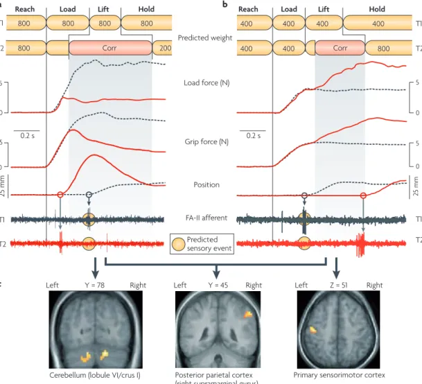
If these predictions are erroneous, corrective action programmes triggered by the mismatch between the pre‑ dicted and the actual sensory events related to lift‑off are launched automatically (fIG. 2a,b). In addition, the
memory supporting the weight prediction for the object is updated. Often, a single lift can efficiently bring about such updating127,129, although repeated interactions with the object may be required for adequate updating under unusual conditions128–131,135,136.
Recent experiments using event‑related functional mRI (fmRI) during lifting tasks involving the right hand suggest that the right inferior parietal cortex has an important role in detecting mismatches between predicted and actual weight137(fIG. 2c). The connectiv‑ ity of this region, which receives inputs from sensory cortices and has reciprocal connections to various motor regions, seems to be suitable for detecting and classify‑ ing performance errors. Results from TmS studies sug‑ gest that the posterior parietal cortex is also crucial for dynamic error detection in visually guided reaching and reach‑to‑grasp actions138,139. fmRI results indicate that both the primary sensorimotor cortex and the cere‑ bellum are engaged in implementing corrective action programmes triggered by poor weight prediction, but in a way that depends on the nature of the mismatch between the predicted and the actual weight137. When
the weight of an object is underestimated (fIG. 2b), the
sensorimotor cortex shows greater activation during the increase in fingertip forces that occurs while probing for tactile events signalling lift‑off than when the weight is accurately predicted (fIG. 2c). There is decreased activa‑
tion in the cerebellum in such instances. The opposite pattern of activity occurs when theweight is overesti‑ mated (fIG. 2c). That is, there is increased activation in the
cerebellum and decreased activation in the sensorimotor cortex. Thus, neural activity in the cerebellum seems to increase during the implementation of corrective actions only when information about the object’s actual weight, obtained at lift‑off (fIG. 2a), is already available (that is,
when the object has been lifted but its weight is less than predicted). This observation agrees with results which suggest that the anterior cerebellar cortex is involved in recruiting internal representations of object properties140 and also agrees with the general notion that transmit‑ ting sensorimotor knowledge from the cerebellum to the motor cortex allows efficient execution of learned motor responses141. TmS‑induced lesions of the primary sensorimotor cortex contralateral to the lifting hand can disrupt weight prediction in object lifting based on som‑ atosensory information acquired in a previous lift142. By contrast, such lesions do not disrupt predictions of object weight based on learned arbitrary colour cues, whereas TmS‑induced lesions of the dorsal premotor area do142. Importantly, these results suggest that anticipatory para‑ metric adaptation of action‑phase controllers to object properties can rely on partly distinct neural networks, depending on the context in which the underlying repre‑ sentation was formed. With respect to predicting object weight on the basis of size, observations in a patient with a left temporoparietallesion suggest that the left parietal cortex is required143. Taken together, these studies pro‑ vide interesting examples of how the different brain areas that support control mechanisms in object manipulation can be teased apart.
Spike timing offers fast afferent information
Rate coding versus relative spike timing.Following Adrian’s discoveries in the 1920s144, most studies have assumed that tactile afferents convey information in their firing rates (rating coding)14–19,145. This requires a given neuron to fire at least two impulses. However, the speed with which the brain detects and classifies prediction errors in the tactile modality and launches corrective actions implies that some information can be transmitted even when most of the afferents recruited have only had time to fire one impulse146. This suggests that the relative timing of impulses in individual tactile afferents in ensembles conveys important information.
Recently, it has been demonstrated that the relative timing of the first spikes that are elicited in ensembles of tactile afferents when objects are contacted provides precise information about the shape of the contacted surface as well as the direction of the force exerted on the hand146, and that it does so fast enough to account for the speed with which tactile signals are used in object manipulation tasks. Changes in either surface shape or force direction can alter the first‑spike latency
Nature Reviews | Neuroscience
25 mm
5 5
0
0
T2 T2
T1
0.2 s 0.2 s
Predicted weight
Corr 200 Corr
Primary sensorimotor cortex T1
Reach Load Lift Hold Reach Load Lift Hold
400 400
400 400 800
400 400
800 800
800 800 800
Posterior parietal cortex (right supramarginal gyrus) Cerebellum (lobule VI/crus I)
Predicted sensory event FA-II afferent Grip force (N) Load force (N)
Position
25 mm
5 5
0
0 T1 T2
T1 T2
Left Y = 78 Right Left Y = 45 Right Left Z = 51 Right
a
c
b of individual afferents without significantly affecting the distribution of first‑spike latencies in the afferent popu‑ lation as a whole. Changes in contact parameters can therefore reliably influence the sequence in which dif‑ ferent members of afferent populations first respond to
tactile events. For a code based on relative spike timing to be effective, sufficient numbers of afferents must be recruited. This is ensured by the high density of affer‑ ents, especially in the fingertips103(TABLE 1), and by the large degree of functional overlap of receptive fields147.
Figure 2 | corrective actions triggered by a mismatch between predicted and actual sensory events. The traces in parts a and b show load force, grip force, object position and afferent responses, as a function of time, as a subject grasps, lifts and holds aloft an object. a | Data for when a participant lifted an 800 g object (T1; grey dashed curves) expecting it to weigh 800 g and then lifted a 200 g object (T2; red curves) expecting it to also weigh 800 g. The top diagram represents the predictions that were fed to the action-phase controllers. When the load-phase controller is primedfor a weight that is greater than the actual weight of the object (T2), FA-II (fast-adapting type II) afferents signal lift-off before the predicted time (circles behind the nerve traces). This unpredicted sensory event triggers abortion of the implemented controller and execution of a corrective action programme (corr) that brings the object back to the intended position. The lift movement becomes faster and higher than intended because the corrective action kicks in after a ~100 ms sensorimotor delay (see position signal, T2). b | The participant also lifted an 800 g object (T2; red curves) while expecting a 400 g weight lifted previously (T1; grey dashed curves). The load phase terminated before the lift-off and the subsequent lift-phase controller was implemented while the object was still standing on its support (T2). In this situation, the absence of an expected sensory event signalling lift-off at the predicted time triggers a corrective action consisting of slow, probing increases in fingertip forces that continue until they are terminated by sensory events signalling lift-off. c | Functional MRI recordings indicate that a mismatch between predicted and actual weight activates the right posterior parietal cortex regardless of whether the weight is lighter or heavier than predicted (middle panel). With the load-phase controller targeted for a heavier weight (as in part a), the corrective action correlates with increased neural activity in the cerebellum (left panel), whereas there is increased activity in the primary sensorimotor cortex if the controller is targeted for a lighter weight (right panel). Traces for load force, grip force and position in parts a and b are modified, with permission, from REf. 127
(1988) Springer Verlag. Afferent recordings in part a are reproduced from REf. 233. Afferent recordings in part b are reproduced, with permission, from REf. 234 (1992) Elsevier. Part c is modified, with permission, from REf. 137 (2006) Society for Neuroscience.
estimates indicate that the FA‑I population can dis‑ criminate different surface curvatures and force direc‑ tions after as few as five afferents have begun firing146. A correspondingly reliable discrimination by the SA‑I population requires approximately twice as many affer‑ ents to fire because of a larger variability in first‑spike latencies. nevertheless, relative spike timing allows much more rapid discrimination than rate coding in ensembles of afferents, providing a time gain of at least 15–20 ms146. SA‑II afferents are much less useful for the fast discrimination of force direction or object shape by either coding scheme, because these afferents have rela‑ tively poor dynamic sensitivity and many of them have relatively high background firing levels. The relative tim‑ ing of impulses presumably also contains information about other crucial initial contact parameters, such as the frictional condition and contact events that occur between held objects and other objects.
Research on the central processing of various sen‑ sory modalities indicates that the precise timing of neural discharges can carry far more information than firing rates alone148,149. The significance of first spikes, in particular, has been emphasized for the auditory150,151, visual152–154 and somatosensory155,156 systems. However, the existence of effective codes based on spike timing does not exclude the possibility that average firing rates also carry information in neural networks157. The lack of a consistent relationship between the latencies for response onsets and firing rates in tactile afferents sug‑ gests that these two codes in fact provide independent information about tactile events146. It is possible that dif‑ ferent codes are used by different processes and by the different pathways that use tactile afferent information. For example, relative spike timing may primarily sup‑ port fast stimulus classification in the control of action, which operates on rapidly varying signals. Firing rates, by contrast, might preferentially support perceptual mechanisms that operate under less time pressure and, often, on steadier signals. Furthermore, the fact that the two codes seem to convey similar information but in apparently independent ways suggests that they repre‑ sent complementary monitoring systems. This might be useful for learning, verifying and upholding the function of various control processes.
A proposed model for the processing of tactile afferent information.If precise spike timing is fundamental to the rapid encoding and transfer of tactile information in object manipulation, a crucial issue is how such informa‑ tion can be decoded. We do not think that the brain can use decoding schemes that quantify spike latency infor‑ mation using an independent and precise internal time reference158, because it is unlikely that the brain can pre‑ dict contact events with millisecond precision in manip‑ ulation tasks. Instead, we propose that the brain exploits one of its most fundamental computational mechanisms — namely coincidence detection, in which central neu‑ rons preferentially respond when receiving synchronous inputs from many sources159–161. Synchronous inputs at a neuron not only evoke larger postsynaptic potentials than asynchronous inputs, according to mechanisms of
spike timing‑dependent synaptic plasticity they can also bring about learning162–168.
We argue that the design of the somatosensory pathways could enable rapid classification of tactile stimuli by temporal‑to‑spatial conversion at the level of second‑order neurons (in the cuneate nucleus and spinal cord), which may function as coincidence detec‑ tors. First, the patterns of divergence and convergence of primary afferents onto second‑order neurons would enable second‑order neurons to uniquely encode a mas‑ sive number of different first‑spike timing patterns. A single primary afferent fibre from the skin may project to ~1,700 cuneate neurons169, and ~ 2,000 tactile affer‑ ents innervate each fingertip103; however, each cuneate neuron receives signals from ~300 cutaneous afferents169, and so ~11,000 second‑order cuneate neurons could be engaged in classifying stimuli at each fingertip. Thus, for one contact‑surface shape, highly synchronous hetero synaptic inputs will occur at one or more second‑ order neurons whereas, for a different contact‑surface shape, synchronous inputs will occur at a different set of second‑order neurons (fIG. 3).
Second, the variation in axonal conduction velocity between afferents of the same functional type20,170,171 is a robust feature of the PnS that might enhance stimulus classification based on coincidence detectors. This vari‑ ation implies that the relative timing of spikes elicited in ensembles of tactile afferents changes when the spikes propagate along the afferent axon from the fingertip to the synaptic terminals at the second‑order neurons
(fIG. 3). With a conduction distance of ~1 m, the disper‑
sion of conduction velocities results in conduction times that range from ~14 to 28 ms among afferents20,170,171. This increases the distribution of spike timing (compared with the periphery) by ~14 ms at the level of the second‑order neurons, and corresponds to an approximate doubling of the latency effects observed on individual afferents with changes in contact parameters146. Indeed, coincidence detectors combined with different transmission delays along separate converging neural pathways have previ‑ ously been implicated as a mechanism that can convert information contained in the relative timing of individual spikes into a place code for further processing148,163,172. Simulation studies using realistic neuron models and afferent signal patterns are needed to obtain credible esti‑ mates of the capacity of the proposed model to classify tactile events in terms of their stimuli at the level of both second‑ and higher‑order neurons.
Multisensory representation of action goals We have argued that the tactile system predicts and mon‑ itors the timing and the physical nature of the discrete mechanical events that represent the outcomes of action phases. Studies of gaze behaviour in object manipulation suggest that predicting and monitoring sensory states also involves the visual modality. When people direct actions towards visible objects, action‑phase control‑ lers provide instructions for task‑ and phase‑specific eye movements so as to acquire visual information that supports the planning and control of hand actions173,174. At the start of most action phases congruent hand and
Nature Reviews | Neuroscience
Wave of spikes
Peripheral nerve fibres
Conduction velocity varies among afferents (~35–75 ms–1; ~14–28 ms conduction delay)
Cuneate neurons (second-order neurons)
Corollary discharge An internal signal, derived in part from motor commands, that can be used to estimate the time-varying afferent input that corresponds to the predicted sensory consequences of the motor command.
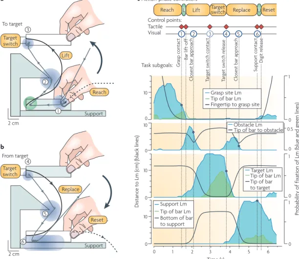
eye movements to the contact location are launched concurrently, based on information from peripheral vision174–178. The high speed of eye movements implies that the gaze reaches the contact location well before the hand (fIG. 4a,b). Importantly, this gaze behaviour develops
gradually during the learning of required action‑phase controllers. That is, during learning the gaze behaviour gradually shifts from pursuing poorly controlled hand movements to a mode in which the gaze predicts forth‑ coming contact locations179, corroborating the notion that learning to predict sensory consequences of motor commands precedes accurate control81.
early fixations of contact locations can support online movement adjustments based on sensed and/or predicted gaze‑position signals180 and on visual feed‑ back of hand movements referenced to the foveated target176,181–183. However, the gaze often remains at the contact location, such that the final part of the action phase, during which visual information can no longer influence the hand movement, is captured in central vision (fIG. 4c). The fact that the gaze often shifts to the
goal of the next action phase around the predicted time of goal completion suggests that the visual system can both predict and monitor contact events representing completion of task subgoals.
Although tactile feedback related to control points is essential for skilled object manipulation, contact events can also be predicted and monitored in the vis‑ ual modality (when visible) as well as the auditory and, presumably, the proprioceptive modalities. multimodal encoding of sensorimotor control points probably allows the sensorimotor system to monitor multiple aspects of task performance and, if errors are detected, respond to the pattern of errors observed in different modalities. Furthermore, because many contact events give rise to salient sensory signals from multiple modalities that are linked in time and space, they provide an opportunity
for sensorimotor integration and intermodal align‑ ment, which are helpful for learning and upholding multimodal sensorimotor correlations that support the prediction of purposeful motor commands. It is indeed well established that the brain can automatically inte‑ grate temporally correlated information occurring in the somatosensory, auditory and visual modalities, and neural activity common to all three stimulus modalities is present in the parietal and frontal cortices184,185 and in the posterior superior temporal sulcus186. Studies in such multisensory areas in monkeys indicate that the relative timing of convergent multimodal inputs is a crucial factor for neuronal responses187,188.
Conclusions and future directions
Dexterous object manipulation serves as an interest‑ ing model system for sensorimotor control because it is explicitly concerned with interactions between the body and the environment, and because manipulation tasks are comprised of sequentially organized distinct move‑ ment phases. These features are key components of most natural actions, ranging from tool use to locomotion. We have proposed that crucial control operations are cen‑ tred on events that mark transitions between consecu‑ tive action phases and that represent task subgoals. At these events, the brain both receives and makes predic‑ tions about sensory information from multiple sources. Thus, these events serve as control points that enable the upholding, adaptation and learning of sensorimotor cor‑ relations that are used to guide future action. In addition, they enable a comparison of context‑specific predictions of the sensory outcome (corollary discharge) of planned
actions and the actual sensory outcome. Through such comparisons, errors in prediction can be detected and used to launch corrective action patterns as required. In general terms, our framework agrees with control theories which imply that control turns desired sensory
Figure 3 | Hypothetical model for the fast processing of afferent information in somatosensory pathways.
Second- (and higher-) order neurons function as coincidence detectors and so are sensitive to specific spatiotemporal properties of impulse patterns in a population of tactile afferents. The first waves of impulses in an ensemble of afferents in response to the fingertip contacting objects with two types of surface shape (flat and curved) are schematically illustrated. The stimulus shape influences the first-spike latencies of the responsive afferents. The substantial divergence and convergence of primary afferents onto second-order neurons and the dispersion of conduction velocities among afferents provide parallel processing of the temporally structured information that aids feed-forward rapid classification of information by temporal-to-spatial conversion.
Nature Reviews | Neuroscience
Dis
ta
nc
e
to
L
m
(c
m
) (
bl
ac
k lines)
Pr
obability of
fix
ation of Lm (blue
and gr
een
lines)
0 0.5
0 0 1
1 0
10
0 10 0 10
1 2
0 3 4 5 6
Time (s)
0 0
1 10
Gr
asp
co
nt
ac
t
C
lo
se
st bar appr
oa
ch
C
lo
se
st bar appr
oa
ch
Tar
get
switch
cont
ac
t
Tar
get
switch
rel
ea
se
Target Lm Tip of bar Lm Tip of bar to target
Tip of bar Lm Support Lm Bottom of bar to support
c Action-phase controllers
Reach L Lift Targetswitch Replace UReset
Task subgoals: Tactile Visual
Target switch
Replace Support 2 cm
3 Target switch
Lift To target
a
2
Support 2 cm
4 From target
b
5
Reset
1 2 3 4 5 6
Bar lif
t-off
Suppo
rt
c
on
ta
ct
Digit
re
le
as
e
Grasp site Lm Fingertip to grasp site Tip of bar Lm
Obstacle Lm Tip of bar to obstacle
6
Control points:
Reach 1
states into motor commands and prediction maps motor commands into expected sensory consequences189–191. Furthermore, the idea that control is centred on dis‑ tinct movement goals is consistent with a recent com‑ putational model of sensorimotor control referred to as optimal feedback control192–194. learned action‑phase controllers correct errors only if they influence the movement goal, and each controller flexibly combines sensory signals and efferent copies of motor commands to optimize its performance.
However, defining the principles for the planning and control of actions based on behavioural observations does not provide direct knowledge of the neural mechanisms
involved. Although numerous studies have examined the central neural mechanisms that support the plan‑ ning and control of reaching, much less is known about object manipulation (for a review see REf. 195). Over the
past two decades, a number of studies have attempted to address this knowledge deficit. Although these studies have generated knowledge about the brain areas that are engaged during manipulation195, an understanding of how defined control operations that support natural manipula‑ tion tasks are implemented in the CnS is just beginning to emerge. There are many challenges involved in develop‑ ing this understanding. For example, it might be difficult to define the embodiment of a given manipulation task
Figure 4 | Visual and tactile control points in a manipulation task. a,b | Gaze and hand movements when a person grasps a bar, moves it around an obstacle to press a target switch (a) and then replaces it on the support surface (b). The dashed black lines represent the path of the tip of the index finger, the solid black lines indicate the path of the tip of the bar and the grey lines indicate the gaze position. The blue and green areas indicate ‘landmark’ (Lm) zones that captured 90% of the fixations recorded during several trials by ten participants. These zones are centred on the grasp site, the tip of the bar (green), the protruding part of the obstacle, the target and the support surface. c | Coordination of gaze and manual actions. The blue and green lines give the time-varying instantaneous probability of gaze fixations in the landmark zones listed, and the black lines indicate the distance between the landmark and either the tip of the index finger, the tip of the bar or the bottom of the bar (indicated for each graph). The red diamonds indicate tactile control points and the blue circles indicate contact events predicted in the visual modality. The spatial locations of these events are indicated by the location of the correspondingly numbered circle in parts a and b. This figure demonstrates that the gaze is directed to key locations that represent task subgoals. The fact that the gaze shifts to the goal of the next action phase around the predicted time of goal completion suggests that the visual system can both predict and monitor contact events representing subgoal completion. Figure is modified, with permission, from REf. 177 (2001) Society for Neuroscience.
1. Macefield, V. G. & Johansson, R. S. Control of grip force during restraint of an object held between finger and thumb: responses of muscle and joint afferents from the digits. Exp. Brain Res.108, 172–184 (1996).
2. Macefield, V. G., Häger-Ross, C. & Johansson, R. S. Control of grip force during restraint of an object held between finger and thumb: responses of cutaneous afferents from the digits. Exp. Brain Res.108, 155–171 (1996).
3. Häger-Ross, C. & Johansson, R. S. Non-digital afferent input in reactive control of fingertip forces during precision grip. Exp. Brain Res.110, 131–141 (1996).
4. Dimitriou, M. & Edin, B. B. Discharges in human muscle receptor afferents during block grasping.
J. Neurosci.28, 12632–12642 (2008).
5. Johansson, R. S. & Flanagan, J. R. in The Senses: a Comprehensive Reference, Volume 6,
Somatosensation (eds Gardner, E. & Kaas, J. H.) 67–86 (Academic, San Diego, 2008).
6. Vallbo, A. B. & Johansson, R. S. Properties of cutaneous mechanoreceptors in the human hand related to touch sensation. Hum. Neurobiol.3, 3–14 (1984).
7. Johansson, R. S., Landström, U. & Lundström, R. Responses of mechanoreceptive afferent units in the glabrous skin of the human hand to sinusoidal skin displacements. Brain Res.244, 17–25 (1982).
8. Löfvenberg, J. & Johansson, R. S. Regional differences and interindividual variability in sensitivity to vibration
in the glabrous skin of the human hand. Brain Res. 301, 65–72 (1984).
9. Brisben, A. J., Hsiao, S. S. & Johnson, K. O. Detection of vibration transmitted through an object grasped in the hand. J. Neurophysiol.81, 1548–1558 (1999).
10. Loewenstein, W. R. & Skalak, R. Mechanical transmission in a Pacinian corpuscle. An analysis and a theory. J. Physiol.182, 346–378 (1966).
11. Westling, G. & Johansson, R. S. Responses in glabrous skin mechanoreceptors during precision grip in humans. Exp. Brain Res.66, 128–140 (1987).
Impulses in single tactile afferents innervating the human fingertips were recorded from the median nerve while small test objects were lifted, held in the air and then replaced. Distinct discharges were observed at various contact events corresponding to the completion of task subgoals.
12. Knibestöl, M. Stimulus-response functions of slowly adapting mechanoreceptors in the human glabrous skin area. J. Physiol.245, 63–80 (1975).
13. Johansson, R. S. Tactile sensibility in the human hand: receptive field characteristics of mechanoreceptive units in the glabrous skin area. J. Physiol.281, 101–125 (1978).
14. Darian-Smith, I. in Handbook of Physiology (eds, Brookhart, J. M., Mountcastle, V. B., Darian-Smith, I. & Geiger, S. R.) 739–788 (American Physiological Society, Bethesda, Maryland, 1984).
15. Johnson, K. O., Yoshioka, T. & Vega-Bermudez, F. Tactile functions of mechanoreceptive afferents innervating the hand. J. Clin. Neurophysiol.17, 539–558 (2000).
16. Goodwin, A. W. & Wheat, H. E. Sensory signals in neural populations underlying tactile perception and manipulation. Annu. Rev. Neurosci.27, 53–77 (2004).
17. Johnson, K. O. & Hsiao, S. S. Neural mechanisms of tactual form and texture perception. Annu. Rev. Neurosci.15, 227–250 (1992).
18. Craig, J. C. & Rollman, G. B. Somesthesis. Annu. Rev. Psychol.50, 305–331 (1999).
19. Sathian, K., Goodwin, A. W., John, K. T. & Darian-Smith, I. Perceived roughness of a grating: correlation with responses of mechanoreceptive afferents innervating the monkey’s fingerpad. J. Neurosci.9, 1273–1279 (1989).
20. Johansson, R. S. & Vallbo, Å. B. Tactile sensory coding in the glabrous skin of the human hand. Trends Neurosci.6, 27–31 (1983).
21. Bisley, J. W., Goodwin, A. W. & Wheat, H. E. Slowly adapting type I afferents from the sides and end of the finger respond to stimuli on the center of the fingerpad. J. Neurophysiol.84, 57–64 (2000).
22. Birznieks, I., Jenmalm, P., Goodwin, A. W. & Johansson, R. S. Encoding of direction of fingertip forces by human tactile afferents. J. Neurosci.21, 8222–8237 (2001).
in the neural networks of the CnS, because the various action‑phase controllers that are brought into play might all use different processes to some extent, in part depend‑ ing on the nature of the sensory information involved. There is indeed evidence that elementary action phases can be organized in various areas of the CnS, from spinal cord networks to frontoparietal cortical net‑ works196–198. Furthermore, a challenging issue is how to distinguish processes involved in detecting mismatches from those involved in triggering corrective actions and those involved in creating, containing and recruiting rel‑ evant internal representations during the progression of manipulation tasks. This requires analysis of the dynamics of the system. new analysis methods applicable to studies in humans might help, such as dynamic causal model‑ ling199, which is used to infer processes and mechanisms at the neuronal level from measurements of brain activity obtained with different techniques.
In the field of motor control, most studies have focused on single actions, such as moving the hand between two points in space, and have therefore overlooked a crucial component of natural tasks: namely that they consist of a series of phases demarcated by discrete events that provide subgoals for task completion. Although there is evidence in monkeys that the prefrontal cortex plays a part in assembling sequences of action‑phase controllers for completing manual tasks200–202, little is known about the mechanisms that are used to coordinate the sensory interactions and predictions across the action‑phase con‑ trollers, as required for smooth linking of action phases. Given that the correct sequence of actions in everyday tasks normally has to be learned or selected through experience, understanding how movement phases are put together is also important for the study of motor learn‑ ing. An important component of skill development prob‑ ably involves combining previously learnt action‑phase controllers in new ways. This ability requires learning to predict event‑related sensory outcomes under new con‑ ditions. Similarly, little is known about how the lateral‑ ized brain flexibly and swiftly allocates complementary
roles to the hands during the sequential action phases that are involved in natural bimanual object manipula‑ tion tasks. Whereas research on bimanual coordination has focused on situations in which the two hands have difficulty acting independently203–205, recent work has started to examine the flexible coordination of the hands in skilled bimanual manipulation206,207.
even at the level of the basic tactile afferent mecha‑ nisms there are numerous unresolved issues. One impor‑ tant problem for future research concerns how the brain deals with the complex interaction effects among the dif‑ ferent parameters of fingertip stimulation, which affect the afferent responses that arise. For example, there are interactions between force direction and object shape that affect the firing rates23 and relative spike timing146 of the afferent responses. In addition, the recent stimulus history can also influence the afferent responses, because of the viscoelastic properties of the fingertips22,49,208. A more general issue concerns how the brain processes tactile afferent signals to detect spatiotemporal events that are conditional on, and specific for, the imple‑ mented action‑phase controller and its current state. In this context, one may speculate that many of the compu‑ tational requirements for decoding tactile messages are satisfied by the somatosensory pathways that function‑ ally model the mechanical state of the fingertips by, in effect, filtering information predictively in a manner that is specific for the active action‑phase controller and its current phase. Indeed, a large proportion of the descend‑ ing information from sensorimotor cortical areas targets nodes of the somatosensory pathways through cortico‑ thalamic, corticocuneate and corticospinal pathways and could thus dynamically control signal processing in ascending somatosensory pathways209–214. Such mecha‑ nisms are in agreement with recent work that has iden‑ tified a pivotal role for corollary discharges at different levels of the CnS in affecting the processing of sensory afferent information215,216, including at low levels of the sensory pathways such as the primary afferent axon terminal and/or first‑order interneurons213,216.