Original Research Article
Pattern distribution of abnormal hemoglobin variants by cation
exchange High Performance Liquid Chromatography: a study
of 9,116 subjects
Sejal Gamit, Arpita Nishal*, Pinal Shah, Archana Patel
INTRODUCTION
Haemoglobinopathies are the group of genetic disorders of haemoglobin in which there is quantitative or qualitative abnormal production or in the structure of haemoglobin molecule.1 These hereditary disorders are major public health problem in many parts of the world including India.1 Beta(β)-thalassaemia and sickle cell disease represents the most frequent haemoglobinopathies.1,2,3,4
The clinical spectrum of the disorders varies from asymptomatic conditions to serious disorders like Thalassaemia major and sickle cell disease that requires regular blood transfusions and extensive medical care.1 The general incidence of thalassaemia trait and sickle cell haemoglobinopathy in India varies between 3-17% and 1-44% respectively.5 But because of consanguinity, caste and area endogamy, some communities show a very high incidence, making the disease a major public health problem in this country.2,3 These haemoglobinopathies Department of Pathology, Government Medical College, Surat, Gujarat, India
Received: 01 September 2019
Revised: 23 September 2019
Accepted: 04 October 2019
*Correspondence:
Dr. Arpita Jitender Pal Nishal, E-mail: [email protected]
Copyright: © the author(s), publisher and licensee Medip Academy. This is an open-access article distributed under the terms of the Creative Commons Attribution Non-Commercial License, which permits unrestricted non-commercial use, distribution, and reproduction in any medium, provided the original work is properly cited.
ABSTRACT
Background: The present study was conducted to identify pattern distribution of abnormal haemoglobin variants by using HPLC method in a tertiary care hospital, Surat, Gujrat, India.
Methods: A cross sectional study of one-year duration was conducted including 9,116 patients screened for the presence of abnormal haemoglobin variants. Blood samples were initially tested for solubility test and run on automated haemoglobin analyzer for complete haemogram. All the suspected and family study cases were processed for HPLC (Bio-Rad Variant II) for conclusive diagnosis. Patients with a history of recent blood transfusion of less than 3 months duration were excluded from the study.
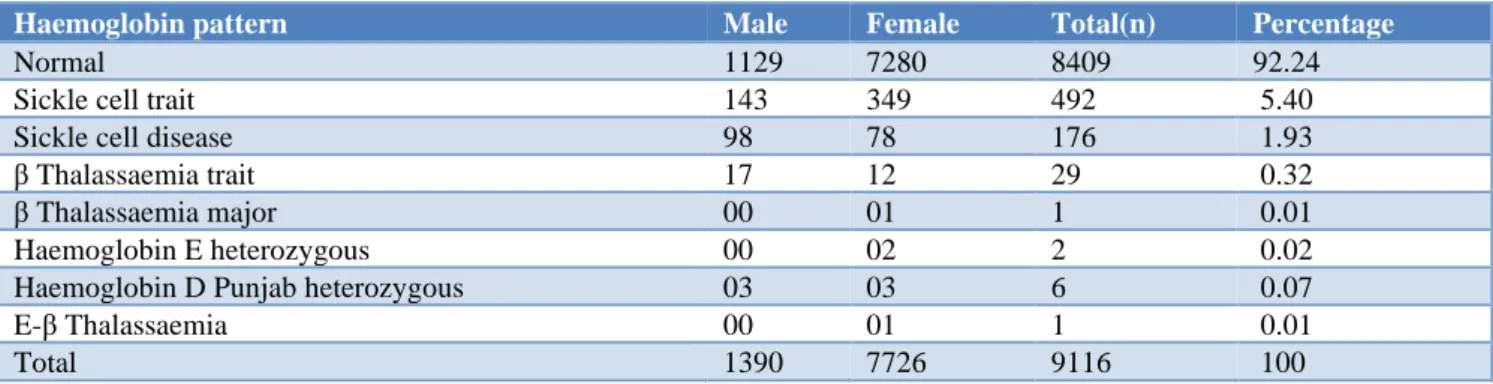
Results: A total of 9,116 cases (1390 males, 7726 females) were included in the present study. The age group of patients ranged from 1 month to 95 years. Solubility test and complete haemogram were performed in all the cases. Out of the 9,116 cases, 8409(92.24%)cases had normal HPLC pattern. 492(5.40%)cases were diagnosed as sickle cell trait, 176(1.93%) cases as sickle cell disease, 29(0.32%) cases as β thalassaemia trait, 1(0.01%) case as β thalassaemia major, 2(0.02%)cases as Hb E heterozygous and 03 (0.07%) cases as Hb D Punjab heterozygous. One case of double heterozygous for Hb E-β thalassaemia was also found.
Conclusions: HPLC is a rapid, accurate and useful method for diagnosing haemoglobinopathies. It serves as an reliable tool in diagnosing the presence of abnormal haemoglobin variants in suspected cases on routine haematology in developing countries like India, where the resources for detection of haemoglobinopathies are limited. Early diagnosis may help in proper management of patients.
are common causes of morbidity and also exert burden on
expenditure. To reduce the burden, accurate and reliable screening procedures should be there. Cation exchange high performance liquid chromatography has become the preferred technique suitable in Indian scenario, as it can detect most of the clinically significant variants. The simplicity of the automated system with internal sample preparation, superior resolution ,rapid assay time and accurate quantification of haemoglobin fractions makes this an ideal methodology for routine clinical laboratory.5,6 Several studies in the literature have reported that Gujarat has higher frequency of β Thalassaemia and Sickle cell disease.7-9 This study was done with the aim to identify pattern distribution of abnormal Hb variants in a tertiary care hospital, Surat, Gujrat India. This type of cross-sectional studies was essential to identify haematological abnormality in pre symptomatic period, preconceptional screening, screening of fetus to offer termination of pregnancy and to confirm a presumptive diagnosis.
METHODS
The Study design was a cross sectional study. The Study period was conducted at tertiary care hospital, Surat, Gujrat, India over a period of 1 year (01/01/2018 to 31/12/018). Study population is a total of 9,116 blood samples received from indoor and outdoor patients attending tertiary care hospital, Surat, Gujrat India.
Inclusion criteria
All clinically suspected cases of haemoglobinopathies and cases with positive family history of haemoglobinopathies from indoor and outdoor department were included in the study.
Exclusion criteria
Patients with history of blood transfusion within 3 months were excluded from the study.
Procedure
5ml of whole blood sample was collected in EDTA which can be stored at 2-8̊ C for maximum 7 days if processing is delayed. No preparation was required. Pre dilution required if sample was in a tube other than the recommended tube or there was less than 500 µl of sample in the tube. HbA2/F calibrator and normal and abnormal controls were analyzed at the beginning of each run on HPLC. Hemoglobin and RBC indices were measured on automated cell counter. Solubility test was performed on all the received samples. All the clinically suspected cases of haemoglobinopathies, solubility positive and grey zone samples were proceeded to be analysed by BIO-RAD VARIANT II machine for confirmative diagnosis. The underlying principle of HPLC is based on haemoglobin separation by an analytic cartridge in cation exchange HPLC using a
pre-programmed buffer gradient with increasing ionic strength to the cartridge. The separated fractions pass through dual wavelength detector, where absorbance of the sample component is measured at 415nm. Background noise is reduced with the use of a secondary wavelength at 690 nm. The absorbance data are transmitted from the detector to the PC and displayed by CDM as a real time chromatogram.
Statistical analysis
Qualitative data are presented as frequencies and percentages, and quantitative data were shown as mean. All the data were analysed using Microsoft excel 2013.
RESULTS
Out of 9116 cases, 8409 cases had normal and 707 cases had abnormal haemoglobin pattern. Presumptive identification of haemoglobin variants was made primarily by analyte identification windows as shown in Table 1; however geographical factors, ethnicity and clinical presentation were also taken into consideration.
Table 1: Analyte identification windows; window time and retention time of predefined parameters of
BIO-RAD VARIANT II.
Peak name Window(min) Retention time(min)
F window 0.98-1.22 1.10 P2 window 1.28-1.50 1.39 P3 window 1.50-1.90 1.70 A0 window 1.90-3.10 2.50 A2 window 3.30-3.90 3.60 D window 3.90-4.30 4.10 S window 4.30-4.70 4.50 C window 4.90-5.30 5.10
In present study, overall patients age ranges from 1 months to 94 years. As shown in Table 2, Out of 9116 cases, 1390 cases (15.25%) were of male and 7726 cases (84.75%) were of female.
Table 2: Sex wise distribution of cases.
Gender No. of cases Percentage(%)
Male 1390 15.25
Female 7726 84.75
Total 9116 100
sickle cell trait and Hb S value >50% with mildly raised
Hb F level for diagnosis of sickle cell disease. Sickle cell trait and sickle cell disease show high level of Hb A2 may be because of Hb S adducts. The most common disorder detected was sickle cell trait (Hb S heterozygous) with 492 cases. Most of the patients had low haemoglobin, reduced MCV, MCHC and raised RBC count. The mean haemoglobin S reported in chromatogram was 28.57%. Authors detected 176 cases of sickle cell disease with mean haemoglobin S reported in chromatogram was 78.3%. Parental study was advised to confirm the double heterozygous status. Solubility test was found to be positive in all these cases.
A total of 29 β Thalassaemia minor cases were diagnosed in this study. The major abnormality was the presence of elevated levels of Hb A2 with RT(3.30-3.90 mins). Authors considered Hb A2 value >3.5% as a cut off for diagnosis of β Thalassaemia minor. In this study, mean percentage of HbA2 was 4.8%.Most of the patients had low haemoglobin, reduced MCV, MCHC and raised RBC
count. One case of β Thalassaemia major was diagnosed. Hb F was raised (91.4%) with variable HbA2 (3.8%). The present case was diagnosed within 1st year of life. Parental study was conducted to find out carrier status and to confirm diagnosis. 06 cases showed peak in the D window (RT 3.90-4.30 mins), indicating presence of structural variant haemoglobin D(Hb D) Punjab. Hb D value between 33-43% considered as a diagnosis of Hb D Punjab heterozygous. These cases showed reduced haemoglobin, MCV and MCH levels.
The Hb E elute into the HbA2 window but differentiated upon the retention time and Hb Fractions %. Authors considered HbA2 value <40% as diagnosis for heterozygous Hb E. In present study, authors found one case of Hb E heterozygous. Hb level was mildly reduced with raised RBC counts and reduced MCV, MCH values. One case of double heterozygous for Hb E/β Thalassaemia was also diagnosed showing peak in HbA2 window (64%)and raised Hb F level (19%). Again, parental study was carried out for confirmation.
Table 3: Pattern distribution of abnormal haemoglobin variants among study subjects.
Haemoglobin pattern Male Female Total(n) Percentage
Normal 1129 7280 8409 92.24
Sickle cell trait 143 349 492 5.40
Sickle cell disease 98 78 176 1.93
β Thalassaemia trait 17 12 29 0.32
β Thalassaemia major 00 01 1 0.01
Haemoglobin E heterozygous 00 02 2 0.02
Haemoglobin D Punjab heterozygous 03 03 6 0.07
E-β Thalassaemia 00 01 1 0.01
Total 1390 7726 9116 100
DISCUSSION
India is an ethnically diverse country with marked regional variation .This diversity is reflected in the presence of different haemoglobin variants in different ethnic groups. Due to migration, there is mixing of people from different regions. Many of these abnormal variants are of little clinical significance in heterozygous state, but when combined with other variants they may give rise to severe disease. Therefore, there is always a need for a screening method which can detect maximum variants. HPLC has the advantage of quantifying Hb F and HbA2 along with detecting other variants in a single screening test.
Besides HPLC, there are other analytical procedures used for detection of thalassaemia and haemoglobinopathies such as alkaline and acid electrophoresis, HbA2 quantification by ion exchange column chromatography and Hb F quantification by alkali denaturation and radial
immunodiffusion.10 Electrophoretic method does not separate all variants from each other and it is recommended by screening programs such that further tests should be carried out to confirm the presumed identify abnormal variant. None of the above-mentioned methods can detect multiple haemoglobin fractions in a single step procedure. HPLC has many advantages over these methods and over the past decades it has evolved as an excellent and powerful diagnostic tool for identification of most of the clinically significant Hb variants. HPLC is sensitive, specific, reproducible and less time consuming and requires less manpower. Hence, it is an ideal tool for a routine clinical laboratory with high workload.
in this study is in concordance with the reported
prevalence.11,12 The distribution of β Thalassaemia is not uniform in the Indian subcontinent. The frequency of β Thalassaemia trait varies for 1-17% in. different population of India.13-15 In this study, the frequency of β Thalassaemia trait is (0.31%.) Borderline HbA2 requires further investigations before reaching out on conclusion. Both iron deficiency and megaloblastic anemia may have an effect on level of HbA2. Iron deficiency anemia can mildly reduce, and megaloblastic anemia can mildly elevate HbA2 level.
HB E trait, homozygous Hb E disease(Hb EE) and double heterozygous Hb E trait and Hb EE are mild disorder. Detection of this variant is very important because when combined with thalassaemia or Hb S, it gives rise to moderate to severe anemia. In present study, the frequency of Hb E is 0.03% with one case diagnosed as double heterozygous for Hb E/β Thalassaemia.
Heterozygous Hb D Punjab mostly presented as asymptomatic heterozygous condition with normal haematological parameter. Hb D Punjab occurs with greatest prevalence that is 2% among Sikhs in Punjab and in Gujarat reported prevalence is 1%. In present study, the frequency of Hb D is 0.07%.
The findings in the present study show HPLC as an excellent, powerful diagnostic tool for the direct identification of Hb variants with a high degree of precision in the quantification of normal and abnomal haemoglobin fractions. With the integration of proper algorithms involving retention time, % Hb and peak characteristics, a clinical laboratory is capable of sidentifying 75% of common variants encounted without the need for confirmatory studies such as alkaline and acid electrophoresis.10 In the light of family study, ethnicity, hemogram and clinical data, authors can detect most of the frequently occurring clinically significant variants by HPLC method alone.
CONCLUSION
To conclude, HPLC findings in correlation with hemogram and family study are sufficient to detect most of the haemoglobin variants prevalent in this study with few inconclusive cases require further genetics and molecular studies.
The present study conducted using HPLC reflects the magnitude of abnormal hemoglobin variants in a tertiary care hospital population which may be in fact the tip of an iceberg, but this type of study can definitely help to increase awareness among both health care givers and general population.
ACKNOWLEDGEMENTS
Authors would like to thank express my gratitude towards Pathology GMCS faculty staff for their valuable
guidance. I am also thankful to sickle cell laboratory technical staff for providing technical help.
Funding: No funding sources Conflict of interest: None declared
Ethical approval: The study was approved by the Institutional Ethics Committee
REFERENCES
1. Vaz FE, Thakur CB, Banerjee MK, Gangal SG. Distribution of beta-thalassaemia mutations in the indian population referred to a diagnostic center. Haemoglobin. 2000;24(3):181-94.
2. Balgir RS. The burden of haemoglobinopathies in india and the challenges ahead. Current Sci Association. 2000;79(11):1536-47.
3. Balgir RS. The genetic burden of haemoglobinopathies with special references to community health in India and the challanges ahead. Indian J Hemat Blood Transfuse. 2002;20:2-7. 4. Baruah MK, Saikia M, Baruah A. Pattern of
haemoglobinopathies and thalassaemia’s in upper Assam region of North Eastern India: high performance liquid chromatography studies in 9000 patients. Ind J Pathol Microbiol. 2014;57(2):236-43. 5. Wild BJ, Stephens AD. The use of automated HPLC
to detect and quantitate haemoglobins. Clin Laborat Haematol. 1997;19(3):171-6.
6. Ou CN, Rognerud CL. Diagnosis of hemoglobinopathies: electrophoresis vs. HPLC. Clinica Chimica Acta. 2001;313(1-2):187-94. 7. Bhatia HM, Shanbagh S.R, Baxi AJ, Bapat J.P,
Sharma R.S. Genetic studies among the endogamous groups of Lohanas of North and West India. Hum Hered.1976;26(4):298-305.
8. Mukherjee MB, Gangakhedkar RR, Sathe MS. Abnormal hemoglobin, G6PD deficiency and their pattern of interaction in the tribal population of Valsad district (Gujarat). Indian J Hematol Blood Transf. 1993;11:227-31.
9. Vyas GN, Bhatia HM, Sukumaran PK, Balkrishnan V, Sanghvi LD. Study of blood groups, abnormal hemoglobins and other genetical characters in some tribes of Gujarat. Am J Phys Anthropol. 1962;20(3):255-65.
10. Joutovsky A, Hadzi-Nesic J, Nardi MA. HPLC retention time as a diagnostic tool for haemoglobin variants and haemoglobinopathies: a study of 60000 sample in a clinical diagnostic laboratory clinical chemistry. 2004;50(10):1736-47.
11. Verma IC, Choudhary VP, Jain PK. Prevention of thalassemia: a necessity in India. Indian J Pediatr. 1992;5(6):649-54.
13. Sharma RS, Parekh JG, Shah KM. Hemoglobin
opathies in western India. J Assoc Physic India. 1963;11:969-973.
14. Misra RC, Ram B, Mohapatra BC, Das SN, Misra SC. High prevalence and heterogenicity of thalassemias in Orissa. Indian J Med Res. 1991;94:391-394.
15. Dash S. Beta thalassemia trait in the Punjab (North India) Br J Haematol. 1985;61(1):185-6.
Cite this article as: Gamit S, Nishal A, Shah P, Patel