DEFINING THE MOLECULAR MECHANISMS OF THE CEREBRAL CAVERNOUS MALFORMATION PROTEINS.
Bryan Timothy Richardson
A dissertation submitted to the faculty of the University of North Carolina at Chapel Hill in partial fulfillment of the requirements for the degree of Doctor of Philosophy in
the Department of Pharmacology
Chapel Hill 2013
Approved by:
ABSTRACT
BRYAN TIMOTHY RICHARDSON: Defining the molecular mechanisms of the Cerebral Cavernous Malformation proteins.
(Under the Direction of Gary L. Johnson, Ph.D.)
Cerebral cavernous malformations (CCM) are the second most common class of cerebrovascular brain malformations affecting .1-.5% of the population. The disease is manifested in endothelial cells as lesions of thin, dilated, and leaky
capillaries lacking normal blood vessel-stromal interactions. Lesions cause varied symptoms ranging from minor headaches to seizure and hemorrhagic stroke. CCMs can be incurred sporadically or inherited in an autosomal dominant manner from loss of function mutations in one of three genes, ccm1/krit1, ccm2/osm, or ccm3/pdcd10. These mutations affect the actin cytoskeleton due to deregulated RhoA/ROCK
signaling, which increases stress fiber incidence, reduces endothelial cell barrier function, and decreases angiogenesis in vitro. We demonstrate through global kinome profiling that numerous kinases controlling the actin cytoskeleton are
TABLE OF CONTENTS
LIST OF TABLES ...vii
LIST OF FIGURES ...viii
LIST OF ABBREVIATIONS ... xi
CHAPTERS I. Introduction ... 1
CCM prevalence and pathophysiology ... 1
CCM inheritance... 1
CCM pathophysiology ... 3
Knudson two hit hypothesis of loss of heterozygosity ... 4
CCM lesion initiating cell(s) ... 5
CCM protein structure, function, and signaling ... 6
CCM1 ... 6
CCM2 ... 11
CCM3 ... 16
Integrated CCM signaling... 20
Small GTPase regulation ... 21
Small GTPase regulation of endothelial junctions... 22
E3 ubiquitin ligase regulation of RhoA levels ... 24
Rho-associated protein kinase (ROCK) ... 27
Simvistatin ... 31
ROCK inhibitors in CCM... 32
Rational for an iPS cell disease model for CCM ... 34
Thesis objectives... 42
II. Materials and methods... 51
Chapter III ... 51
Chapter IV ... 57
Chapter V ... 59
III. Global kinome profiling of deregulated kinases in CCM ... 64
Introduction ... 64
Results ... 67
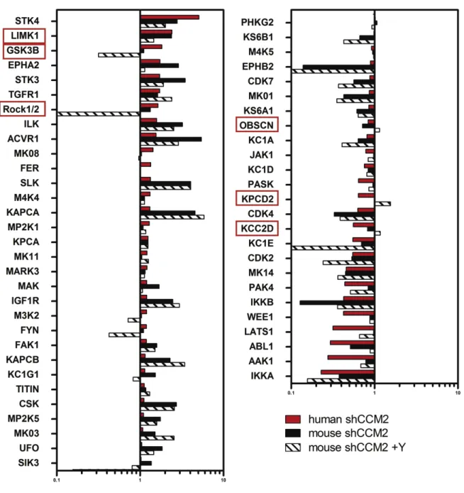
Kinome profiling of CCM protein deficient human and mouse endothelial cells ... 67
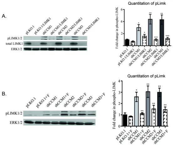
LIMK and cofilin phosphorylation decreases tube Formation rescuable by LIMK1 knockdown ... 71
Phospho-cofilin levels are increased in surgically resected human CCM lesions ... 72
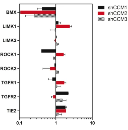
CCM proteins regulate the expression of Tie2 and BMX upstream of ROCK ... 73
Discussion... 75
IV. Ubiquitin ligase mediated degradation of RhoA as a molecular mechanism deregulated in CCM protein deficient ECs ... 97
Introduction ... 97
Results ... 99
Forskolin stimulates longer term RhoA degradation
And Mst3/4 kinases phosphorylate Smurf1... 101
In vitro loss of Smurf1 and Cullin E3 ligases increases F-actin stress fibers and decreases endothelial cell tube formation ability ... 103
Smurf2 binds CCM2 and is required for proper endothelial tube formation through regulation of Rap1 ... 105
Discussion... 106
V. Induced pluripotent stem cells as a new patient specific model for CCM ... 119
Introduction ... 119
Results ... 126
hESCs differentiate to the endothelium and can be used to model CCM phenotypes ... 126
Isolation and characterization of endothelial progenitor derived endothelial cells as a model for CCM ... 131
Generation of iPS cells from EPC derived ECs... 133
Discussion... 136
VI. Concluding remarks... 152
Summary... 152
Future directions ... 155
LIST OF TABLES TABLE
1.4: CCM protein phenotypes reported in the literature ... 49 1.5: CCM protein binding interactors reported in the literature ... 50 3.10: Number of kinases shared between CCM1, -2, or -3
LIST OF FIGURES FIGURE
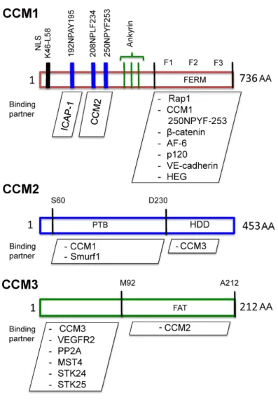
1.0: Core CCM signaling circuitry in endothelial cells ... 45 1.1: Defined structural domains and interacting proteins for
CCM1, -2, and -3 ... 46 1.2: Strategy for developing and utilizing a CCM patient
specific iPS disease library ... 47 1.3: Differentiation strategy of pluripotent stem cells to endothelium... 48 3.0: CCM protein deficient Huvecs have increased stress fiber formation ... 78 3.1: Strategy for assessing global kinome activation status in
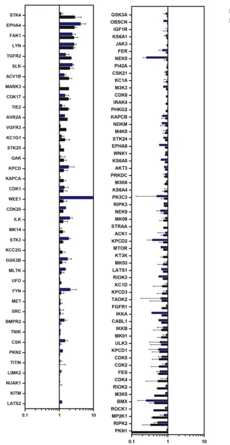
CCM protein deficient cell culture ... 79 3.2: CCM2 protein loss affects the kinome ... 80 3.3: Cytoskeletal regulating kinases are both over and under
represented... 81 3.4: Subset of over or under represented kinases are conserved
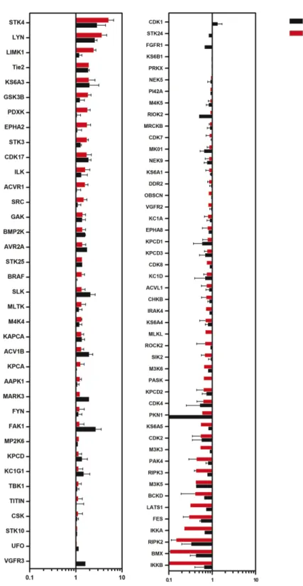
across mouse and human cells ... 82 3.5: CCM1 protein loss affects the kinome ... 83 3.6: CCM3 protein loss affects the kinome ... 84 3.7: A subset of over or under represented kinases are conserved across CCM1 and CCM2 deficient Huvecs ... 85 3.8: A subset of over or under represented kinases are conserved across CCM2 and CCM3 deficient Huvecs ... 86 3.9: A subset of over or under represented kinases are conserved across CCM1 and CCM3 deficient Huvecs ... 87 3.10: A subset of over or under represented kinases are conserved across CCM1, -2, and -3 CCM3 deficient Huvecs... 88 3.11: Kinases important for endothelial function are deregulated in
3.12: pLIMK1 is increased in stable CCM1, -2, or -3 knock down
MEECs... 90 3.13: pCofilin levels are increased by LIMK1 following CCM
protein loss... 91 3.14: Knock down of LIMK1 is sufficient to rescue CCM phenotypes
in vitro ... 92 3.15: Elevated pCofilin staining is observed in surgically resected
human CCM1, -2, and -3 lesions ... 93 3.16: Knock down of LIMK1 is sufficient to rescue CCM phenotypes
in vitro ... 94 3.17: Tie2 and BMX are increased both at protein and mRNA
levels in Huvecs after CCM1, -2, or -3 loss ... 95 3.18: Tie2 and BMX message levels are not significantly affected
by ROCK inhibition... 96 3.19: qRT-PCR analysis of knock down lines used for
chapter III ... 97 4.0: Smurf1 binds to CCM2 and not CCM1 or CCM3 ... 110 4.1: Ubiquitinated GTP bound RhoA is decreased after CCM
protein loss... 111 4.2: Adenylyl cyclase activation by Forskolin promotes
RhoA degradation ... 112 4.3: Loss of Smurf1 increases stress fiber formation and decreases
tube forming ability of Huvecs... 113 4.4: Cullin inhibitor MLN 4924 increases stress fibers, decreases tube formation, and increases RhoA protein... 114 4.5: MLN 4924 treatment decreases the ubiquitination of total
4.8: qRT-PCR and western blotting analysis of knock down lines
used for chapter IV... 118 5.0: H9 hESCs do not randomly differentiate efficiently to CD31+ ECs .... 119 5.1: Mesodermal inducing cytokines followed by TGF-β inhibition
promotes H7 hESCs to efficiently differentiate to
CD31+CDH5+ ECs ... 120 5.2: hESC derived ECs grow best in the vascular specification media... 121 5.3: CCM protein knock down does not affect hESC pluripotency
or differentiation to the endothelium... 122 5.4: WT but not CCM knock down EBs sprout tube-like
structures from differentiating EBs... 123 5.5: CCM proteins regulate endothelial function through
RhoA in hESC derived ECs ... 124 5.6: Endothelial progenitor cell derived ECs can be derived
from peripheral blood ... 125 5.7: CCM1, -2, and -3 deficient endothelial progenitor cells are
unable to form tube like structures... 126 5.8: Two independent sources of EPC ECs demonstrate
elevated RhoA signaling ... 127 5.9: EPC ECs form iPS colonies after retroviral transduction
with Oct4, Sox2, Klf4, and cMyc ... 128 5.10: EPC EC derived iPS cells express a panel of pluripotent
stem cell markers... 129 5.11: EPC EC derived iPS cells are able to differentiate to all
three germ layers ... 130 5.12: Endothelial derived iPS cells differentiate differently to
LIST OF ABBREVIATIONS
AQUA: Advanced Quantitative Analysis bEND.3: Mouse brain Endothelial cell bFGF: Basic fibroblast growth factor BMP4: Bone morphogenic protein 4 CCM: Cerebral cavernous malformations
CCM1: Cerebral Cavernous Malformation 1 gene
CCM1: Protein encoded by Cerebral Cavernous Malformation 1 gene, also known as KRIT1
CCM2: Cerebral Cavernous Malformation 2 gene
CCM2: Protein encoded by cerebral cavernous malformation 2 gene; also known as OSM, malcavernin
CCM2L: CCM2-Like protein
CCM3: Cerebral Cavernous Malformation 3 gene
CCM3: Protein encoded by cerebral cavernous malformation 3 gene; also known as PDCD10
EC: Endothelial Cell
EPC: Endothelial Progenitor Cell
EP-EC: Endothelial progenitor-derived endothelial cell FAT: Focal Adhesion Targeting
FDA: Food and Drug Administration
GCKIII: Germinal Center Kinase 3
GDI: Guanine nucleotide dissociation inhibitor GEF: Guanine nucleotide exchange factor GEMM: Genetically engineered mouse model GFP: Green fluorescent protein
H1/H7/H9: Human embryonic stem cell lines (1,7,9) HEG1: Heart of glass
hESC: Human embryonic stem cell.
Huvec: Human umbilical vein endothelial cell ICAP1: Integrin cytoplasmic adapter protein-1 IHC: Immunohistochemistry
IPS: Induced pluripotent stem cell KRIT1: Krev Interaction trapped 1 LIMK: LIM kinase
LOH: Loss of heterozygosity
MEEC: Mouse embryonic endothelial cell
MEKK3: Mitogen-activated protein kinase kinase kinase 3 MLC2: Myosin light chain 2
MLCK: Myosin light chain kinase
MLCP: Myosin light chain phosphatase mM: millimolar
MSH2: MutS homolog 2 protein
MST4: Mammalian Ste20-like Kinase 4 NuDiX: Nucleoside Diphosphate linked to X OMIM: Online Mendelian inheritance in man
OSM: Osmosensing scaffold for MEKK3; also known as CCM2, malcavernin PECAM1: Platelet endothelial cell adhesion molecule 1
PCR: Polymerase chain reaction
PDCD10: Programmed cell death 10 protein PKA/C: Protein kinase A or C
PTB: Phosphotyrosine binding RNA: Ribonucleic acid
RNAi: RNA interference ROCK: Rho kinase
RT-PCR: Reverse transcription polymerase chain reaction
SDS-PAGE: Sodium dodecyl sulfate polyacrylamide gel electrophoresis shRNA: short hairpin RNA
siRNA: small interfering RNA
SMURF1/2: Smad ubiquitination regulatory factor 1/2 STK24/5: Serine/threonine kinase 24/25
TGF-β: Tumor derived growth factor beta VECadherin: Vascular endothelial cadherin VEGF: Vascular endothelial growth factor
I. Introduction
CCM prevalence and pathophysiology
Stroke is a leading cause of death in the United States behind heart disease
and cancer. Cerebral cavernous malformation (CCM; OMIM 116860) is the second
most prevalent intracranial vascular malformation (IVM), which has symptoms
ranging from mild headaches to epileptic seizure to hemorrhagic stroke [1]. Lesions
appear principally in the central nervous system vasculature, but there have been
reports of peripheral lesions in the retina, liver, and spinal cord [2]. Pathologically,
CCM lesions are clusters of leaky capillaries, and the only treatment option for CCM
is through invasive surgery or radiation therapy. Recent in vitro and in vivo
experiments have attributed lesion generation to a break down of normal vascular
remodeling and the blood brain barrier (BBB) linked to deregulation of the actin
cytoskeleton of endothelial cells (EC)s through aberrantly high levels of the small
GTPase RhoA [3-8].
CCM inheritance
CCM can be inherited in an autosomal dominant fashion or incurred
sporadically [9]. In nearly all cases, loss of function gene mutations that lead to
non-sense mediated mRNA decay have been mapped to three genes, krit1 (ccm1), osm
prevalence in Hispanic populations due to the presence of a founder mutation
[10-12]. Mutation rates of the three genes have been estimated at 40%, 20%, and 40%
for CCM1, CCM2, and CCM3, respectively [13-16]. Both genders are affected
equally, and lesions are detected at a mean age of 35 with 25% of the cases
presenting in childhood [17-19]. However, broad ranges, 10-40% of patients with
CCM lesions remain asymptomatic [1, 20]. CCM symptom severity is a function of
lesion location, size, and likelihood of hemorrhage [21]. CCM disease occurs
sporadically and familially in an inherited form with 10-40% and over 50% in the
Caucasian and Hispanic populations, respectively [2, 9]. There is some debate on
the prevalence of familial CCM lesions as a study determined that 75% of reported
sporadic CCMs were actually familial [22]. These numbers are skewed to more
sporadic reporting of CCM lesions because CCM patients typically aren’t genetically
tested for germ-line mutations and typically do not receive genetic counseling.
Interestingly, 22% of CCM patients that have multiple lesions have no mutation in
any of the CCM genes, suggesting that other deregulated genes may cause CCM
lesions to form [23]. This finding may also result from inadequate older generation
PCR gene mutation identification strategies, which do not identify potential point
mutations that can inhibit CCM gene function without complete protein loss. The
identification of familial patients is important with over 50% of familial patients
accruing multiple lesions, which are larger and more severe. In contrast, only 12%
of sporadic patients have multiple lesions [1, 24]. Early lesion identification allows
for the careful MRI monitoring of CCM lesions, which improves morbidity. Thus,
mutation identification more commonplace and accurate, which will expand current
knowledge of familial vs. sporadic CCM formation and which CCM gene is mutated.
CCM pathophysiology
CCMs are characterized as well-circumscribed lesions that have a
mulberry-like appearance ranging in size from one millimeter up to nine centimeters [25, 26].
Most all of CCM lesions are located within the subcortical cerebrum [27, 28].
Histologically, the endothelium has thin dilated walls with an intact basal lamina
lacking any intervening brain parenchyma often with signs of prior
microhemorrhages and hemosiderin deposition [25, 29, 30]. Typically, lesions have
clots of blood in the endothelial lumen [30]. Endothelial cells maintain contacts with
pericytes and astrocytes for the establishment of the blood brain barrier (BBB).
However, CCM protein deficient ECs are void of these intracellular contacts, which
decreases BBB stability and increases vascular leak [30]. Lesions are stratified into
two stages; stage 1 lesions have enlarged blood vessels with hemosiderin deposits,
and stage 2 lesions have more clusters of tangled blood vessels with calcification
and astrogliosis [31]. Lesions increase both in size and severity with age, and they
principally occur in areas of de-novo angiogenesis following surgical intervention [2,
32, 33].
CCM lesions are diagnosed through MRI as multi-lobule structures with a
peripheral hemosiderin ring and are monitored yearly; they are observed until
symptoms require treatment. Epilepsy and seizure symptoms are pharmacologically
not respond to anti-epileptics [34]. In these cases, surgery is recommended, but
many CCM lesions are surgically inaccessible. Radiation surgery is an alternative,
but this method has increased complication rates with 16% of patients displaying
permanent neurological deficits [35, 36]. These facts highlight the importance of
understanding the basic signaling mechanisms contributing to CCM, which will
promote the generation of non-invasive pharmacological agents for the treatment of
CCM.
Knudson two hit hypothesis of loss of heterozygosity.
Specific inactivation of the mutated CCM protein in endothelial cells from
patient lesion samples but not in surrounding normal brain tissue has given rise to
the notion of CCM as a disease defined by a loss of heterozygosity, similar to the
Knudson “two-hit” hypothesis for neoplastic cancer progression [37, 38]. Further
evidence for LOH as a mechanism for inherited CCM phenotypes comes from
mouse models, which show that homozygous knock out of CCM1, 2, or 3 are
embryonically lethal [3, 39-41]. Furthermore, studies with CCM1 and CCM2
heterozygous mice in a p53 or Msh2 null background developed normally but
displayed lesion formation with increasing age, phenocopying the human disease [8,
42, 43]. Two studies, however, have demonstrated that there is a CCM protein
haplo-insufficiency related increase in vascular leak in the brain and lung in CCM1
and CCM2 heterozygous mice [3, 7]. This vascular leak was described post mortem
through Evan’s blue dye extravasation and suggests that there is a defect in
histological analysis of human sections and tissue specific inactivation of CCM
proteins in endothelial cells, neurons, and smooth muscle cells in mouse models
have demonstrated that CCM phenotypes are isolated to endothelial cells [3, 37, 41].
However, A recent study, utilizing a tissue specific conditional knock out of CCM3
specifically in astrocytes from mice results in CCM lesion generation [44]. Therefore,
it is widely accepted that the LOH mechanisms contribute to CCM lesion
development; however, whether there are cells other than ECs affected by CCM
protein loss remains controversial, and new models to describe these differences
are needed.
CCM lesion initiating cell(s)
The cell of origin for the development of CCM lesions is unknown, and lesion
formation most likely is a multifactorial process. It is possible that CCMs arise from
the loss of CCM-1, -2, or -3 in an endothelial cell which undergoes uncontrolled
proliferation and sprouting angiogenesis or groups of endothelial cells accrue
mutations concomitantly or slowly over time. Increased proliferation remains
controversial with several reports both in cell culture and in mouse models
demonstrating an increased and decreased cell proliferative effect after loss of the
CCM proteins [8, 40, 41, 45, 46]. These conflicting differences, at least in vivo, may
be associated with the lesion stage and developmental timing [8, 46] (Table 1). A
new mouse model, which investigated the developmental timing loss of CCM2,
showed that lesions only formed during times of active angiogenesis in murine
vessels [46]. This indicates there is a peripheral signaling cue that may promote
lesion generation in cells lacking CCM proteins. An attractive hypothesis, which has
not been tested, is whether a circulating endothelial progenitor stem cell could lead
to CCM lesion formation in adult tissues. Along these lines, there have been
numerous reports of circulating endothelial progenitor cells leading to adult
angiogenesis and recruited bone marrow-derived circulating cells that can lead to
adult neovascularization [47]. Thus, it would be possible that a circulating stem cell,
which has accrued a mutation in one of the CCM proteins, could hone to a site of
active adult angiogenesis and form a CCM lesion. While speculative, this
hypothesis could explain why CCM lesions form and from what cell they originate.
The identification of the CCM cell(s) of origin will present an important discovery for
the CCM field and may shed light into specific treatment paradigms for the
prevention of lesions in familial CCM patients.
CCM protein structure, function, and signaling
CCM1
The three genes CCM1, CCM2, and CCM3 are associated with global
cytoskeletal mediated cell shape and polarity regulation by regulating RhoA and
ROCK signaling (Fig 1.0 and 1.1). CCM1 was mapped as the first known gene to
cause CCM through linkage analyses to 7q21.2 [48, 49]. CCM1 knock out is
developmentally lethal from improper primary branchial arch artery formation [40].
There have been over 100 independent germ line mutations that have been mapped
discovered in a yeast-2-hybrid screen as an interactor with the small G-protein
Rap1A [58]. This interaction was direct and occurred through its FERM (band 4.1
Ezrin, Radixin, Moesin) domain, which was specific for Rap1A and not Ras [58]. The
presence of four ankrin repeat domains within CCM1 are thought to establish
additional protein-protein interactions. Interestingly, there are differently spliced
isoforms of CCM1, which abrogate binding of CCM1 to Rap1A, and the expression
of these different isoforms have been sequenced in familial CCM1 patients [59-61].
This CCM1/Rap1A interaction led researchers to investigate whether CCM1
was a regulator of Rap1A signaling. It was shown that CCM1 associates with the
membrane proteins β-catenin, AF-6, and p120catenin, which was dependent upon
the FERM domain dependent interaction of activated Rap1A and CCM1 [62].
Functionally, the loss of this membrane protein association with CCM1 led to the
delocalization of β-catenin, increased membrane permeability, and increased F-actin
stress fiber formation. It was further shown that CCM1 moves through the cell
through an association with microtubules, and it is subsequently displaced from the
microtubule network at the membrane by GTP bound Rap1A [63]. An additional
yeast two-hybrid screen demonstrated that CCM1 binds to the integrin binding
protein ICAP-1 through an n-terminal NPXY motif within CCM1 [63]. Binding of
ICAP-1 to CCM1 converts CCM1 from a closed to open form by displacing an
intramolecular interaction between the CCM1 C-terminal FERM domain and an
N-terminal NPAY motif, promoting a ternary interaction between CCM1, ICAP-1, and
Rap1A [63]. Overall, these experiments have delineated an important role for CCM1
CCM1 was further established as a scaffolding protein after it was
demonstrated that CCM1 directly binds to the CCM2/MEKK3 complex endogenously
[65]. This binding is dependent upon the functional CCM2 PTB domain as a single
point mutation abrogates the interaction [65]. Functionally, the CCM1/ICAP1/CCM2
complex promotes cytoplasmic accumulation of CCM1 and ICAP1, which is further
enhanced during hyperosmotic stress [65]. This phenotype has ascribed CCM2 as
having a nuclear shuttling role for CCM1. The CCM1 scaffolding complex may also
be functionally restricted to the endothelium. Data supporting this idea was
generated after the discovery of the mammalian ortholog to the zebrafish heart of
glass receptor 1 (HEG1) [39]. HEG1 is required for the formation of a patent blood
vascular network and is only expressed in endothelial cells [39]. The malformation
following HEG1 knock out phenotypically copied CCM lesions with increased
aberrant endothelial junctions [39, 66]. The CCM1/CCM2 complex
co-immunoprecipitates with HEG1 with CCM1 binding directly and CCM2 indirectly
through its interaction with CCM1 [39]. These data suggest that CCM1 is further
regulating the endothelial junctions through its association with HEG1, CCM2, and β
-catenin.
CCM is a multifactorial disease process; therefore, it is not surprising that
there have been reports of CCM1 function outside of the more investigated
Rap1A/CCM2/HEG signaling (Table 1). Recently, CCM1 has been described as
promoting low reactive oxygen species (ROS) through FoxO1 mediated upregulation
of the antioxidant protein SOD2 [67]. This functionally resulted in a transition from
growth has remained a point of contention with some groups reporting positive or
negative growth promoting effects of CCM proteins (Table 1). Recently, it was
reported that CCM1 functions to activate NOTCH signaling through increased
PIP2/3 induced AKT phosphorylation signaling to reduce aberrant angiogenesis and
cell proliferation through inhibition of phospho-ERK1/2 [45]. Because CCM1 loss
increased phospho-ERK1/2 the multi-kinase inhibitor Sorafenib, which is a potent
ERK1/2 inhibitor, was used as a potential therapeutic to reduce aberrant endothelial
sprouting [45]. In contrast to these results, our lab has consistently observed
decreased proliferation after shRNA knock down of CCM1; these results may differ
based upon the type of RNAi knock down technology employed. Thus, a better
approach may be to assess the in vitro proliferation potential of endothelial cells from
CCM1 null mouse embryos
Loss of CCM1 protein increases F-actin stress fiber formation, which is one of
the hallmarks of an epithelial to mesenchymal transitions (EMT). EMT is a normal
developmental process, which becomes aberrantly occurs in metastatic cancer and
fibrosis. In general, an EMT involves a loss in cell polarity and increased migratory
capability accompanied by a loss in epithelial cell markers. These epithelial markers
include adherens and tight junction proteins such as E-cadherin, ZO-1, Occludin,
and Laminin. Mesenchymal cells gain expression of the EMT promoting transcription
factors Snail, Slug, Twist, Goosecoid, Lef-1 and FOXC2 with a concomitant increase
in the expression of mesenchymal markers α-SMA, N-cadherin, and Vimentin. A
major upstream driver behind EMT is through tumor derived growth factor beta
kinases, which elicit a cellular response through phosphorylation of the Smad
proteins. Smads function as transcription factors promoting EMT gene expression.
Endothelial to mesenchymal transition (EndMT) is a highly related process, and
during embryogenesis endothelial cells undergo an EndMT to form the endocardial
cells and heart valves [68].
Interestingly, an inducible murine CCM1 knock out model generated lesions,
which had highly disorganized VE-cadherin staining and an increase in the EMT
markers N-cadherin, Slug, ID1, α-SMA CD44 and stem cell markers Sca1, CD44,
and Klf4. The expression of these markers were proportional to lesion size and
were not present in the normal surrounding brain vascular tissue [69]. TGF-β
signaling through BMP6 was found to be the upstream driver of this process
selectively in brain endothelial cells. BMP6 is a strong mesenchymal inducer and in
line with previous results, CCM1 loss reduced Notch signaling, which functioned to
inhibit BMP6 expression [45, 69]. Importantly, small molecule inhibition of TGF-β
signaling with either SB-431542 or LY-364947 reduced the number of lesions,
prevented vascular leak, and restored correct astrocyte endothelial connections.
This EndMT phenotype was further visualized in mouse CCM3 lesions and in human
CCM lesions from CCM1 and CCM2 patients [69]. These data suggest that the
CCM1 is playing an important role in promoting the Notch pathway, which
antagonizes mesenchymal differentiation programs by blocking TGF-β signaling.
With CCM2 and CCM3 also giving similar EndMT phenotypes it will be of importance
to understand at a molecular level if CCM2 and CCM3 are also regulating Notch
whether the EndMT is a driver of lesion formation or if it is a developmental artifact
detected during developmental angiogenesis in mice. It will be interesting to
determine if this EndMT is driven by RhoA/ROCK signals and whether it is reversible
by ROCK inhibition.
CCM2
The second protein responsible for CCM (CCM2) was originally characterized
as the Osmosensing Scaffold for MEKK3 (OSM). Sorbitol induced hyperosmotic
shock causes dynamic actin polymerization and membrane ruffles that are regulated
by Rac1 and MEKK3 mediated activation of p38. MEKK3 was used as a bait to
identify potential unknown scaffolding proteins that may be important regulators of
the cellular response to hyperosmotic shock [70]. The OSM/CCM2 gene product
was identified in this screen and was shown to bind to MEKK3 and Rac1 at sites of
active membrane ruffles following sorbitol treatment. Furthermore, OSM/CCM2
co-immunoprecipitated with actin in vitro and was required for activation of p38. Knock
down of CCM2 or MEKK3 alone led to decreased p38 activation and the double
knock down synergistically decreased p38 activation, suggesting that CCM2
coordinates MEKK3 subcellular localization. This study suggested that the
Rac1-OSM-MEKK3-p38 signaling cascade regulates cellular adaptation to osmotic stress
similarly to the Hog1 stress signaling pathway in yeast [70]. Interestingly, MEKK3
and p38 knock out animals die in utero from defective vascularization [71, 72]. It is
Concomitant with this work, OSM was genetically mapped as a novel PTB domain
containing protein, which was the second gene responsible for CCM [73].
The establishment of CCM2 as a scaffolding protein closely resembled the
function of CCM1. Hilder et al. 2007 utilized nanoelectrospray mass spectrometry
and multidimensional protein identification technology (MudPIT) to map out all
potential CCM2 interactors in mouse macrophage cells [74]. Importantly, in this
unbiased proteomic approach the previously identified CCM interactors CCM1,
MEKK3, Rac1, ICAP-1, and PDCD10 (CCM3) was identified as a novel CCM2
interactor (Table 2 and Fig 1.3). Follow up co-immunoprecipitation assays
demonstrated that CCM3 binds CCM2 but not CCM1, and the three are found in a
complex when overexpressed. Interestingly, a designed CCM2 F217A PTB domain
mutant, which mimics CCM2 point mutations found in patients, abrogated the
binding of many of the interacting proteins. The PTB domain was not found to be
essential for CCM3 binding as the crystal structure of CCM3 and CCM2
demonstrated that binding was between the CCM3 FAT domain and CCM2
C-terminal Karet domain [75]. This concept of the CCM1, -2, and -3 proteins working
in complex helps to explain how the loss of three structurally different proteins yields
indistinguishable clinical presentation and identical in vitro cell phenotypes. Much
work remains to describe how each of the CCM proteins function in this complex to
regulate EC homeostasis.
CCM2 knock out mice die mid gestation due to improper heart patterning and
branchial arch artery formation, which phenocopies CCM1 knock out animal models
CCM2 gene by cre recombinase in neurons and smooth muscle had no effect [3].
This loss of CCM2 decreased endothelial tube morphogenesis, increased
endothelial cell permeability, and increased F-actin stress fiber formation [3].
However, in contrast to CCM1, the CCM2 knock out model did not affect β-catenin
membrane localization, cellular proliferation, or phospho-Erk levels [3]. One of the
major regulators of F-actin formation is the small GTPase RhoA. Indeed, it was
shown that CCM2 binds to RhoA and Rac1 but not CDC42 and loss of CCM2 only
increases the basal activity of RhoA [3]. Importantly, direct inhibition of RhoA
prenylation and membrane association by Simvistatin decreased actin stress fibers
and membrane permeability. Deregulated RhoA signaling concomitant with F-actin
stress fibers and changes in cellular morphogenesis suggests that there is a major
cytoskeletal defect component to the pathology of CCM.
In CCM deficient endothelial cells, increased RhoA levels are due to the
decreased degradation of RhoA as no changes in message levels have been
detected [4, 5]. Crose et al. demonstrated that CCM2 binds to Smurf1 in a PTB and
HECT domain dependent fashion, which functions to localize Smurf1 to the cell
periphery where it ubiquitinates RhoA, leading to its proteasomal degradation [5].
CCM2 was neither a substrate of Smurf1 nor did CCM2 affect the ability of Smurf1 to
ubiquitinate other targets [5]. It was further shown that CCM2 functions to degrade
RhoA in a dose dependent fashion, which was specific as the levels of other Smurf1
substrates, such as MEKK2 were unchanged [5]. This role of CCM2 as a molecular
shuttle for Smurf1 is analogous to the recruitment of Smurf1 to the membrane of
selectively of the active form of RhoA in the regulation of protrusion formation [76].
It is unknown whether CCM2 functions within the Par6-PKCς complex. However, it
was shown that loss of CCM2 endogenously stabilizes total RhoA protein [4]. Given
that loss of CCM2 increases active RhoA, and Smurf1 increases active RhoA in
other cell systems we hypothesized that the loss of Smurf1 would also increase
active RhoA in endothelial cells through its interaction with CCM2. Furthermore, it is
unknown mechanistically how the loss of CCM1 and CCM3 increases RhoA levels,
but it is likely that they work in conjunction with CCM2 to regulate Smurf1 dependent
RhoA degradation. Thus, degradation, in addition to GEF and GAP regulation of
RhoA signaling, is likely a major pathway that underlies the etiology of CCM.
Recently the CCM2 like or CCM2L paralog of CCM2 has been discovered,
which developmentally antagonizes CCM2 function [77]. CCM2L is expressed
solely in endothelial cells of the developing embryo at sites of active angiogenesis,
and mice lacking CCM2L were severely retarded in their ability to form xenografted
tumors due to lack of neovascularization [77]. Thus, CCM2L functions as a positive
regulator of angiogenesis during development and tumor progression. CCM2L
competes with CCM2 for binding with CCM1 [77]. In contrast to CCM2, CCM2L
cannot bind CCM3 and decouples the endothelial cell stabilizing effects of the CCM1,
-2, and -3 complex [77]. Expression of CCM2L mimics the loss of CCM2 by
increasing RhoA activation, and total RhoA protein, while also decreasing
lumenogenesis in vitro [77]. These effects on RhoA protein levels could be through
competition for Smurf1 binding and provides another example of where the
receptor, CCM2L mutations or overexpression have not been described in patients;
however, with agreement in the field on the importance of sequencing CCM patient
mutations, the identification additional genes involved in CCM pathology, such as
HEG and CCM2L may be realized.
In addition to its endothelial cell autonomous functions with CCM1 and CCM3,
CCM2 regulates TrkA receptor tyrosine kinase dependent apoptosis [78, 79].
Normally the TrkA receptor is involved in prosurvival signaling; however, in the case
of pediatric neuroblastomas it functions as a pro-apoptotic protein [78]. Intriguingly,
CCM2 was found to interact with the TrkA receptor with the requirement of both the
PTB domain and Karet domain [78]. Loss of CCM2 in TrkA sensitive neuroblastoma
cells increased cell survival, where as the overexpression of CCM2 decreased cell
survival in a dose dependent fashion in TrkA insensitive cells [78]. Mechanistically,
CCM2 functioned as a scaffolding protein, which bound to the germinal center
kinase III (GCKIII) Stk25 and to the TrkA receptor [79]. CCM2 also bound to the
GCKIII kinases Mst4 and Stk24, but this interaction had no effect on cell survival [79].
The interaction between Stk25 and CCM2 is essential as knock down of Stk25
protected neuroblastoma cells from TrkA- dependent cell death [79]. Furthermore,
an intact kinase domain of Stk25 is also essential for TrkA- dependent cell death, as
mutations in the active site abrogate its protective functions [79]. Interestingly,
CCM2 has numerous phosphorylation sites and was shown to be phosphorylated by
Stk25 [79]. The functional consequences of this phosphorylation is still unknown.
Previously, CCM3 had been shown to directly bind to Mst4, Stk24, and Stk25 [6]. It
whether the phosphorylation of CCM2 by Stk25 differentially affects CCM2 function.
These studies will be of importance to further understanding the molecular
mechanisms of how CCM2 contributes to CCM pathology.
CCM3
CCM3 appears to have more diverse functionality than that of CCM1 or
CCM2. The gene product of CCM3 (PDCD10) was discovered as a protein that was
up regulated in fibroblasts in response to pro-apoptotic stimuli and was later defined
as the third gene responsible for CCM through mutational analysis [13, 80]. CCM3
is a 25 Kd protein with a focal adhesion targeting (FAT) domain that is required for
binding to CCM2 and paxillin [75]. Through proteomic analysis it has been shown to
interact with the GCKIIIs Stk23, Stk25, Mst4, and the striatin-interacting
phosphatase and kinase (STRIPAK) complex [81]. This association with the
GCKIIIs Mst4, Stk24, and Stk25 has led to numerous potential functions of CCM3.
CCM3 was found to associate in a complex with GCKIII kinases and the GM130
Golgi protein at the cis side of the Golgi apparatus [82]. Functionally CCM3 led to
the correct orientation of the Golgi during wound healing [82]. Golgi re-orientation is
reflective of the cells ability to polarize correctly. Interestingly, lack of polarization
due impart to cytoskeletal defects in endothelial cells is one of the hallmarks of CCM
[83]. It will be important to confirm these findings on CCM3 regulation of the Golgi in
endothelial cells as CCM proteins are expressed in all cell types and may function
Developmentally, the knock out of CCM3 by specific morpholinos in zebrafish
results in branchial arch artery defects that are identical to that of CCM1 and CCM2
[6]. Moreover, morpholino knock down of all Stk24 and Stk25 phenocopies the loss
of CCM1, -2, or -3 in zebrafish, suggesting that there may be some functional
redundancy between these two kinases; independent knock down had no
developmental effect [6]. This single knock down effect is in contrast to the knock
down of CCM3 alone, which has a profound defect on heart development [6]. In
vitro, knock down of Stk25 increases endothelial monolayer permeability and F-actin
stress fibers [6]. At a molecular level this interaction with GCKIII kinases promotes
the STK24/25 mediated phosphorylation of moesin both in in vitro kinase assays and
in vivo cell immunoflourescence [6]. Phospho-moesin negatively regulates RhoA,
and promotes cell junction protein interactions and stability [6]. These data strongly
link CCM3 both developmentally in vivo and in cell culture to the CCM specific
phenotypes observed in CCM1 and CCM2 deficient cells.
Similar to data generated in zebrafish, the murine CCM3 knock out mouse
exhibited global primary vascularization defects with no surviving embryos past
embryonic day 8.5 [31]. This defect was found to be due to decreased VEGFR2
signaling as both phospho-VEGFR2 and total VEGFR2 levels were decreased as
well as the VEGFR2 downstream targets phospho-PLC- γ and phospho-AKT [84].
Similar observations were seen in an independent study, which described CCM3
positively regulates the Notch pathway and subsequent VEGFR2 signaling [85].
CCM3 and downstream VEGFR2 loss decreased endothelial cell proliferation,
localization, and disrupted VEGF dependent in vitro tubulogenesis [31, 85].
Mechanistically, CCM3 was found to bind to the VEGFR2 protein, which led to its
stabilization by preventing receptor endocytosis [31]. This effect was specific to
CCM3 as CCM2 overexpression was unable to increase VEGF [31]. These data
point to a potential differing role for CCM3 in the generation of CCM3 lesions;
however, it is unknown whether loss of CCM1 or CCM2 also decreases VEGFR2
dependent signaling due to the disruption of the CCM1, -2, and -3 complex. While,
there are many examples of VEGFR2 activation increasing the activation state of
RhoA and promoting aberrant angiogenesis in cancer, it is unknown what the effect
of VEGFR2 loss upstream on the RhoA protein is in the CCM signaling environment
[86].
There remains some controversy about whether CCM3 functions in the same
mechanistic pathway of CCM1 and CCM2 (Table 1). Chan et al. generated a
separate CCM3 knock out mouse model, which growth arrests at embryonic day
E8.0 before circulation is required and are embryonic lethal at day 13 [87]. This
observation is in contrast to the CCM3 mouse model generated by He et al., which
was lethal at day 8.5, failed to vascularize properly, and had noticeable cardiac
structural defects [31]. In addition, CCM1 or CCM2 knock out mice die because of
ineffective circulation and disrupted branchial arch artery development [3, 40]. The
Chen et al. CCM3 knock out mouse developed both normal patent branchial arch
arteries and cardiac structures but died due to venous rupture. In vitro, loss of
CCM3 was shown to not increase actin stress fibers or phospho Myosin Light Chain
did not bind to VEGFR2, effect VEGFR2 levels, or effect VEGFR2 downstream
signaling molecules [87]. These differing in vitro phenotypes could be explained by
residual CCM3 present in cell culture.
Further obfuscating the CCM3 mouse model phenotypic consensus, Louvi et
al. described neural cell autonomous phenotypes in a third CCM3 knock out mouse
model [44]. This study generated tissue conditional knock out mice under the
control of the Nestin, GFAP, and Emx1 promoters [44]. All three neural specific
knock out mice gave rose to enlarged brains and cerebrovascular defects [44].
These observations are in contrast to the mouse models generated by Chen et al.
and He et al, which had no observed neural phenotypes. The developmental time
points used to assess these neural effects could account for these conflicts. The
first two CCM3 mouse models described differences in vascular development during
embryogenesis and assessed that there were no relevant phenotypes because mice
were born. These studies failed to examine whether brain tissue abnormalities
existed where CCM lesions are the most symptomatic in patients. The neural
specific knock out mice generated by Louvi et al. were born but died at P3; tissue
examination was restricted to the brain. Thus, the knock out mice from the first two
studies does not exclude the possibility of a neural cell autonomous function of
CCM3. Importantly, the GFAP CCM3 knock out mouse described by Louvi et al.
developed CCM lesions that pathologically are similar but not identical to human
lesions. One characteristic of these lesions was an increased proliferation of
astrocytes and astrogliosis. Global cytoskeletal deregulation was detected through
proteins in the RhoA signaling pathway were more abundant than in controls [44].
These data suggest that CCM3 may be functioning in neural tissues in a similar way
to CCM1 and CCM2 in the endothelium by regulating RhoA signaling. Moreover, the
alternative CCM3 mouse models do no exclude CCM3 deficient neural tissues from
promoting CCM lesion formation. To establish the clinical relevancy of CCM3 loss in
neurons it will be important to determine whether patient CCM3 lesions occur when
CCM3 is absent in neural tissues and present in endothelial cells. CCM lesions
exhibit astrogliosis and those that exhibit the highest levels may result from
mutations in CCM3 in neural tissues. These experiments are now possible with
CCM tissue banks and CCM3 antibodies that can be used for immunohistochemistry
[87].
Integrated CCM signaling
All signaling work to date has described the three proteins as adapter-like
proteins that lack enzymatic activity. Clinically, mutations in the CCM proteins are
indistinguishable and are only determined upon genetic sequencing analysis. This
finding has given rise to the notion that the CCM proteins function in a similar way.
This hypothesis is supported by data from Hilder et al. where all three CCM proteins
form a stable ternary complex in the cell [74]. It has also become clear that the CCM
proteins have dynamic independent functions. Therefore, it is likely that CCM1,
CCM2, and CCM3 have both independent and dependent functions that regulate a
common endothelial cell pathway. Clinically, it would be advantageous to find
as a single entity. Currently, the only way to definitively distinguish between CCM1,
-2, and -3 mutations is through expensive whole exome illumina DNA sequencing.
Thus, there may be numerous independent molecular pathways that can lead to
CCM lesion formation in mouse models and cell culture, but establishing the most
accepted common pathway that is present in both CCM models and in patient
lesions is paramount to establishing the first CCM therapy. This concept would limit
the time for drug development and FDA approval, which is both time and cost
intensive.
Small GTPase regulation
With this goal in mind, the most phenotypically relevant hallmarks of CCM
pathology are increased endothelial cell monolayer permeability with deregulated
angiogenesis attributable to global changes in the actin cytoskeleton. Work in the
CCM field has conclusively shown that these hallmarks are due principally to
deregulated RhoA signaling [83]. The role of deregulated RhoA signaling in CCM
was first established from the experiments demonstrating that siRNA mediated loss
of CCM1 led to an increase in F-actin stress fiber formation, a major in vitro
phenotype occurring from overactive RhoA [62]. The Rho GTPases (RhoA, RhoB,
and RhoC) are members of the Ras-related super family of small GTPases. They
function as molecular switches that regulate many cellular processes including cell
cycle progression, migration, gene expression, and cytoskeletal dynamics. This
class of proteins cycles between an active GTP-bound state and an inactive
exchanging GDP for GTP, and GTPase activating proteins (GAPs) inactivate them
by promoting their intrinsic GTPase hydrolysis activity. The third type of regulation is
through guanine nucleotide dissociation inhibitors, which block spontaneous GDP to
GTP exchange [86].
Small GTPase regulation of endothelial junctions
CCM phenotypes can be effectively narrowed down to the deregulation of
important small GTPases in angiogenesis. Part of the CCM protein complex
function is to tightly regulate RhoA and Rap1 small GTPases. Therefore it is
important to understand how the RhoA, Rap1, CDC42, and Rac1 small GTPases
control EC migration, angiogenic patterning, barrier function, and capillary stability.
In response to an angiogenic cue such as VEGF, vessels sprout from preexisting
vessels and migrate into new tissue through a RhoA, Rac1, and CDC42 mediated
up regulation of matrix metallo proteins [88]. The tip cells, which lead this sprout,
form lamellipodia and filopodia through activation of Rac1 and Cdc42, respectively
[89]. Cdc42 and Rac1 then activate p21 activated kinase (PAK), which activates
Lim kinase, which phosphorylates and inactivates the actin de-polymerization factor
cofilin [90]. This process leads to F-actin polymerization and protrusion formation.
When neighboring endothelial cell lamellipodia and filopodia contact each other they
form adherens junction interactions that have been shown to be mediated by nectin
proteins [91]. This junction formation results in a positive feed back loop leading to
further leading edge activation of Cdc42 and Rac1, which interact with the WASP
polymerizing proteins, leading to actin polymerization, branching, and fortification at
sites of interendothelial junctions [91]. During initial migration, RhoA works
coordinately with this system by activating several downstream effectors. One is
Rho associated kinase (ROCK1/2). One branch of the RhoA Rock pathway results
in a similar activation of Lim kinase and phosphorylation of cofilin. This leads to
F-actin polymerization and stress fiber formation. Concomitantly, active Rock also
directly phosphorylates myosin light chain and myosin light chain phosphatase,
which cumulatively promotes the association of actin and myosin II. In addition to
ROCK, RhoA activates mDia, which results in actin polymerization and stress fiber
formation [92]. Overall, this cascade results in cell contraction along the cell
cytoskeleton. When cell contacts are made there is both inactivation and
degradation of RhoA [91, 93]. With aberrant spatial and temporal activation of RhoA,
the cell experiences aberrant contractile forces that physically separate junctional
proteins [94]. These complex cytoskeletal dynamics in angiogenesis is tightly
regulated in endothelial cells and recent studies suggest that CCM proteins may
function to scaffold these regulators of the cytoskeletal GTPases.
In addition to limiting opposing contractile forces through regulating RhoA
degradation and activity, CCM proteins play a direct role in junctional stabilization.
CCM1 binds to Rap1 through its FERM domain and recruits it to the membrane [62,
95]. Once at the membrane, Rap1 functions to activate VAV2, a GEF for both
CDC42 and Rac1, which promotes the stabilization of adherens junctions through
VEcadherin [96]. In endothelial cells, RNAi knock down of CCM1 was able to
Concomitantly, cell contraction and junctional instability is inhibited by the
inactivation of RhoA through Rac1 mediated activation of p190RhoGAP [97].
Interestingly, CCM1 co-immunoprecipitates with p120CTN, which also has
redundant functions with Rac1 in activating p190RhoGAP [98]. This deregulation of
the actin cytoskeleton and EC barrier through increased and overactive RhoA likely
accounts for CCM pathology of leaky and aberrantly clustered capillaries.
E3 ubiquitin ligase regulation of RhoA levels
An emerging role in small GTPase regulation in addition to GEF, GAP, and
GDI regulation, is through the ubiquitin proteasomal system (UPS) protein
degradation pathway. This pathway has been recognized as a major regulator of
cellular function and has been shown to be an important mechanism for the
regulation of the small GTPases Rac1, RhoA, and Rap1 and their respective GEFs
and GAPs [99]. The signal for this pathway is carried out through addition of the
ubiquitin modifier protein, a small peptide tag that binds covalently to acceptor lysine
residues of specific substrates. Following ubiquitination with a string of four or more
ubiquitin proteins, the substrate is transported to the 26S proteasome where the
substrate protein is degraded to peptide fragments and the ubiquitin protein tags are
recycled. This process occurs through three sequential ubiquitin activating enzymes
(E1, E2, and E3). A series of biochemical reactions occurs resulting in the
recruitment of ubiquitin by E1 and the subsequent passing of ubiquitin from E1 to E2
to E3 and then covalently to the substrate protein. The number of ubiquitin ligases
drastically increased number of E3 ubiquitin ligases yields an important dynamic
range in substrate specificity [99]. The HECT domain family and RING/U-box family
are the two families of E3 ligases shown to regulate the small GTPases. RhoA
levels are regulated by the SMAD ubiquitin regulatory factor (Smurf) family and the
Cullin E3 ubiquitin ligases, HECT domain and RING/U-box family members,
respectively [99].
Smurf1 is a widely found E3 ubiquitin ligase, which has important roles in
cellular growth, differentiation, and migration. It has a C2 domain, WW1 and 2
domains, and a catalytic HECT domain. Smurf1 originally was discovered as an
important regulator of TGF-β signaling during osteogenesis [100-102]. Specifically,
it ubiquitinates the SMADs 1 and 5 (receptor SMADs), 4 (common SMAD), and 7
(inhibitory SMAD) [103-105]. It also ubiquitinates type 1 and 2 BMP receptors and
the TGF-β receptor 1[106, 107]. When phosphorylated at T306, Smurf1 is induced
to ubiquitinate RhoA and has been shown to induce membrane protrusions, reduce
actin stress fibers, and decrease cellular mobility in numerous cell systems [108].
Smurf1 ubiquitination is inhibited by the protein synaptopodin and reduces stress
fibers [109]. PKCζ localizes Smurf1 to the membrane where it interacts with Par6
and degrades active RhoA at active sites of cellular protrusions. CCM2 also
relocates Smurf1 to the membrane at sites of active actin polymerization [5, 70]. It
has been shown that CCM1 regulates the membrane localization of PKCζ and
mPAR3 for correct establishment of cellular polarity [110].
Cellular polarity is carefully maintained through the coordinate regulation of
been shown to regulate the function and localization of the small GTPase Rap1A
[95]. Similar to RhoA, Rap1A is degraded by the E3 ubiquitin ligase Smurf2.
Functionally, this results in the establishment of correct neuronal polarity during
development by the selective degradation of Rap1 in retracting neurites [111]. The
Par polarity complex is responsible for localizing proteins for the establishment of
proper cell polarity. Just as the Smurf1 interaction with PKCζ, Smurf2 interacts with
mPar3, which localizes Smurf2 properly [112]. Interestingly, Smurf2 has been
shown to ubiquitinate and degrade Smurf1, and these two E3 ligases have opposing
functions in development [113]. Thus, it is clear that the localization of the E3
ubiquitin ligases and post translational modifications are paramount to their proper
function. Smurf1 and Smurf2 are highly similar; therefore, an outstanding question
in the CCM field is whether Smurf2 regulates Rap1A levels in ECs and whether
CCM proteins function to localize Smurf2.
The Cullin ring ligase (CRL) family of ubiquitin ligases have broad functions
and regulate the ubiquitination of many cell proteins [114]. There are six members
of this family (Cul1, Cul2, Cul3, Cul4, Cul5, and Cul7); they function as scaffolds that
link RING finger domain containing proteins, which bind to E2 ligases, and specific
BTB domain containing substrate adapters that bind target proteins [115]. The cullin
3 family member previously has been shown to ubiquitinate total RhoA protein
leading to its degradation [116]. In cell culture, loss of either cullin 3 or the RhoA
substrate adapter Bacurd increases RhoA protein, stress fibers, and inhibits cellular
migration [116]. This process has been implicated in vascular smooth muscle
model [117]. Currently, there have been no investigations into cullin function in ECs
and whether CCM proteins have any roles in modulating cullin regulation
Rho-associated protein kinase (ROCK)
The main effector of RhoA that results in broad cytoskeletal changes that
underlies CCM specific phenotypes occurs through ROCK. ROCK was initially
identified as a RhoA effector that was involved in stress fiber and focal adhesion
formation [118]. Currently, two highly similar isoforms of ROCK have been found
(ROCK1 and ROCK2) [119]. Both ROCK1 and ROCK2 are required for normal
development as knock out results in death soon after birth and in utero in ROCK1
and ROCK2 knock out mice [119]. Both isoforms of ROCK are up regulated by the
cytokines angiotensin II and interleukin-1B. Interestingly, an isoform difference in
angiotensin converting enzyme (ACE) in patients with familial CCM has been found
[84, 120]. Structurally, ROCKs contain a catalytic kinase domain, coiled coil domain
that includes a Rho binding domain, and a pleckstrin-homology domain with a
cysteine rich domain [121]. ROCK1 and ROCK2 are inactive until RhoA in its active
GTP bound form binds to the Rho binding domain of either Rock1 or Rock2
releasing their autoinhibited state [122]. ROCKs are ubiquitously expressed with
ROCK II expression being the highest in the brain and in muscle tissue and ROCK I
expression highest in the liver, spleen, lung, kidneys, and testis [118, 123]. ROCK
proteins are found principally in the cytosol with a small fraction being membrane
localized [118]. This membrane localization only occurs after activation of RhoA and
phosphorylates and regulates a myriad of substrates [121]. There is no empirical
evidence that ROCK1 and ROCK2 phosphorylate alternate substrates, which is
expected based upon their 92% kinase domain similarity [121].
ROCK substrates include, myosin light chain phosphatase, myosin light chain,
adducin, ezrin-radixin-moesin (ERM proteins), and LIM kinase [121]. These
substrates are directly involved in regulating actin filament polymerization and actin
cytoskeletal dynamics. ROCKs provide a mechanism for smooth muscle contraction
independently of calcium. ROCK2 dependent phosphorylates myosin light chain on
serine 19, which results in the association of actin and myosin [124]. In addition to
phosphorylating MLC directly, ROCKs phosphorylate myosin light chain
phosphatase, which increases the stoichiometry of phosphorylated myosin to
non-phosphorylated myosin [125]. This association results in the ATP dependent myosin
cross bridge cycle of contraction. ROCK1 is involved in phosphorylating LIM kinase
(LIMK) leading to subsequent actin stabilization through LIMK mediated
phosphorylation and in activation of the actin severing protein cofilin [126-128].
Aberrant RhoA signaling through its effector ROCK is a major driver behind
CCM and numerous aspects of human disease, including multiple aspects of
cardiovascular disease, tumor metastasis, vasospasm, edema, glaucoma, and CNS
neurological disorders [129]. The important role of ROCKs in the pathogenesis of
CCM lesions is just now being realized to be the underlying molecular basis to the
pathology of CCM. To this extent, positive pMLC2 staining has been reported in
patient lesion samples that lack CCM1 or CCM2 expression but not in the normal
in CCM patient lesion samples, consistent with the role of ROCKs in antagonizing
junction stability [130, 131]. In vitro, positive pMLC2 immunoflourescent staining and
immunoblotting has been shown in primary cell lines isolated from CCM1 and CCM2
haplo-insufficient mice and in shRNA knock down endothelial cells for CCM1, CCM2,
and CCM3 [7] [4]. These high levels of pMLC2 result in F-actin stress fiber
formation indicating that ROCK activity is a physiological readout for CCM both in
vivo and in vitro. In addition, ROCK down regulates eNOS expression in endothelial
cells through destabilization of eNOS mRNA, which leads to the activation of
pro-inflammatory pathways and reactive oxygen species (ROS) generation [132].
Changes in redox state has been implicated in multiple facets of cardiovascular
disease, including atherosclerosis, aortic aneurysm, and vascular stenosis [133]. In
patient CCM lesions it has been shown that macrophages and lymphocytes enter
into lesion sites and contribute to local inflammation around the lesion [134].
ROCKs are known to activate the expression of pro-inflammatory cytokines, such as
NF-ΚB that lead to the infiltration of inflammatory cells [135]. Thus, it will be
important to determine if the inflammation found in CCM lesions is due directly to
overactive ROCK or secondarily because of the decreased barrier protection, which
allows for immune cell extravasation.
In vitro assays that have been used to mimic in vivo phenotypes include
permeability assays, tube formation assays, and migration assays. Loss of the CCM
proteins and the subsequent increase in ROCK activity results in decreased
endothelial barrier that has been reproduced by both transwell permeability assay
and stress fiber formation could also be obtained by overexpressing a mutant form
of CCM2 (CCM2 F217A) that is unable to bind to CCM1, suggesting that CCM
proteins must work in a complex to regulate RhoA and ROCK [7]. In addition to
decreased barrier, high ROCK activity due to the loss of CCM proteins results in
deregulated endothelial cell morphogenesis, cell shape programs, and migration [4,
7]. With the loss of CCM proteins, endothelial cells are unable to properly form
capillary like tube structures in both Matrigel and a 3D collagen matrix [4]. This
phenotype has been replicated in two separate studies utilizing time-lapse
microscopy and is thought to be due to an inability of the cell to form proper shape
by sending out filopodial protrusions of the proper length or number [4, 7].
Endothelial cell migration is also effected with the loss of CCM proteins, which is in
line with the important role of RhoA and ROCK signaling in cell migration [4]. In a
wound healing assay, CCM2 deficient cells are unable to migrate and reform a
monolayer, and CCM1, CCM2, and CCM3 deficient ECs demonstrate a decreased
invasive ability in Matrigel invasion assays [4, 5]. However, the distance of
haptotactic migration towards fibronectin is increased in an active RhoA dependent
manner as simvastatin was able to rescue this increased migration [3]. These
differences could be assay and substrate dependent effects, and more experiments
are needed to elucidate the effects of CCM protein loss on ROCK mediated
endothelial cell migration that may play roles in the dysangiogenesis and vascular
Therapeutic avenues for CCM
Simvastatin
A promising avenue for the pharmacological treatment of CCM is through
statins. Statins are safe, effective, and have been used in treating
hypercholesterolemia since 1987 [136]. Their efficacy comes from inhibiting
3-hydroxy-3-methyl-glutaryl-CoA reductase (HMG-CoA Reductase), which is an
enzyme that controls the rate-limiting step in cholesterol biosynthesis [136]. Statins
have been shown to decrease endothelial cell tone, reduce hypertension, and
decrease arteriosclerosis [137]. Some of these “pleiotropic effects” come through
the inhibition of RhoA localization through preventing its posttranslational prenylation.
This inhibition has been shown in vitro to affect endothelial cell angiogenesis [138].
Relevant to CCM, Whitehead et al. demonstrated that Simvistatin could rescue CCM
phenotypes in vitro and in vivo. The physiologically relevant EC50 Simvistatin to
inhibit RhoA function, and whether this would be safe in patients needs to be
determined. Therefore, while feasibly challenging, it would be very interesting to
initiate a retrospective clinical study on CCM patients that take statins and whether
the overall outcome of these patients is improved.
A phase 0 clinical trial was recently initiated for the use of statins in CCM
titled, “Permeability MRI in Cerebral Cavernous Malformations Type 1 in New
Mexico: Effects of Statins” (NCT01764451). The study will follow CCM patients by
MRI who were treated with placebo vs. Simvastatin. Because statins are already
FDA approved, the repurposing of this class of drugs for a new indication in the
initiated for CCM, and it highlights the importance of how understanding the basic
biology of CCM over the past decade has led to a potential therapeutic avenue.
ROCK inhibitors in CCM
Inhibition of ROCK is able to rescue in vitro and in vivo CCM phenotypes and
constitutes a promising avenue for the pharmacological treatment of CCM. Inhibition
of RhoA and the activation of ROCKs by simvastatin rescues VEGF stimulated
vascular leak in CCM2 deficient heterozygous mice [7]. This rescue was shown to
be ROCK dependent as the ROCK selective inhibitor Y27632 was able to decrease
stress fiber formation in CCM2 knock down cells [7]. Importantly, shRNA mediated
ablation of ROCK2 was able to lower pMLC levels by immunoblotting in CCM1,
CCM2, and CCM3 deficient endothelial cells [4]. Phenotypically, both knock down of
ROCK2 and inhibition with Y27632 rescued Matrigel tube formation [4]. This effect
is likely do to the rescue of cell shape by normalizing actin cystoskeleton dynamics
as cells treated with Y27632 or ROCK2 stable knock down cells were able to extend
multiple protrusions [4]. In addition to tube formation, ROCK inhibition by two
structurally distinct inhibitors, H-1152 and Fasudil, reversed permeability and stress
fiber defects in vitro in CCM1 and CCM2 knock down cells and decreased in vivo
vascular leak stimulated by LPS in CCM1 and CCM2 heterozygous mice,
respectively [7].
Recently, two mouse models for CCM1 and CCM2 were generated that
model the two hit hypothesis for loss of heterozygosity that lack either the mismatch
the loss of the remaining CCM allele because of heightened genomic instability.
These loss of function mutations result in CCM lesion generation, which
pathologically mimics human CCM lesion development. Importantly, when treated
with a median dosage of Fasudil, these mice demonstrate a decrease in both lesion
burden and severity as well as decreased hemosiderin deposition, a principle factor
behind the seizures in CCM patients [8]. Due to phenotypes unrelated to CCM in
the Msh2 deficient strain, namely lymphoma, mice were sacrificed at 5 months of
age; therefore, no long term morbidity studies following Fasudil treatment can be
done with these mice [8]. Regardless, the in vitro and in vivo completed at this point
points to ROCK inhibition as a promising therapeutic option for CCM.
ROCK inhibition represents and important avenue for the treatment of CCM.
In Japan, the ATP competitive inhibitor, Fasudil has been successfully used for the
treatment of cerebral vasospasm after subarachnoid hemorrhage since 1995, and
displays positive outcomes with little to no adverse side effects [139]. Therefore,
Fasudil has been used successfully in small-scale clinical trials for the treatment of
multiple cardiovascular diseases, including hypertension, coronary artery disease,
coronary vasospasm, ischemia, and infarction [140]. The principal effect of Fasudil
has been to inhibit ROCK, resulting in decreased smooth muscle contraction,
increased eNOS expression, and reduced inflammation [140]. However, first
generation ROCK inhibitors such as Fasudil, Y-27632, and H1152 have been shown
to have off target effects and inhibit other kinases as well [140]. Thus, the success
in these trials has led to the development of isoform specific and more selective
Rationale for an iPS cell disease model for CCM
The work from current models has allowed for the understanding of many
complex phenotypes; however, it is unknown how these findings will translate to
patients. Thus, most cell culture models of CCM entail the use of finite endothelial
cells, which have limited usefulness in fully understanding the development of CCM
lesions. Mouse models are now beginning to be utilized; however, generating an
accurate animal model of spontaneous CCM, which encompasses the genetic
diversity of human patients, is not possible. This basic disconnect underlines the
importance of generating new patient specific models of CCM. Thus, understanding
CCM in the context of patient cells will verify current disease mechanisms, allow for
the discovery of new deregulated pathways in CCM, and test therapeutics for
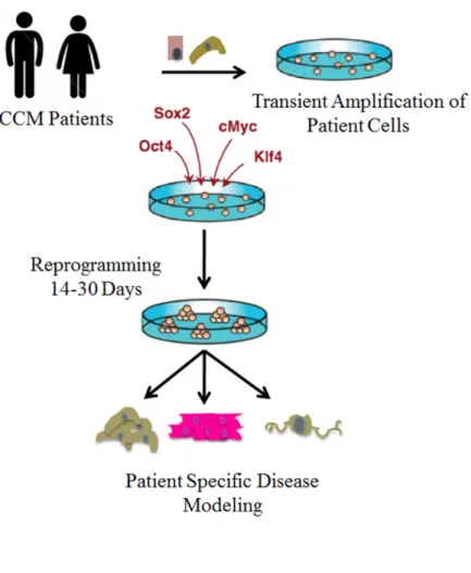
efficacy and toxicity (Fig 1.2).
Recent advances in cell biology have made the study of patient cells from
rare or hard to study diseases possible. This importance of studying diseased cells
from patients has been fully recognized for many years. However, the isolation and
propagation procedures are often complicated by access, purity after isolation and
slow cellular growth rates. Seminal work from the laboratory of Shinya Yamanaka
demonstrated that fully differentiated fibroblasts can be induced to a pluripotent state
or become induced pluripotent stem cells (iPS) through the ectopic introduction of
transcription factors, which officially initiated the “reprogramming” era in cell biology