Direct detection of blood nitric oxide reveals a burn
injury-dependent decrease of nitric oxide in response to Pseudomonas
aeruginosa infection
Julia L.M. Dunn1,Ŧ, Rebecca A. Hunter2,Ŧ, Karli Gast2, Robert Maile1,3, Bruce A. Cairns1,3, and Mark H. Schoenfisch2
Julia L.M. Dunn: [email protected]; Rebecca A. Hunter: [email protected]; Karli Gast: [email protected]; Robert Maile: [email protected]; Bruce A. Cairns: [email protected]; Mark H. Schoenfisch:
1Department of Chemistry, University of North Carolina, Chapel Hill, NC 27599
2Department of Microbiology and Immunology, University of North Carolina, Chapel Hill, NC
27599
3North Carolina Jaycee Burn Center, Department of Surgery, University of North Carolina, Chapel
Hill, NC 27599
Abstract
Purpose—Burn injury is associated with severe immune dysfunction, including an
anti-inflammatory state that occurs late after burn injury. While increased nitric oxide (NO) production is associated with severe infection and sepsis, the effect of burn trauma on these levels during a non-lethal infection remains unknown. We hypothesized that in a mouse model, 1) NO levels would be increased after infection without trauma and 2) burn injury would lead to decreased NO production even during infection.
Methods—Mice were infected via intra-tracheal inoculation with Pseudomonas aeruginosa 14 d following a 20% total body surface area contact burn. At 48 h following infection, blood was drawn to quantify NO concentrations using a microfluidic electrochemical sensor.
Significant findings—In uninjured mice, infection caused a significant increase in blood NO levels. Increases in NO occurred in a dose-dependent response to the bacterial inoculum. Following burn injury, an identical infection did not elicit increases in NO.
Conclusions—While increases in NO are expected over the course of an infection without prior trauma, burn injury and subsequent immune suppression decreases NO levels even in the presence of infection.
Correspondence to: Mark H. Schoenfisch, [email protected].
ŦCo-first authors contributing equally to this work.
Publisher's Disclaimer: This is a PDF file of an unedited manuscript that has been accepted for publication. As a service to our
HHS Public Access
Author manuscript
Burns
. Author manuscript; available in PMC 2017 November 01.Published in final edited form as:
Burns. 2016 November ; 42(7): 1522–1527. doi:10.1016/j.burns.2016.05.005.
A
uthor Man
uscr
ipt
A
uthor Man
uscr
ipt
A
uthor Man
uscr
ipt
A
uthor Man
uscr
Keywords
Nitric oxide; burn injury; Pseudomonas aeruginosa; pneumonia; electrochemistry; microfluidic sensor; compensatory anti-inflammatory response syndrome; sepsis
Introduction
Physical trauma such as burn injury causes severe immune dysfunction, often resulting in infection, sepsis, multiple organ dysfunction, and death [1]. Historically, this immune response has been characterized by an initial pro-inflammatory period and a subsequent anti-inflammatory phase [1, 2]. These periods have been referred to as the systemic anti-inflammatory response syndrome (SIRS), during which pro-inflammatory mediators (e.g., tumor necrosis factor, interleukin-6, interleukin-1β) are released [3], and a compensatory anti-inflammatory response syndrome (CARS) thought to limit damage due to chronic inflammation [1–6]. More recent studies have illustrated a more complex scenario, wherein pro- and anti-inflammatory cytokines may be secreted simultaneously, suggesting that the SIRS/CARS paradigm may be insufficient to characterize the immune response to trauma [4–6]. Despite this paradigm shift, it remains true that late after trauma patients exhibit elevated
susceptibility to nosocomial infection [7–9]. Burn patients are especially prone to ventilator-associated pneumonia and wound infections [7, 10], and the leading cause of death
following burn injury is related to infection and sepsis [7, 11]. In particular, pulmonary infections by Gram-negative Pseudomonas aeruginosa are quite common [8]. Novel methods are needed that will enable healthcare providers and researchers to monitor the immune response during trauma and infection.
Nitric oxide (NO) is a free radical species that is intricately involved with the innate immune response [12–27], and as such, in vivo levels will likely reflect the immune status of an individual. Indeed, the up-regulation of inducible nitric oxide synthase (iNOS) during SIRS has been observed, in addition to accumulation of NO byproducts in blood and tissue [8, 13, 18, 28–32]. Until recently, direct detection of NO in whole blood was not feasible, requiring either the measurement of its byproducts (i.e., nitrate and nitrate) or the use of complex instrumentation (i.e., electron paramagnetic resonance spectroscopy) [33]. Studies measuring NO byproducts in sheep have found that serum NO is increased in sheep after burn and smoke inhibition, and that this increase can be blocked with iNOS inhibitors [34].
Direct measurement of NO is likely to sensitively detect changes in patient status; therefore, such measurements should be incorporated into clinically relevant models. Because of the availability of genetically modified mice, confirming the usefulness of NO as a readout in mouse models of trauma will enable elucication of disease mechanisms that impact NO production among other phenotypes. Recently, a microfluidic amperometric sensor was developed and used to directly measure increases in NO levels during a lethal murine model of sepsis[29].
We expect that NO levels may be decreased during an infection following burn injury. Because patients demonstrate susceptibility to infection late after trauma, we focused on NO levels in a nonlethal infection model at 14 days after burn injury. Quantifying changes in NO
A
uthor Man
uscr
ipt
A
uthor Man
uscr
ipt
A
uthor Man
uscr
ipt
A
uthor Man
uscr
concentration may allow for the elucidation of immune dysfunction, indicated by either elevated levels during systemic infection or decreased levels during immune suppression.
Materials and Methods
Murine model of burn injury and infection
Nine week-old female C57BL/6 mice weighing ~18 g underwent a 20% total body surface area (TBSA) burn injury as previously described [35]. Briefly, mice were anesthetized with gaseous isofluorane, their dorsal flanks were shaved, and they received a subcutaneous injection of morphine sulphate prior to receiving a full-thickness burn with 4 applications of a copper rod heated in boiling water. Following burn injury, mice were resuscitated via an intraperitoneal injection of lactated Ringer’s solution. Throughout the experiment, mice were monitored and received morphine in their drinking water (0.02 mg mL−1; 4 mg kg−1 body weight per day) ad libitum. Sham (0% TBSA) mice also underwent these treatments as described, except the application of the copper rod.
At 14 d following burn injury, pneumonia was induced via the intratracheal administration of 50 μL Pseudomonas aeruginosa (PAK strain) following sedation with Avertin. Uninfected groups were administered 50 μL PBS with 1% protease peptone in the same manner. At 48 h following infection, ~300 μL blood was drawn into EDTA-coated microcentrifuge tubes via submandibular puncture. This blood was immediately injected into the microfluidic device and analyzed amperometrically to determine NO concentrations.
Microfluidic amperometric sensor for nitric oxide measurement
Devices were fabricated as previously described [29]. Working electrodes were 100 μm wide and consisted of 150 nm thick platinum with a 10 nm titanium seed layer, coated with a selective film (adhesion layer of 1% v/v (3-aminopropyl)triethoxysilane and a
fluoroalkoxysilane xerogel) to impart selectivity to NO. The fluoroalkoxysilane membrane solution was prepared via the acid catalyzed hydrolysis and condensation of (heptadeca uoro-1,1,2,2-tetrahydrodecyl)trimethoxysilane and methyltrimethoxysilane as reported previously [36, 37]. Reference electrodes consisted of a 10 nm titanium adhesion layer followed by a ~1 μm silver layer, chemically oxidized by reaction with 50 mM ferric chloride for 10 s to create a silver/silver chloride pseudo-reference/counter electrode. The ~90 μm microfluidic channel was formed using Kapton® tape.
The working and reference/counter electrodes of the microfluidic device were connected to a CH Instruments 1030A 8-channel potentiostat (Austin, TX). Prior to sample analysis, the device was polarized at +800 mV vs. the Ag/AgCl pseudo-reference/counter electrode for at least to 1 h in PBS. To calibrate the device, a saturated NO standard solution (prepared by purging deaerated PBS with NO gas for ~10 min to yield a 1.9 mM solution of NO) was diluted with PBS and introduced into the inlet reservoir.
A
uthor Man
uscr
ipt
A
uthor Man
uscr
ipt
A
uthor Man
uscr
ipt
A
uthor Man
uscr
Statistical analysis
Where appropriate, either an unpaired, two-sided Student’s t-test or two-way ANOVA with Bonfennori post-test was used to determine statistical significance between groups, with p <0.05 considered to be significant.
Results
Nitric oxide levels are increased during pneumonia without prior burn injury
Blood samples were drawn 48 h following infection with 1 × 10⁶ CFU of P. aeruginosa and immediately analyzed using the microfluidic sensor. As shown in Figure 1A, blood NO levels were significantly increased in the infected versus uninfected mice at this time point (810 ± 180 nM and 370 ± 40 nM, respectively). As expected, these data suggest that activation of the immune response following infection leads to increased production of NO.
Increased infectious dose corresponds with elevated serum NO
Blood samples were drawn 24 h and 72 h following infection with either 5 × 10⁵ or 5 × 10⁶ CFU of P. aeruginosa. As indicated in Figure 1B, blood NO levels were higher in uninjured mice exposed to a higher infectious dose at 24 h (3.0 ± 0.2 μM vs. 6.2 ± 0.7 μM) and 72 h (7.0 ± 1.4 μM vs. 15.2 ± 4.7 μM) following infection. At both high- and low-dose inocula, blood NO concentrations were higher at 72 vs. 24 h. We also harvested lungs and quantified bacterial load in these mice at 72 h after infection. A significant difference in bacterial load was observed between mice with high and low dose bacterial inoculum, with a
corresponding difference in plasma NO levels (Figure 1B).
Burn injury inhibits nitric oxide release following infection
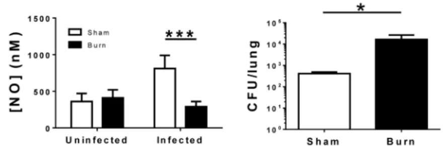
Blood samples from infected (1 × 10⁶ CFU) and uninfected mice 14 d after burn injury were analyzed using the electrochemical sensor. Concentrations of NO in blood of infected burned mice were not significantly elevated compared to uninfected burn mice (290 ± 80 nM vs. 410 ± 110 nM). We also harvested lungs and quantified bacterial load in these mice at 72 h post infection, and observed both reduced NO and pulmonary bacterial clearance in burn versus sham mice (Figure 2). These data indicate a late-stage effect of burn injury on immune function and NO production during an infection. Also of note, the NO
concentrations observed in uninfected burn mice were equivalent to those in the uninfected sham mice.
Blood NO levels were measured at 24 and 72 h following a low-dose (5 × 10⁵ CFU) infection with P. aeruginosa. We observed that the NO measured was lower in burn infected mice compared to unburned infected mice at both 24 h (1.1 ± 0.4 vs. 3.0 ± 0.2 μM) and 72 h (6.0 ± 0.6 vs. 7.0 ± 1.4 μM) following the infection (Figure 3A). In contrast, we observed elevated blood NO following inoculation with a high-dose (5 × 10⁶ CFU) of P. aeruginosa in burned mice compared to unburned mice at 24 h (7.5 ± 0.7 μM vs. 6.2 ± 0.7 μM). However, the measured NO was not increased in burned mice at 72 h (4.2 ± 2.0 μM vs. 15.2 ± 4.7 μM) post infection (Figure 3B) while blood NO continued to rise in unburned mice.
A
uthor Man
uscr
ipt
A
uthor Man
uscr
ipt
A
uthor Man
uscr
ipt
A
uthor Man
uscr
Discussion
In this study, we evaluated the effect of infection and burn injury on endogenous NO levels in a murine model. While it is well understood that NO, along with other reactive oxygen and nitrogen species (ROS and RNS), are produced at higher levels during infection, direct detection of NO has not been possible and analysis of its byproducts (nitrate and nitrite) has been required to estimate NO production. The microfluidic electrochemical sensor utilized herein allowed for the immediate and direct detection of NO in a small (~250 μL) volume of whole blood, without the addition of external reagents. As hypothesized, during an infection with no previous trauma, NO levels were increased relative to control mice. This result is not surprising, as NO is known for its antibacterial activity [38] and has been previously
demonstrated to increase during systemic infection [29]. We therefore conclude that our novel monitor sensitively detects changes in endogenous NO during nonlethal infection in mice.
However, this trend was not observed or was attenuated during infection that occurred 14 d following burn injury. Indeed, NO concentrations in infected burn mice were equivalent to those of uninfected burn and sham mice, indicating immune suppression as a result of the burn injury. Even following high dose infection, when at 24 h NO concentrations were higher in burn than unburned mice, the injured mice were unable to sustain a robust response and NO concentrations diminished by 72 h compared with unburned mice.
The dysfunctional immune response following burn injury has been well documented [2]. Cairns et al. demonstrated that while expression of TLR2 and TLR4 by macrophages is increased early (3 d) following burn injury, these levels are significantly decreased at 14 d [39]. Numerous immune defense systems are linked to TLR, including the induction of iNOS [40]. Other studies have linked TLR with the induction of iNOS and concomitant release of NO by innate immune cells (e.g., macrophages) [41–43]. For example, TLR is directly involved in microbe recognition by innate immune cells (TLR2 for Gram-positive peptidoglycan and TLR4 for Gram-negative lipopolysaccharide) and thus mediates subsequent inflammatory signals, including NO [40]. Future studies should determine whether TLR signaling is directly linked to endogenous NO following infection.
While increased levels of endogenous NO may indicate development of a severe infection in otherwise healthy animals, the immune dysfunction that occurs late after trauma (e.g., burn injury) significantly alters NO production. As such, monitoring in vivo NO production in real time may provide insight into emerging infection as well as immune dysfunction. Reduction in NO production by innate immune cells, along with other aspects of immune suppression (e.g., shifts in T cell phenotype [44], altered cytokine profiles[45]), contribute to the increased infection susceptibility of burn patients. The clinical utility of NO
measurement can be further evaluated by monitoring concentration changes throughout the course of infection/sepsis and throughout the dynamic immune response following trauma. In murine models, differences in NO concentration caused by infection with more virulent strains and during antibiotic treatment must also be evaluated.
A
uthor Man
uscr
ipt
A
uthor Man
uscr
ipt
A
uthor Man
uscr
ipt
A
uthor Man
uscr
Acknowledgments
Funding
The authors acknowledge research support from the National Institutes of Health (NIAID AI094719).
References
1. Mannick JA, Rodrick ML, Lederer JA. The immunologic response to injury. Journal of the American College of Surgeons. 2001; 193:237–244. [PubMed: 11548792]
2. Hotchkiss RS, Coopersmith CM, McDunn JE, Ferguson TA. The sepsis seesaw: tilting toward immunosuppression. Nature Medicine. 2009; 15:496–497.
3. Dinarello CA. Proinflammatory cytokines. Chest Journal. 2000; 118:503–508.
4. Liu T, Qian WJ, Gritsenko MA, Xiao W, Moldawer LL, Kaushal A, Monroe ME, Varnum SM, Moore RJ, Purvine SO, Maier RV, Davis RW, Tompkins RG, Camp DG 2nd, Smith RD.
Inflammation and the Host Response to Injury Large Scale Collaborative Research P. High dynamic range characterization of the trauma patient plasma proteome. Mol Cell Proteomics. 2006; 5:1899– 1913. [PubMed: 16684767]
5. Finnerty CC, Jeschke MG, Herndon DN, Gamelli R, Gibran N, Klein M, Silver G, Arnoldo B, Remick D, Tompkins RG. Investigators of the I the Host Response Glue G. Temporal cytokine profiles in severely burned patients: a comparison of adults and children. Mol Med. 2008; 14:553– 560. [PubMed: 18548133]
6. Xiao W, Mindrinos MN, Seok J, Cuschieri J, Cuenca AG, Gao H, Hayden DL, Hennessy L, Moore EE, Minei JP, Bankey PE, Johnson JL, Sperry J, Nathens AB, Billiar TR, West MA, Brownstein BH, Mason PH, Baker HV, Finnerty CC, Jeschke MG, Lopez MC, Klein MB, Gamelli RL, Gibran NS, Arnoldo B, Xu W, Zhang Y, Calvano SE, McDonald-Smith GP, Schoenfeld DA, Storey JD, Cobb JP, Warren HS, Moldawer LL, Herndon DN, Lowry SF, Maier RV, Davis RW, Tompkins RG. Inflammation and Host Response to Injury Large-Scale Collaborative Research P. A genomic storm in critically injured humans. J Exp Med. 2011; 208:2581–2590. [PubMed: 22110166]
7. Weinstein RA, Mayhall CG. The Epidemiology of Burn Wound Infections: Then and Now. Clinical Infectious Diseases. 2003; 37:543–550. [PubMed: 12905139]
8. Driscoll J, Brody S, Kollef M. The Epidemiology, Pathogenesis and Treatment of Pseudomonas aeruginosa Infections. Drugs. 2007; 67:351–368. [PubMed: 17335295]
9. Adib-Conquy M, Cavaillon J-M. Compensatory anti-inflammatory response syndrome. Thrombosis and Haemostasis. 2009; 101:36–47. [PubMed: 19132187]
10. Wurtz R, Karajovic M, Dacumos E, Jovanovic B, Hanumadass M. Nosocomial infections in a burn intensive care unit. Burns. 1995; 21:181–184. [PubMed: 7794498]
11. Cioffi WG. What’s new in burns and metabolism. Journal of the American College of Surgeons. 2001; 192:241–254. [PubMed: 11220727]
12. Armstrong R. The physiological role and pharmacological potential of nitric oxide in neutrophil activation. International immunopharmacology. 2001; 1:1501–1512. [PubMed: 11515815] 13. Assreuy J, Barja-Fidalgo C, Tavares-Murta BM. Inflammatory and Vascular Alterations in Sepsis:
The Role of Nitric Oxide-Dependent Mechanisms. Anti-Inflammatory & Anti-Allergy Agents in Medicinal Chemistry (Formerly Cu. 2006; 5:35–44.
14. Beckman JS, Koppenol WH. Nitric oxide, superoxide, and peroxynitrite: The good, the bad, and the ugly. American Journal of Physiology-Cell Physiology. 1996; 271:C1424–C1437.
15. Bogdan C. Nitric oxide and the immune response. Nat Immunol. 2001; 2:907–916. [PubMed: 11577346]
16. Boscá L, Zeini M, Través PG, Hortelano S. Nitric oxide and cell viability in inflammatory cells: a role for NO in macrophage function and fate. Toxicology. 2005; 208:249–258. [PubMed: 15691589]
17. Coleman JW. Nitric oxide in immunity and inflammation. International Immunopharmacology. 2001; 1:1397–1406. [PubMed: 11515807]
A
uthor Man
uscr
ipt
A
uthor Man
uscr
ipt
A
uthor Man
uscr
ipt
A
uthor Man
uscr
18. Fortin CF, McDonald PP, Fülöp T, Lesur O. Sepsis, leukocytes, and nitric oxide (NO): an intricate affair. Shock. 2010; 33:344–352. [PubMed: 19789465]
19. Frances R, Munoz C, Zapater P, Uceda F, Gascon I, Pascual S, Perez-Mateo M, Such J. Bacterial DNA activates cell mediated immune response and nitric oxide overproduction in peritoneal macrophages from patients with cirrhosis and ascites. Gut. 2004; 53:860–864. [PubMed: 15138214]
20. Hierholzer C, Kalff JC, Billiar TR, Bauer AJ, Tweardy DJ, Harbrecht BG. Induced nitric oxide promotes intestinal inflammation following hemorrhagic shock. American Journal of Physiology-Gastrointestinal and Liver Physiology. 2004; 286:G225–G233. [PubMed: 14715517]
21. Hollenberg SM, Broussard M, Osman J, Parrillo JE. Increased microvascular reactivity and improved mortality in septic mice lacking inducible nitric oxide synthase. Circulation research. 2000; 86:774–778. [PubMed: 10764411]
22. Kirkebøen KA, Strand ØA. The role of nitric oxide in sepsis–an overview. Acta Anaesthesiologica Scandinavica. 1999; 43:275–288. [PubMed: 10081533]
23. Knight J. Review: Free radicals, antioxidants, and the immune system. Annals of Clinical & Laboratory Science. 2000; 30:145–158. [PubMed: 10807157]
24. Lowenstein CJ, Dinerman JL, Snyder SH. Nitric Oxide: A Physiologic Messenger. Annals of Internal Medicine. 1994; 120:227–237. [PubMed: 8273987]
25. MacMicking J, Xie QW, Nathan C. Nitric oxide and macrophage function. Annual Review of Immunology. 1997; 15:323–350.
26. Snyder SH, Bredt DS. Biological roles of nitric oxide. Scientific American. 1992; 266:74–77. 27. Vincent J-L, Zhang H, Szabo C, Preiser J-C. Effects of Nitric Oxide in Septic Shock. American
Journal of Respiratory and Critical Care Medicine. 2000; 161:1781–1785. [PubMed: 10852744] 28. Carraway MS, Piantadosi CA, Jenkinson CP, Huang Y-CT. Differential expression of arginase and
iNOS in the lung in sepsis. Experimental lung research. 1998; 24:253–268. [PubMed: 9635249] 29. Hunter RA, Privett BJ, Henley WH, Breed ER, Liang Z, Mittal R, Yoseph BP, McDunn JE, Burd
EM, Coopersmith CM, Ramsey JM, Schoenfisch MH. Microfluidic amperometric sensor for analysis of nitric oxide in whole blood. Analytical Chemistry. 2013; 85:6066–6072. [PubMed: 23692300]
30. Carcillo JA. Nitric oxide production in neonatal and pediatric sepsis. Critical care medicine. 1999; 27:1063–1065. [PubMed: 10397205]
31. Dhillon SS, Mahadevan K, Bandi V, Zheng Z, Smith CW, Rumbaut RE. Neutrophils, nitric oxide, and microvascular permeability in severe sepsis. CHEST Journal. 2005; 128:1706–1712.
32. Kao CC, Bandi V, Guntupalli KK, Wu M, Castillo L, Jahoor F. Arginine, citrulline and nitric oxide metabolism in sepsis. Clin Sci. 2009; 117:23–30. [PubMed: 19105791]
33. Hetrick EM, Schoenfisch MH. Analytical Chemistry of Nitric Oxide. Annual Review of Analytical Chemistry. 2009; 2:409–433.
34. Soejima K, Schmalstieg FC, Traber LD, Szabo C, Salzman A, Traber DL. Role of nitric oxide in myocardial dysfunction after combined burn and smoke inhalation injury. Burns. 2001; 27:809– 815. [PubMed: 11718983]
35. Hultman CS, Cairns BA, deSerres S, Frelinger JA, Meyer AA. Early, complete burn wound excision partially restores cytotoxic T lymphocyte function. Surgery. 1995; 118:421–430. [PubMed: 7638760]
36. Shin JH, Weinman SW, Schoenfisch MH. Sol-gel derived amperometric nitric oxide microsensor. Analytical Chemistry. 2005; 77:3494–3501. [PubMed: 15924380]
37. Shin JH, Privett BJ, Kita JM, Wightman RM, Schoenfisch MH. Fluorinated xerogel-derived microelectrodes for amperometric nitric oxide sensing. Analytical Chemistry. 2008; 80:6850– 6859. [PubMed: 18714964]
38. Carpenter AW, Schoenfisch MH. Nitric oxide release: Part II. Therapeutic applications Chemical Society Reviews. 2012; 41:3742–3752. [PubMed: 22362384]
39. Cairns BA, Barnes CM, Mlot S, Meyer AA, Maile R. Toll-like Receptor 2 and 4 Ligation Results in Complex Altered Cytokine Profiles Early and Late After Burn Injury. Journal of Trauma and Acute Care Surgery. 2008; 64:1069–1078.
40. Underhill DM, Ozinsky A. Toll-like receptors: key mediators of microbe detection. Current opinion in immunology. 2002; 14:103–110. [PubMed: 11790539]
41. Baumgarten G, Knuefermann P, Schuhmacher G, Vervölgyi V, von Rappard J, Dreiner U, Fink K, Djoufack C, Hoeft A, Grohé C. Toll-like receptor 4, nitric oxide, and myocardial depression in endotoxemia. Shock. 2006; 25:43–49. [PubMed: 16369185]
42. Hoshino K, Takeuchi O, Kawai T, Sanjo H, Ogawa T, Takeda Y, Takeda K, Akira S. Cutting edge: Toll-like receptor 4 (TLR4)-deficient mice are hyporesponsive to lipopolysaccharide: evidence for TLR4 as the Lps gene product. The Journal of Immunology. 1999; 162:3749–3752. [PubMed: 10201887]
43. Brightbill HD, Libraty DH, Krutzik SR, Yang R-B, Belisle JT, Bleharski JR, Maitland M, Norgard MV, Plevy SE, Smale ST. Host defense mechanisms triggered by microbial lipoproteins through toll-like receptors. Science. 1999; 285:732–736. [PubMed: 10426995]
44. Daniel T, Alexander M, Hubbard WJ, Chaudry IH, Choudhry MA, Schwacha MG. Nitric oxide contributes to the development of a post-injury Th2 T-cell phenotype and immune dysfunction. Journal of Cellular Physiology. 2006; 208:418–427. [PubMed: 16642464]
45. Ulloa L, Tracey KJ. The ‘cytokine profile’: a code for sepsis. Trends in molecular medicine. 2005; 11:56–63. [PubMed: 15694867]
A
uthor Man
uscr
ipt
A
uthor Man
uscr
ipt
A
uthor Man
uscr
ipt
A
uthor Man
uscr
Highlights
• Microfluidic nitric oxide sensor is used to directly measure nitric oxide levels in fresh whole blood
• Increased nitric oxide production measured in mice infected with Pseudomonas aeruginosa
• Significantly diminished nitric oxide levels observed in infected animals that had suffered a burn 14 d prior
A
uthor Man
uscr
ipt
A
uthor Man
uscr
ipt
A
uthor Man
uscr
ipt
A
uthor Man
uscr
Figure 1. Nitric Oxide (NO) levels are elevated following infection without prior injury in a dose-dependent fashion
Mice (n=4) were infected with of P. aeruginosa and blood NO analyzed. A) NO levels were significantly increased 48 h following infection with1 × 106 CFU compared to uninfected counterparts. Statistical significance is indicated by *, p < 0.05 by a Student’s t test. B) NO levels in blood and bacterial recovery from lungs were higher in mice inoculated with 5 × 10⁵ CFU vs. 5 × 10⁶ CFU at 72 hours post infection. Statistical significance is indicated by *, p<0.05 and ***, p < 0.001 by Two-way ANOVA with Bonfennori post-test. Data are given as mean ± standard deviation.
A
uthor Man
uscr
ipt
A
uthor Man
uscr
ipt
A
uthor Man
uscr
ipt
A
uthor Man
uscr
Figure 2. Relative to sham mice, burn injury causes decreased blood NO concentrations and increased pulmonary bacterial load following a 14 d post-burn infection
Mice (n=4) were infected with 1 × 10⁶ CFU of P. aeruginosa 14 d after injury and blood NO analyzed 48 h after infection. Statistical significance is indicated by *, p<0.05 and ***, p < 0.001 by a Two-way ANOVA followed by Bonfennori post-test. Data are given as mean ± standard deviation.
A
uthor Man
uscr
ipt
A
uthor Man
uscr
ipt
A
uthor Man
uscr
ipt
A
uthor Man
uscr
Figure 3. Inoculation dose impacts blood NO concentration in burn mice following infection
Mice (n=3–4) were infected with A) 5 × 10⁵ or B) 5 × 10⁶ CFU of P. aeruginosa and blood NO analyzed after 24 h or 72 h. Statistical significance is indicated by ***, p < 0.001 by a Two-way ANOVA followed by Bonfennori post-test. Data are given as mean ± standard deviation.