Shah Vidhi et al IJSRE Volume 05 Issue 11 November 2017 Page 7721 Volume||5||Issue||11||November-2017||Pages-7721-7731||ISSN(e):2321-7545 Website: http://ijsae.in
Index Copernicus Value- 56.65 DOI: http://dx.doi.org/10.18535/ijsre/v5i11.03
Measurement of Elbow Range of Motion in Full Term Neonates
Authors
Shah Vidhi1, Lata Parmar2, Shilpa Khandare3, Tushar Palekar4
1
Assistant Professor, Dr. D.Y.Patil College of Physiotherapy, Dr. D.Y.Patil Vidyapeeth Pune, India
2
PhD, Principal, College of Physiotherapy, Sumandeep Vidyapeeth ,waghodia, India
3
Associate Professor, Dr. D.Y.Patil College of Physiotherapy, Dr. D.Y.Patil Vidyapeeth Pune, India
4
PhD, Principal, Dr. D.Y.Patil College of Physiotherapy, Dr. D.Y.Patil Vidyapeeth Pune, India *Corresponding Author
Vidhi Shah
Email- [email protected]
ABSTRACT
Introduction: This cross sectional study was done to establish normal values of range of motion (ROM) of elbow joint. Since normative values for elbow flexion passive range of motion (PROM) and extension limitation of the elbow joint in neonates are not well-documented in literature, the present study was undertaken.
Methods: The PROM and extension limitation of elbow joint measured using universal goniometer. It was based on 160 elbow joints of neonates.
Observations And Results: The mean elbow flexion PROM was 159.3 degrees in neonates. The extension limitation was 19.2 degrees in neonates. The results of the present study showed that there was no significant statistical difference of PROM and extension limitation between male and female neonates and between right and left elbow joints.
Conclusions: The findings of the present study will be helpful to the clinicians, therapists and researchers as ready references to PROM and extension limitations of elbow joint in infants. Since there was no statistically significant difference between right and left knee joint, in the presence of a lesion of one elbow joint, the movement on the healthy side of the joint can be used for routine comparison with the affected limb. In the presence of bilateral lesion, the present data can be used for comparison. The advantage of using these data is that, they provide the best available estimates of normal elbow joint PROM and extension limitation.
Keywords: Elbow range of motion, Gestational Age, Goniometry and Neonate
INTRODUCTION:
The full-term newborn comes into this world after 38 to 42 weeks of fetal development which has been influenced primarily by genetic coding and nervous system maturation. The neonate has had minimal experience with extra uterine environmental factors (such as gravity, sound, and light). These will now begin to have a potent effect on development.1
Shah Vidhi et al IJSRE Volume 05 Issue 11 November 2017 Page 7722
extended.2, 3. Von Hofsten (1990) suggests that the arm movements of a neonate are often coordinated with visual fixation on a target and thus beginning of eye-hand coordination.4 In neonates elbow, wrist, and finger movements of flexion and semi extension occurs, but rarely is full elbow extension or full shoulder external rotation seen.
Limitation of elbow extension is physiologically normal in infants and the term that is often used to describe this condition is elbow flexion contracture (EFC). However, the term “contracture” usually refers to a pathological condition, which is not the case in the pediatric population when limitation of elbow extension is within the normal ranges.5 Early diagnosis of an abnormality would seem to have certain implications on early treatment. Physical therapists are increasingly being asked to evaluate developing infants.
The measurement of passive joint range of motion in infants and children differ from adult values,6,7,8 and that specific sequential changes in the range of joint motion occur during the first 15 months of normal development.7,8
Goniometric measurements of joint range are used clinically with infants to aid in detecting pathological joint and muscle conditions, to assess and document changes in response to therapeutic regimens, and to provide useful information about muscle tone and motor development. For example, in assessing newborns, the popliteal angle and the amount of passive dorsiflexion are both used as indices of gestational age.2, 9,10. In the pediatric population, the assessment of range of motion (ROM) can contribute to early detection of hidden pathologies, 11,12,13 therefore the knowledge of normal limits for ROM of the extremities is important in young subjects. The inter-tester reliability of goniometry was higher for upper extremity motions than lower extremity(r =.86)The intra-tester reliability for flexion and extension of the elbow joints was high (r = .91 to .99). Intertester reliability was also high (r = .88 to .97)14
Understanding normal motion, strength, and physiologic flexion will assist with the diagnosis of pathology. The main focus of this study understands the normal examination and values to appreciate an abnormal finding.
METHOD:
The cross-sectional study was done in the Neonatal unit of Paediatric department and Obstetric department of Dhiraj General Hospital, Piparia, The project was started with official referral from department of Paediatrics of DGH.
All neonates were screened by the Paediatrician and subjects were recruited for the study after checking for selection criteria. The neonates with Small for gestational Age (SGA), Medical illness, Neurological orthopaedic and genetic disorder were excluded.
Total 146 Full term neonates (Neonates with gestational age >37 weeks) were screened and from that 80 neonates included in this study who met the selection criteria as well as the willingness of parents were considered.
Prior to the measurement, the infant’s medical records were checked for Apgar scores and Neuro-motor behaviour assessment to rule out those who have serious complications which are likely to affect the further development. Infants with similar Apgar scores were included in the study.
The neonate should be in an alert, non crying state, if neonates are crying vigorously the recording will be difficult and unreliable. For all measurements, infants were placed in a supine position with their heads maintained in midline to diminish any effects of neonatal neck reflexes, such as the asymmetrical tonic neck reflex (Fig). As the head position can alter muscle tone; eliciting asymmetrical tonic neck reflex,so that head turning during the examination is prevented .
Shah Vidhi et al IJSRE Volume 05 Issue 11 November 2017 Page 7723
standardized approach based on the international neutral zero method and different basic planes designation. SFTR: S = Sagittal; F = Frontal; T = Transverse; R = Rotation.
A standard plastic goniometer (180°) was used to measure joint range of motion. The elbow range of motion was measured with the Neonate in supine lying position and the shoulder was stabilized in neutral position along the side of the body. The axis of the goniometer was placed at the lateral aspect of the elbow. The stationary arm was aligned parallel to the lateral side of the arm (humerus) and the movable arm was parallel to the lateral side of the forearm (radial), if needed the arm was placed on a soft roll for proper alignment. Then the elbow joint gently passively flexed and extended. The angle formed at the elbow was recorded. Both right and left side of elbow flexion-extension limitation range of motion was tested and recorded with the same method. The value for right elbow flexion noted as RTFL and for left elbow flexion as LTFL. The value for right elbow extension limitation noted as RTEXTL and for left elbow extension limitation as LTEXTL.
Fig.1 Measurement of elbow flexion
Fig.2 Measurement of elbow extension
DATA ANALYSIS AND INTERPRETATION:
Each subject was coded and all data were statistically analyzed with the SPSS 20.0 statistical package in a password protected computer. Mean difference scores and standard Deviation of these scores were calculated for each variable. The Kolmogorov-smirnov goodness of fit Test was used for normal distribution of the data. The Pearson’s correlation coefficient was calculated to assess the relationship between Birth Weight (BW) and Elbow Passive range of motion (Flexion and Extension limitation)
A significant level of p=0.05 was set for all analysis.
Shah Vidhi et al IJSRE Volume 05 Issue 11 November 2017 Page 7724 RESULT:
Result was obtained after measurement of 80 full term neonates (160 elbow joint - right and left side). Total 122 full term neonates were born during the duration of December 2012 and January 2013.
Table 1- The mean and SD of demographic data was mentioned Variables Mean SD Gestational age 37.87 1.18 Chronological age 2.96 1.85 Gender
Male 39
Female 41
Mode of delivery
Vaginal 51
LSCS 29 0.33
Birth weight 2.71 Low Birth Weight 24 Normal Birth Weight 56 APGAR
1st min 7/10 5thmin 9/10
Table 2- The Mean and Standard Deviation (SD) of all Elbow ROM
Variable Minimum Maximum Mean SD RTFL 140 152 147.12 3.03 LTFL 140 152 147.22 3.14 RTEXTL 14 30 19.5 2.97 LTEXTL 14 30 19.5 2.97
(RTFL-right flexion, LTFL- left flexion, RTEXTL- right extension limitation and LTEXTL- left extension limitation)
Graph 1 The Mean Score and SD of Elbow joint Range of Motion
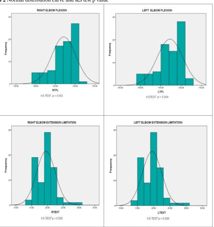
The Kolmogorov- Smirnov Test for normal distribution shows that all the data of Elbow Range of Motion are fall in normal distribution
MEAN 0
50 100 150
RTFL LTFL RTEXT LTEXT
147.12 147.22
Shah Vidhi et al IJSRE Volume 05 Issue 11 November 2017 Page 7725 Graph 2 Normal distribution curve and KS test p value
The significance value of each measurement is listed below the graph (i.e. KS TEST p =)
The independent t-test was used for comparison of elbow flexion and extension limitation in right and left side of elbow joint.
Table 3 comparison of right and left side of elbow flexion range of motion
FLEXION
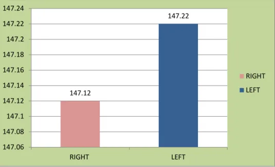
MEAN SD T -Value p Value RIGHT 147.12 3.03
Shah Vidhi et al IJSRE Volume 05 Issue 11 November 2017 Page 7726 Graph 3 Comparisons between Right and Left Elbow Flexi
It shows that there is no significant difference between flexion of right and left side of elbow joint.
Table 4 Comparison of Right and Left side of Elbow Extension Limitation Range of Motion
EXTENSION LIMITATION
MEAN SD T-Value p Value RIGHT 19.50 2.97
0.00 1.00 LEFT 19.50 2.97
Graph 4 Comparisons between Right and Left Elbow Extension Limitation
It shows that there is no significant difference between extension limitation of right and left side of elbow joint.
The independent t-test was used to compare the difference between male and female elbow joint range of motion.
Table 5 Comparison of Right Elbow Flexion between Male and Female
Right Elbow
Flexion MEAN SD T-Value
p-value
MALE 147.13 2.78
0.009 0.10 FEMALE 147.12 3.28
19.5 19.5
0 5 10 15 20 25
RIGHT LEFT
RIGHT
LEFT
147.12
147.22
147.06 147.08 147.1 147.12 147.14 147.16 147.18 147.2 147.22 147.24
RIGHT LEFT
RIGHT
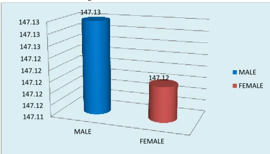
Shah Vidhi et al IJSRE Volume 05 Issue 11 November 2017 Page 7727 Graph 5 Comparisons between Mean Right Elbow Flexion in Male & Female
This shows that there is no significant difference of right elbow flexion between male and female.
Table 6 Comparison of Left Elbow Flexion between Male and Female
Left Elbow Flexion MEAN SD T-Value p-value MALE 147.33 2.87
0.29 0.11 FEMALE 147.12 3.40
Graph 6 Comparisons between Mean of Left Elbow Flexion in Male & Female
There is no significant difference of left elbow flexion between male and female.
Table 7 Comparison of Right Elbow Extension Limitation between Male and Female Right Elbow
Extension Limitation MEAN SD T-Value p-value MALE 19.79 3.48
0.86 0.12 FEMALE 19.21 2.40
147.11 147.12 147.12 147.12 147.12 147.12 147.13 147.13 147.13
MALE
FEMALE 147.13
147.12 MALE
FEMALE
147 147.05 147.1 147.15 147.2 147.25 147.3 147.35
MALE
FEMALE 147.33
147.12 MALE
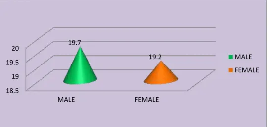
Shah Vidhi et al IJSRE Volume 05 Issue 11 November 2017 Page 7728 Graph 7 Comparisons of Mean of Right Elbow Extension Limitation in Male & Female
There is no significant difference of right elbow extension limitation between male and female.
Table 8 Comparison of Left Elbow Extension Limitation between Male and Female LEFT Elbow
Extension Limitation MEAN SD T-Value p-value
MALE 19.79 3.48
0.86 0.12 FEMALE 19.21 2.40
Graph 8 Comparisons between Mean of Left Elbow Extension in Male & Female
This test shows that there is no significant difference of left elbow extension limitation between male and female.
The Pearson’s correlation analysis was used to find any relation between Birth Weight (BW)and Elbow Joint Range of Motion
Table 9 Pearson’s correlation analysis between Birth Weight and Right and Left Elbow Flexion – Mean,
Standard Deviation, coefficient correlation (r), p value
Variable MEAN SD r-value p-value
BW 2.71 0.33
Elbow Flexion Right 147.12 3.03 0.023 0.83 Left 147.22 3.14 0.029 0.80 18.5
19 19.5 20
MALE FEMALE
19.7
19.2 MALE
FEMALE
18.8 19 19.2 19.4 19.6 19.8
MALE
FEMALE 19.7
19.2 MALE
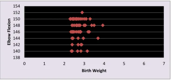
Shah Vidhi et al IJSRE Volume 05 Issue 11 November 2017 Page 7729 Graph 9 Scatter Plot for Correlation between BW and ELBOW FLEXION
Scatter plot for correlation between BW and Right and Left Elbow Flexion - No significant correlation between BW and Elbow Flexion (right and left)
Table 10-Pearson’s correlation analysis between Birth Weight and Right and Left Elbow Extension
Limitation – Mean, Standard Deviation, coefficient correlation (r), p value
Variable MEAN SD r-value p-value
BW 2.71 0.33
Elbow Extension Limitation
Right 19.50 2.97 0.43 0.00 Left 19.50 2.97 0.44 0.00
Graph 10- Scatter Plot for Correlation between BW and ELBOW EXTENSION LIMITATION
Scatter plot for correlation between BW and Right and Left Elbow Extension Limitation – Significant moderate correlation between BW and Elbow Extension Limitation (right and left)
DISCUSSION
The goal of this study was to establish normal values of elbow joint motions in neonates that could be reproduced and used clinically. Joint range limitations at the elbow in newborns have often been referred to as "contractures" or "deformities,"15but the characteristic elbow joint limitations observed in newborns do not necessarily reflect pathological conditions as these terms imply.
The term "extension limitation" rather than "flexion contracture" was therefore used in this study to describe the lack of full passive extension at the elbow in neonates. Values reported as "elbow flexion contracture" in
138 140 142 144 146 148 150 152 154
0 1 2 3 4 5 6 7
El
b
ow
Fl
e
xi
on
Birth Weight
0 5 10 15 20 25 30 35
Shah Vidhi et al IJSRE Volume 05 Issue 11 November 2017 Page 7730
the studies by Harris and colleagues16and by Hoffer15 represent the same angle as the values reported as "elbow extension limitation" in this study.
Hoffer reported elbow flexion contractures ranging from 0 to 30 degrees, but he did not include mean values in his study,15while the mean of elbow extension limitation reported in the present study is 19.50. Neither any study had reported the normal elbow flexion values in full term neonate, though it has equal importance to know in orthopedic disorders like Arthrogryposis multiplex Congenita, and Brachial Plexus injury.
The reliability of goniometric measurements being affected by the testing procedures.17,18 several sources of error such as incorrect identification of bony landmarks, faulty testing positioning and inaccurate placement and reading of the goniometer will affect reliability. 17
Results show that there was no difference between Right and Left Elbow joint Rang of Motion (ROM). In comparison between right and left elbow flexion t value (-0.21) and p=0.84 and In Comparison between right and left elbow extension limitation t value (0.00) and p=1.00 which indicates that the right and left side elbow joint ROM is same.
Also there was nodifference observed between Male and Female Elbow joint Range of Motion.In comparison between male and female right elbow flexion the t value (0.009) and p=0.10, In comparison between male and female left elbow flexion the t value (0.29) and p=0.11, In Comparison between male and female right elbow extension limitation the t value (0.86) and p=0.12, and In Comparison between male and female left elbow extension limitation the t value (0.86) and p=0.12 which proves that the elbow joint ROM is same in male and female.
Schwarze DJ, Denton JR had also found thatthere were no difference between right and left values of lower limb joints as well as boys and girls had essentially the same values. Both studies support the result of present research though it was done on upper limb (elbow) joint.
A significant correlation was found in this study between Birth Weight and elbow joint range of motion. This correlation indicated that there was more elbow extension limitation in neonates with increased weight at birth.
The elbow extension limitation at birth was noted as 10 to 30 degree in full term neonate. Results of this study are consistent with other reports describing elbow extension limitations at birth of0 to 30 degrees.15,19
CONCLUSION:
The 80 full-term neonates were assessed in the study and the passive ranges of motion of elbow joint (elbow flexion and elbow extension limitation) were assessed. Results revealed mean of elbow flexion was 147.12 and the mean of elbow extension limitations was 19.50. There was no significant difference noted between right and left side range of motion of elbow joint. Also the male and female had essentially same value. The birth weight and elbow extension limitation shows significant moderate correlation.
REFERENCES:
1. Lois BLY-The neonate: Birth to Ten days: Motor Skills Acquisition in the First Year; An illustrated guide to normal development:1994,1-9
2. Saint Anne Dargassies.Neurological development in the full term and premature neonate New York: Elsevier North Holland; 1977.
3. Heathcock JC, Bhat AN, Lobo MA, Galloway JC. The performance of infants born preterm and full term in the mobile paradigm: learning and memory. Physical Therapy 2004; 84 (9).
4. Von Hofsten.Neurological evaluation of the maturity of the newborn infant. Arch Dis Child 43:89-93, 1968
Shah Vidhi et al IJSRE Volume 05 Issue 11 November 2017 Page 7731 6. Griffin P: Orthopedics in the newborn. In Avery GB(ed): Neonatology: Pathophysiology and
Management of the Newborn, ed 2, Philadelphia, PA, JB Lippincott Co, 1981, pp 890-909
7. Haas SS, Epps CH Jr, Adams JP: Normal ranges of hip motion in the newborn. Clin. Orthop 91:114-118, 1973
8. Barnes,4 C. J., Van Steyn, S. J., & Fischer, R. A. (2001). The effects of age, sex, and Shoulder dominance on range of motion of the shoulder. Journal of Shoulder and Elbow Surgery, 10, 242-246. doi:10.1067/mse.2001.115270
9. Nancy Berryman Rees, Willium D. Landy: Joint Range of Motion And Muscle Length Testing: 2009
10.Boone, D. C., &Azen, S. P. (1979). Normal range of motion of joints in male subjects. TheJournal of Bone and Joint Surgery, 61, 756-759.
11. Richard L. Gajdosik and Richard W. Bohannon, clinical measurement of range of motion; Review of Goniometry Emphasizing Reliability and Validity: PHY THER.
12. 1987; 67:1867-1872
13. Evans, A. M., &Scutter, S. D. (2006). Sagittal plane range of motion of the pediatric anklejoint: a reliability study. Journal of the American Podiatric Medical Association, 96, 418-422
14. Broughton, N. S., Wright, J., & Menelaus, M. B. (1993). Range of knee motion in normal neonates. Journal of Pediatric Orthopedics, 13, 263-264.
15. Forero, N., Okamura, L. A., & Larson, M. A. (1989). Normal ranges of hip motion in 16. neonates. Journal of Pediatric Orthopedics, 9, 391-395.
17.Hoffer M: Joint motion limitation in newborns. ClinOrthop rel. re. 148:94-96, 1980
18. Harris, M. B., Simons, C. J. R., Ritchie, S. K., Mullett, M. D., &Myerberg, D. Z. (1990). Jointrange of motion development in premature infants. Pediatric Physical Therapy, 2, 185-191.
19. Kilgour, G., McNair, P., & Stott, N. S. (2003). Intrarater reliability of lower limb sagittal range-of motion measures in children with spastic diplegia. Developmental Medicine and Child Neurology, 45, 391-399. doi: 10.1111/j.1469-8749.2003.tb00418.
20. Gajdosik, R. L., & Bohannon, R. W. (1987). Clinical measurement of range of motion: Review of goniometry emphasizing reliability and validity. Physical Therapy, 67, 1867-1872.