Published online 2015 November 2. Research Article
Simultaneous Detection of
Escherichia coli
,
Salmonella enterica
,
Listeria
monocytogenes
and
Bacillus cereus
by Oligonucleotide Microarray
Meysam Sarshar,
1Nader Shahrokhi,
2,*Reza Ranjbar,
3and Caterina Mammina
41Department of Public Health and Infectious Diseases, Sapienza University of Rome, Rome, Italy 2Molecular Biology Unit, Pasteur Institute of Iran, Tehran, IR Iran
3Molecular Biology Research Center, Baqiyatallah University of Medical Sciences, Tehran, IR Iran
4Department of Sciences for Health Promotion and Mother Child Care ‘G. D’Alessandro’, University of Palermo, Palermo, Italy
*Corresponding author: Nader Shahrokhi, Molecular Biology Unit, Pasteur Institute of Iran, P. O. Box: 13164, Tehran, IR Iran. Tel: +98-9123847794, Fax: +98-2188759622, E-mail: [email protected]
Received 2015 May 23; Revised 2015 June 21; Accepted 2015 July 8
Background: Traditional laboratory methods to detect pathogenic bacteria are time consuming and laborious. Therefore, it is essential to use powerful and reliable molecular methods for quick and simultaneous detection of microbial pathogens.
Objectives: The current study aimed to evaluate the capability and efficiency of 23S rDNA sequence for rapid and simultaneous detection of four important food-borne pathogens by an oligonucleotide microarray technique.
Materials and Methods: The 23S rDNA sequences of Escherichia coli, Salmonella enterica, Listeria monocytogenes and Bacillus cereus were obtained from GenBank databases and used to design the oligonucleotide probes and primers by Vector NTI software. Oligonucleotide probes were placed on a nylon membrane and hybridization was performed between probes and 23S rDNA digoxigenin-labeled polymerase chain reaction (PCR) products. Hybridization signals were visualized by NBT/BCIP color development.
Results: Positive hybridization color was produced for Escherichia coli, Salmonella enterica, Listeria monocytogenes and Bacillus cereus. The oligonucleotide microarray detected all bacterial strains in a single reaction in less than five hours. The sensitivity of the performed microarray assay was 103 cfu/mL for each species of pathogen. No cross reaction was found between the tested bacterial species.
Conclusions: The obtained results indicated that amplification of 23S rDNA gene followed by oligonucleotide microarray hybridization is a rapid and reliable method to identify and discriminate foodborne pathogens tested under the study.
Keywords: Oligonucleotide Microarray, Pathogenic Bacteria, Hybridization, 23S rDNA
Copyright © 2015, Alborz University of Medical Sciences. This is an open-access article distributed under the terms of the Creative Commons Attribution-NonCom-mercial 4.0 International License (http://creativecommons.org/licenses/by-nc/4.0/) which permits copy and redistribute the material just in noncomAttribution-NonCom-mercial us-ages, provided the original work is properly cited.
1. Background
Pathogenic bacteria are responsible for life-threaten-ing diseases in humans and pose a serious challenge to public health worldwide (1, 2). The clinical syndromes caused by different food-borne and intestinal patho-gens are usually not distinguishable. Therefore, the identification of pathogens greatly depends on the help provided by clinical laboratories (3, 4). There are several traditional microbiological methods to detect patho-genic bacteria, which rely on culture followed by stan-dard biochemical and serological methods (5, 6). These methods are very time consuming, onerous and not sen-sitive enough. Furthermore, these methods are unable to detect several pathogens simultaneously. Therefore it is essential to use powerful and reliable molecular meth-ods for quick and simultaneous detection of microbial pathogens (7-9). Over the past ten years, several molecu-lar methods were increasingly used to improve the sen-sitivity and speed of diagnosis in clinical microbiology. Most of them have several advantages over traditional microbiological methods including shorter time need-ed for data analysis, low detection limits, higher speci-ficity and sensitivity (2, 10, 11). DNA hybridization using
oligonucleotide microarray is a specific technique to detect microbial pathogens. In this technique, probes are designed and synthesized for different genes and deposited on glass slides or nylon membranes, or may be directly synthesized on them. The arrays have the ability to perform a series of simultaneous hybridiza-tion assays that can be easily interpreted. Hybridizahybridiza-tion patterns of DNA sequences with specific probes indicate the presence or absence of specific microorganisms in the sample (12-14). DNA microarrays are developed to detect and identify a large number of bacteria present in food, blood, stool and urine (15-17). Miller et al. (18) re-ported that microarray technology and its application to diagnose infectious diseases, is vastly grew from 2000 to 2008. Among the rDNA genes in bacterial genomes, the 23S rDNA genes are the most frequently used mark-ers and are repeatedly applied to detect and identify in-testinal pathogens by DNA microarray (5, 7, 12). Wilson et al. (19) described the multi pathogen identification (MPID) microarray for high confidence identification of eighteen pathogens. Using this detection system, these eighteen pathogens were screened for their presence by
the examination of rDNA specific regions. Anthony et al. (16), introduced a rapid detection and identification system that uses universal polymerase chain reaction (PCR) primers to amplify a variable region of 23S rDNA
by reverse hybridization of the products to a panel of oligonucleotides from a wide range of clinically signifi-cant bacterial species in blood cultures. Several studies report that 23S rDNA based microarray using specific sequences for each bacterial species can easily identify and discriminate bacterial pathogens (16, 20, 21).
2. Objectives
The current study aimed to evaluate the capability and efficiency of 23S rDNA sequences for rapid and simultane-ous detection of Escherichia coli, Salmonella enterica, Liste-ria monocytogenes and Bacillus cereus by oligonucleotide microarray.
3. Materials and Methods
3.1. Preparation and Cultivation of Bacterial Strains
Bacterial reference strains used in the current study were Escherichia coli (ATCC 11303), Salmonella spp. (ATCC 14028), Listeria monocytogenes (ATCC 19114) and Bacillus cereus (ATCC 11778) provided by the Centers for microbial culture collection (CMCC), Pasteur Institute of Iran. These strains were transferred to brain heart infusion broth (BHI; Sigma Chemical Co., St Louis, MO, USA) and culti-vated at 37˚C for 24 - 36 hours. Pure culture of each strain was diluted from 106 to 1 cfu/mL. Two mixed samples were prepared as follows: No.1 contained E. coli, S. enterica andB. cereus; No. 2 contained S. enterica, L. monocytogenes and
B. cereus. Freshly cultured bacterial samples (109 cfu/mL) were serially diluted tenfold to 1 cfu/mL, as determined by colony forming unit (CFU) count, in 0.1 M PBS buffer (pH 7.5). DNA was extracted and amplified by PCR and after that microarray was performed for the lower detec-tion limit of the diludetec-tion.
3.2. DNA Extraction and PCR
Bacterial DNA was extracted using the genomic DNA purification kit (Fermentas, Lithuania) according to the manufacturer protocol. Five microliters of the DNA tem-plate was amplified by PCR in 25 µL of 1 × PCR buffer con-taining 200 µmol/L of each dNTP, 2 U Taq DNA polymerase (Fermentas, Lithuania) and 1 µmol/L of each forward and reverse primers. Amplification conditions were one cycle at 94˚C for four minutes, then 35 cycle consisting of de-naturation (94˚C for 45 seconds), annealing (60˚C for 45 seconds), and extension (72˚C for 45 seconds), with a final extension step at 72˚C for three minutes. Quality of PCR products Digoxigenin- (DIG) labeling were tested with DIG nucleic acid detection kit (Roche, Mannheim, Ger-many) according to the manufacturer protocol.
3.3. Primers and Probes
Four bacterial 23S rDNA sequences were obtained from the GenBank database. The GenBank accession numbers for the 23S rDNA sequences are provided in Table 1. Align-ment of the 23S rDNA sequences of different species were performed by the Clustal W algorithm with Align X (a component of Vector NTI Advance 11.0) and areas dis-playing sequence divergence among species were used for probe selection. The selected variable regions of the alignment were checked for self-binding, GC content, secondary structure, and melting temperature (Tm) by the Vector NTI (Invitrogen Corporation, Carlsbad, CA, US) software and screened for homology with other bac-terial sequences using National Center for Biotechnol-ogy BLAST (Basic Local Alignment Search Tool). To detect
23S rDNA gene, the primers were designed from the con-servation region of the two ends of this gene fragment as follows: 23S-F 5’-ACCAGGATTTTGGCTTAGAAG-3’ (cor-responding to E. coli 1051 - 1071 nucleotide sequence of
23S rDNA gene) and 23S-R 5’-digoxigenin-CACTTACCCC-GACAAGGAAT-3’ (corresponding to E. coli 1938 - 957 nu-cleotide sequence of 23S rDNA gene) (11). The forward primer was labeled with a digoxigenin dye (Metabion, Germany).
3.4. Preparation of the Oligonucleotide Array
The probes listed in Table 1 were suspended at a con-centration of 20 µmol/L, and 1 µL of each probe was spotted at a specific position of the SensiBlot Plus Ny-lon Membrane (Fermentas, Lithuania). To facilitate the hybridization analysis of the different bacterial species, the 12 oligonucleotide probes were arrayed in suitable grids on the nylon membrane (Table 1). The oligonucleotide probes were fixed on the membrane, and cross linked by UV crosslinker for 30 seconds to al-low binding of probes onto the nylon membrane. After cross linking, any unbounded oligonucleotides were removed by two times washing in 0.5x SSC, 0.1% sodium dodecyl sulfate (SDS) for two minutes at 37˚C (5, 11). The strips were dried and stored at room temperature. The layout of the probes is listed in Figure 1. The numbers in the layout are listed in Table 1.3.5. Hybridization
All reagents except buffers were included in the DIG nu-cleic acid detection kit (Roche, Mannheim, Germany). In brief, each coated membrane was pre-hybridized for one hour with 1 mL of hybridization solution (5x SSC, 0.2% SDS, 0.1% blocking reagent in a petri dish. 10 µL of DIG-labeled PCR products were denatured by heating at 95˚C for five minutes and quick cooling in an ice bath. Then the dena-tured PCR product was diluted with 0.5 mL of hybridiza-tion soluhybridiza-tion and added to the pre-hybridized membrane. Hybridization was carried out at 50˚C for 60 minutes with gentle shaking. The membranes were washed twice in 1
mL of washing buffer (2x SSC, 0.1% SDS) for five minutes. Approximately 0.5 mL of alkaline phosphatase conjugated sheep anti-digoxigenin antibodies (diluted 1:5000 in block-ing solution) was added and the plate was incubated at 37˚C for one hour. NBT/BCIP color development was clearly visible between 30 minutes and one hour after the start of the reaction (11, 13).
4. Results
4.1. Amplification of 23S rDNA Gene Fragment
Universal primers on the basis of the previously de-scribed conserved regions of the bacterial 23S rDNA were successful to amplify the region of interest. The PCR prod-ucts showed bands at approximately 900 base pair, as expected (Figure 2). Quality control of PCR products DIG labeling showed that all of them were labeled with DIG.4.2. Hybridization Results for the Pure Bacterial
Culture and Mock Samples
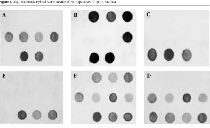
PCR products from each bacterial sample were hybrid-ized with oligonucleotide probes attached to nylon mem-branes. The results showed high specificity of hybridiza-tion with the four bacterial species under investigahybridiza-tion (Figures 3A - D). Hybridization results of mock samples including bacterial species of S. enterica, E. coli, and B. ce-reus (Sample 1), and S. enterica, L. monocytogenes, and B. cereus (Sample 2) are shown in Figures 3E and 3F.
4.3. Sensitivity of Oligonucleotide Microarray
PCR assays were randomly performed to test the lower detection limit of the DNA microarrays. Positive signal could be obtained from dilutions between 109 and 103 cfu/mL. The results showed that the sensitivity of oligo-nucleotide microarray was 103 cfu/mL.Table 1. Oligonucleotide Probes Used in the Study
Probe No. Target Access No.GenBank Sequence (5’ to 3’) References
1 Shuffled GFP a (negative control) - CAGCGAGTGTGATATGAGTGATGAGG (13)
2 Eschrichia coli V00331 CTGATATGTAGGTGAGGTCCCT (11)
3 Eschrichia coli AJ278710 CTGATATGTAGGTGAAGCGACTTGC This study
4 Escherichia coli AF053968 CACGCTGATATGTAGGTGAAGTCCC This study
5 Salmonella enterica U77919 AAATCCGGTTCACTTTAACACTGAGGCGTG (11)
6 Salmonella spp. U77919 GAAGTGATTTACTCATGGAGCTGAAGTC This study
7 Salmonella spp. AL627282 TGAAGTCAGCCGAAGATACCAGC This study
8 Enterbacteriaceae V00331 GATGTAACGGGGCTAAACCA (11)
9 Listeria monocytogenes X64533 CGTCCAAGCAGTGAGTGTGAGAAGT This study
10 Bacillus cereus X94448 GTGCTGGAAGGTTAAGGAGAGGG This study
11 Bacterial universal probe V00331 ACGGTCCTAAGGTAGCGAAA (11)
12 Aequorea victoria GFP b (positive control) AB103336 CAGAGTGTGCGATATTGATGAAAGTG (13)
aShuffled GFP that contains no sequence similarity to the known genes in GenBank database. bGreen Fluorescent Protein.
1 2 3 4
5 6 7 8
9 10 11 12
Figure 1. Layout of the Oligonucleotide Probes for 23S rDNA
Figure 2. Amplification of 23S ribosomal DNA Gene from the DNA of Four Bacterial Species
Lane 1, the negative control; Lanes 2 - 5, PCR products from Escherichia coli,
Listeria monocytogenes, Salmonella enterica, and Bacillus cereus; Lane M, 1000 bp molecular size DNA marker.
Figure 3. Oligonucleotide Hybridization Results of Four Species Pathogenic Bacteria
A, Salmonella enterica; B, Escherichia coli; C, Listeria monocytogenes; D, Bacillus cereus; E and F are hybridization results of two mock samples; E, E. coli, S. enterica and B. cereus; F, S. enterica, L. monocytogenes and B. cereus.
5. Discussion
From the late 20th century till now, bacterial infectious diseases are remarkably responsible for morbidity and mortality in both humans and animals (1, 10, 22). Routine detection of bacterial pathogens in clinical laboratories is commonly based on microscopic observation, enrich-ment culture and biochemical and serological methods. All these routine techniques have several limitations such as long culture time, reliance on enrichment and selective culture, and difficulty of quantitative analysis (23, 24).
Oligonucleotide microarray coupled with PCR can serve to enhance rapidity and effectiveness of microbial detec-tion and identificadetec-tion (22-25). Selecting proper target genes is very critical in oligonucleotide microarray meth-ods (14). Various target regions are used to identify patho-gens in oligonucleotide microarray methods. These re-gions include 16S rDNA, 23S rDNA and 16S-23S rDNA internal transcribed spacer region (ITS) (14, 16, 26, 27). The 23S rDNA
genes appear to be the best performing regions as target sequences. The 23S rDNA has highly specific sequences and can be used easily as a marker to differentiate bacterial pathogens. Recently, there have been several reports on ap-plication of 23S rDNA to identify bacterial species (16, 20, 21). Anthony et al. (16) reported that the 23S rDNA genes show more variation between species of medically important than the 16S rDNA genes. They concluded that the accuracy and discriminating power of the assay can be continually
extended by adding further oligonucleotides to the panel without significantly increasing complexity or cost.
For an ideal array, sequences should be selected as a probe and primer hybridized with only one target gene without cross-hybridization. The bacterial members of Enterobac-teriaceae family have multiple similar 23S rDNA sequences which makes it difficult to differentiate them. Therefore, in order to design additional oligonucleotides to identify species of this group of organisms, more strains should be studied (7, 16). In order to distinguish Salmonella spp., and
E. coli, several probes were applied. However, the specific probe for E. coli had cross reaction with Salmonella spp., but it did not affect the discrimination because of using more than one probe to detect E. coli and Salmonella spp. In order to determine the ability of this method to detect several bacteria simultaneously, three mixtures of bacteria were combined together and examined with specific probes. The results indicated that each bacterium reacts with its strain specific probes. Using fluorescent labeled probes in microarray technique are not used as a routine approach in clinical laboratories for bacterial identification. In such methods, complex technical experiments such as regular stringency over the array chip, data analysis, and high cost are needed (28). Hence in the current study DIG-labeled primers were used to evaluate the hybridization reaction by naked eyes or conventional scanners. Wang et al. (29)
developed a membrane-array method to detect human intestinal bacteria by DIG-labeled primers without expen-sive microarray-arrayer and laser-scanner. They concluded that the method shares the same principle with micro-array method, but does not need any expensive micro-arrayer and scanner. DIG-labeled probes are not as expensive as fluorescence-labeled probes. Furthermore, the procedure is non-radioactive and can therefore be performed in any routine laboratory (28). For this reason, authors believe that diagnosis laboratories can easily perform this rapid and accurate test. The present study investigated an ef-ficient tool for rapid, accurate and specific detection of four bacterial pathogens, and the results showed unique hybridization in combination with the oligonucleotide probes. In conclusion, the employed oligonucleotide mi-croarray method was reliable and accurate to detect the four food-borne bacteria under study. Regarding the per-formance, the technique appears to be comparable with other reported methods, but is less expensive.
Acknowledgments
Authors are highly indebted to all the staff of Molecular Biology Unit, Pasteur Institute of Iran, for their valuable cooperation. Authors are very much thankful to Negar Souod for her valuable suggestions and to Abbas Doosti for his help during study.
Footnotes
Authors’ Contribution:Meysam Sarshar and Nader Shahrokhi: Designing the study, obtaining samples and writing the manuscript; Reza Ranjbar: Drafting the man-uscript; Caterina Mammina: Making substantial contri-butions to the conception of the study and interpreta-tion of the data. All authors read and approved the final manuscript.
Funding/Support:This work was supported in part by a grant-in-aid from the Molecular Biology Unit, Pasteur Institute of Iran.
References
1. Tang YW, Procop GW, Persing DH. Molecular diagnostics of infec-tious diseases. Clin Chem. 1997;43(11):2021–38.
2. Millar BC, Xu J, Moore JE. Molecular diagnostics of medically im-portant bacterial infections. Curr Issues Mol Biol. 2007;9(1):21–39. 3. Sakallah SA. Molecular diagnostics of infectious diseases: state of
the technology. Biotechnol Annu Rev. 2000;6:141–61.
4. Krzyszycha R, Bielak J. Food pathogens as the most com-mon cause of bacterial food poisoning. Ann Univ Mariae Curie Sklodowska Med. 2004;59(2):354–6.
5. Wang XW, Zhang L, Jin LQ, Jin M, Shen ZQ, An S, et al. Develop-ment and application of an oligonucleotide microarray for the detection of food-borne bacterial pathogens. Appl Microbiol Bio-technol. 2007;76(1):225–33.
6. Wang J, Luo Y, Zhang B, Chen M, Huang J, Zhang K, et al. Rapid label-free identification of mixed bacterial infections by surface plasmon resonance. J Transl Med. 2011;9:85.
7. Mao Z, Zheng H, Wang X, Lin S, Sun Y, Jiang B. DNA microarray for direct identification of bacterial pathogens in human stool samples. Digestion. 2008;78(2-3):131–8.
8. Kostic T, Weilharter A, Rubino S, Delogu G, Uzzau S, Rudi K, et al. A microbial diagnostic microarray technique for the sensitive detection and identification of pathogenic bacteria in a back-ground of nonpathogens. Anal Biochem. 2007;360(2):244–54. 9. Muldrew KL. Molecular diagnostics of infectious diseases. Curr
Opin Pediatr. 2009;21(1):102–11.
10. Liu YT. A technological update of molecular diagnostics for infec-tious diseases. Infect Disord Drug Targets. 2008;8(3):183–8. 11. Hong BX, Jiang LF, Hu YS, Fang DY, Guo HY. Application of
oligo-nucleotide array technology for the rapid detection of patho-genic bacteria of foodborne infections. J Microbiol Methods.
2004;58(3):403–11.
12. Pariset L, Chillemi G, Bongiorni S, Romano Spica V, Valentini A. Microarrays and high-throughput transcriptomic analysis in species with incomplete availability of genomic sequences. N Biotechnol. 2009;25(5):272–9.
13. Lee DY, Shannon K, Beaudette LA. Detection of bacterial patho-gens in municipal wastewater using an oligonucleotide mi-croarray and real-time quantitative PCR. J Microbiol Methods.
2006;65(3):453–67.
14. Yoo SM, Lee SY, Chang KH, Yoo SY, Yoo NC, Keum KC, et al. High-throughput identification of clinically important bacterial patho-gens using DNA microarray. Mol Cell Probes. 2009;23(3-4):171–7. 15. Kim HJ, Park SH, Lee TH, Nahm BH, Kim YR, Kim HY. Microarray
de-tection of food-borne pathogens using specific probes prepared by comparative genomics. Biosens Bioelectron. 2008;24(2):238–46. 16. Anthony RM, Brown TJ, French GL. Rapid diagnosis of bactere-mia by universal amplification of 23S ribosomal DNA followed by hybridization to an oligonucleotide array. J Clin Microbiol.
2000;38(2):781–8.
17. Anthony RM, Brown TJ, French GL. DNA array technology and di-agnostic microbiology. Expert Rev Mol Diagn. 2001;1(1):30–8. 18. Miller MB, Tang YW. Basic concepts of microarrays and
poten-tial applications in clinical microbiology. Clin Microbiol Rev.
2009;22(4):611–33.
19. Wilson WJ, Strout CL, DeSantis TZ, Stilwell JL, Carrano AV, An-dersen GL. Sequence-specific identification of 18 pathogenic microorganisms using microarray technology. Mol Cell Probes.
2002;16(2):119–27.
20. Amann R, Ludwig W. Ribosomal RNA-targeted nucleic acid probes for studies in microbial ecology. FEMS Microbiol Rev.
2000;24(5):555–65.
21. Hunt DE, Klepac-Ceraj V, Acinas SG, Gautier C, Bertilsson S, Polz MF. Evaluation of 23S rRNA PCR primers for use in phylo-genetic studies of bacterial diversity. Appl Environ Microbiol.
2006;72(3):2221–5.
22. Momtaz H, Karimian A, Madani M, Safarpoor Dehkordi F, Ranjbar R, Sarshar M, et al. Uropathogenic Escherichia coli in Iran: sero-group distributions, virulence factors and antimicrobial resis-tance properties. Ann Clin Microbiol Antimicrob. 2013;12:8. 23. Yoshikawa TT. Epidemiology and unique aspects of aging and
infectious diseases. Clin Infect Dis. 2000;30(6):931–3.
24. Gabig-Ciminska M. Developing nucleic acid-based electrical de-tection systems. Microb Cell Fact. 2006;5:9.
25. Xing JM, Zhang S, Du Y, Bi D, Yao LH. Rapid detection of intesti-nal pathogens in fecal samples by an improved reverse dot blot method. World J Gastroenterol. 2009;15(20):2537–42.
26. Pei A, Nossa CW, Chokshi P, Blaser MJ, Yang L, Rosmarin DM, et al. Diversity of 23S rRNA genes within individual prokaryotic ge-nomes. PLoS One. 2009;4(5):e5437.
27. Kim CM, Song ES, Jang HJ, Kim HJ, Lee S, Shin JH, et al. Devel-opment and evaluation of oligonucleotide chip based on the 16S-23S rRNA gene spacer region for detection of patho-genic microorganisms associated with sepsis. J Clin Microbiol.
2010;48(5):1578–83.
28. Mothershed EA, Whitney AM. Nucleic acid-based methods for the detection of bacterial pathogens: present and future consid-erations for the clinical laboratory. Clin Chim Acta. 2006;363 (1-2):206–20.
29. Wang RF, Kim SJ, Robertson LH, Cerniglia CE. Development of a membrane-array method for the detection of human intestinal bacteria in fecal samples. Mol Cell Probes. 2002;16(5):341–50.