THE IMMUNOLOGICAL CONSEQUENCES OF OBESITY ON PRIMARY AND SECONDARY IMMUNE DEFENSES TO THE 2009
PANDEMIC H1N1 INFLUENZA VIRUS
J. Justin Milner
A dissertation submitted to the faculty of the University of North Carolina at Chapel Hill in partial fulfillment of the requirements for the degree of Doctor of Philosophy in the
Department of Nutrition in the Gillings School of Global Public Health.
Chapel Hill 2014
Approved by: Melinda A. Beck
Patricia A. Sheridan Liza Makowski
iii
ABSTRACT
J. JUSTIN MILNER: The Immunological Consequences of Obesity on Primary and Secondary Immune Defenses to the 2009 Pandemic H1N1 Influenza Virus.
(Under the direction of Melinda A. Beck)
Obese individuals are more susceptible to hospitalization and death from infection with
the 2009 pandemic H1N1 influenza virus (pH1N1). Greater pH1N1 severity in the obese is a global public health concern given the persistent threat of influenza outbreaks and the current
obesity epidemic. In this dissertation, the consequences of obesity on pH1N1 immunity were investigated in mice to uncover mechanisms by which obesity enhances pH1N1 illness. During a primary pH1N1 infection, 80% of obese mice died compared with 40% of lean, low fat diet fed
mice and no mortality in lean, chow fed mice. Further, a genetic model of obesity was generated in which leptin signaling was conditionally disrupted in hypothalamic neurons to confirm that
obesity, independent of diet, enhances pH1N1 mortality. Both diet- and genetic-induced obese mice exhibited greater lung damage during infection, likely due to fewer lung regulatory T cells and impaired regulatory T cell function. We extended our analysis to include a secondary
heterologous pH1N1 infection model. Obese mice had fewer cross-reactive, non-neutralizing pH1N1 antibodies, overactive CD8+ effector memory T cell responses and greater lung damage
in this model.
During the primary pH1N1 infection, obese mice had greater serum and bronchoalveolar lavage leptin concentrations compared with lean mice. Given that leptin regulates T cell
iv
obesity-induced pH1N1 mortality. However, obese mice lacking leptin signaling in T cells were not protected from pH1N1 mortality compared with control, obese mice. The pathophysiological
complications of obesity are diverse and complex. Therefore, we also extended our analysis to include 1H NMR-based metabolic profiling of urine, feces, serum, lungs, bronchoalveolar lavage
fluid, mesenteric white adipose tissue, and livers to obtain a more comprehensive examination of infection responses in obese mice. We uncovered a number of metabolites and metabolic
signatures uniquely altered in obese mice that, ultimately, may facilitate early prediction of
influenza infection outcomes and help to identify mechanisms for impaired. In summary, novel immunologic and metabolic techniques were integrated in this dissertation to establish that
v
vi
ACKNOWLEDGEMENTS
I would like to acknowledge my dissertation committee for their incredible guidance and support: Melinda A. Beck (chair), Patricia A. Sheridan, Liza Makowski, P. Kay Lund and Ilona Jaspers. During my graduate studies, Dr. Beck provided me with the perfect balance of
guidance and independence that has allowed me to develop to my maximum potential as a researcher and independent thinker. Her unwavering support has encouraged me to take risks
and pursue difficult research questions, which ultimately has given me the confidence that I can be successful in approaching and solving any research question. Additionally, she has been a great mentor in terms of demonstrating that integrity, hard work and fun are critical components
of being a successful scientist. Additionally, I would like to acknowledge previous and past members of the Beck lab for all of their help, especially Erik A. Karlsson and Patricia A.
Sheridan. I would also like to thank all my professors over the years for all their dedication, hard work and help, especially Liza Makowski and Rosalind Coleman. I am also grateful to the extremely helpful staff members of the Department of Nutrition, including Joanne Lee.
I would like to acknowledge the funding sources that made this research possible: NIH ROI AI078090 to Melinda A. Beck and NIK DK056350 to the University of North Carolina
Nutrition Obesity Research Center.
vii
TABLE OF CONTENTS
LIST OF TABLES ... x
LIST OF FIGURES ... xii
LIST OF ABBREVIATIONS ... xiiii
CHAPTER I: OVERVIEW AND SPECIFIC AIMS ... 1
Overview ... 1
Specific Aims ... 3
CHAPTER II: BACKGROUND AND SIGNIFICANCE ... 4
Introduction ... 4
The obesity epidemic ... 4
Evidence for greater infection susceptibility in obese individuals ... 5
Obesity and host defense in rodents ... 7
Dietary considerations in mouse models of obesity ... 9
Obesity and influenza infection in humans and mice ... 11
Influenza virus biology ... 12
Antigenic drift, antigenic shift and highly pathogenic influenza viruses ... 14
The 2009 pandemic H1N1 influenza virus ... 16
Innate defenses to influenza virus infection in the lung ... 16
Adaptive immunity to influenza virus ... 17
viii
Influenza virus vaccination ... 19
Heterologous immunity ... 21
Mechanisms by which obesity may impair immune cell function ... 22
Leptin and immunity in the obese ... 23
Additional considerations of the consequences of obesity on immunity ... 25
Metabolic profiling and infectious disease ... 25
CHAPTER III: DIET-INDUCED OBESE MICE EXHIBIT ALTERED HETEROLOGOUS IMMUNITY DURRING A 2009 PANDEMIC H1N1 INFECTION ... 27
Introduction ... 27
Materials and methods ... 30
Results ... 37
Discussion ... 45
Tables and Figures ... 52
CHAPTER IV: DIET- AND GENETIC-INDUCED OBESITY RESULTS IN GREATER LUNG DAMAGE AND MORTALITY DURING A PRIMARY 2009 PANDEMIC H1N1 INFLUENZA VIRUS INFECTION IN MICE ... 61
Introduction ... 61
Materials and methods ... 64
Results ... 67
Discussion ... 76
Tables and Figures ... 80
CHAPTER V: 1H NMR-BASED PROFILING REVEALS DIFFERENTIAL IMMUNE-METABOLIC NETWORKS DURING INFLUENZA VIRUS INFECTION IN OBESE MICE... 88
ix
Results ... 94
Discussion ... 98
Tables and Figures ... 103
CHAPTER VI: SYNTHESIS ... 119
Overview of research findings ... 119
How does obesity alter pH1N1 immunity? ... 121
Does hyperleptinemia underlie the consequences of obesity on pH1N1 immunity? ... 122
Mechanisms of greater lung damage during pH1N1 infection ... 124
Recommendations for future research ... 127
Tables and Figures ... 129
x
LIST OF TABLES
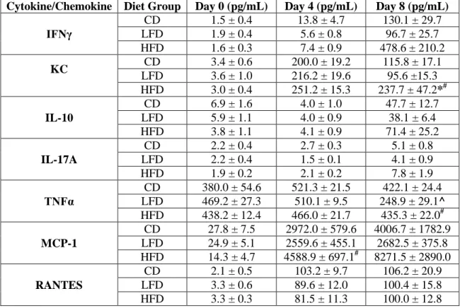
Table 3.1 Similar levels of lung cytokine and chemokine expression during
a secondary heterologous pH1N1 challenge in lean and obese mice ... 52 Table 4.1 BALF cytokine concentrations during a primary pH1N1 infection ... 80 Table 5.1 Metabolic biomarkers recovered from urine and fecal extracts
during influenza infection in lean and obese mice. ... 103 Table 5.2 Discriminatory metabolites between lean and obese mice at 9 days
post-infection in liver, serum and white adipose tissue samples ... 104 Table 5.3 Lung metabolite correlation patterns with BAL T cell populations ... 105 Table 5.4 Lung metabolite correlation patterns with mLN T cell populations ... 106 Table 5.5 Correlation patterns between 1H NMR data and BAL or mLN T
xi
LIST OF FIGURES
Figure 3.1 Sublethal influenza A/PR/8/34 infection induces heterologous protection against a lethal pH1N1 challenge in lean and diet-
induced obese mice ... 53 Figure 3.2 Obese mice exhibit delayed antibody production and impaired
antibody maintenance following a sublethal PR8 infection ... 54 Figure 3.3 Obese mice have lower levels of cross-reactive anti-pH1N1
nucleoprotein antibodies and exhibit greater lung viral titers
during a secondary pH1N1 infection ... 55 Figure 3.4 Obese mice exhibit a greater inflammatory and pathological
response in the lungs following a secondary heterologous pH1N1
challenge ... 56 Figure 3.5 Exposure to PR8 elicits a rapid and robust memory CD4+ T-cell
response following pH1N1 infection in both lean and obese mice ... 57 Figure 3.6 Obese mice have enhanced cross-reactive CD8+ T-cell responses
at 5 dpi after a secondary heterologous pH1N1 challenge ... 58 Figure 3.7 Obese mice have a greater number of Tregs in the lung airways
during a heterologous secondary pH1N1 challenge ... 59 Figure 3.8 Tregs isolated from obese mice are significantly less suppressive
than Tregs isolated from lean mice ... 60 Figure 4.1 Diet-induced obesity results in greater pH1N1 infection mortality
in mice ... 81 Figure 4.2 Genetically obese mice are more susceptible to pH1N1 mortality ... 82 Figure 4.3 Obese mice exhibit greater lung damage during pH1N1 infection ... 83 Figure 4.4 Obese mice did not exhibit differences in lung pathology during
pH1N1 infection... 84 Figure 4.5 Obese mice have fewer BAL macrophage during pH1N1 infection ... 85 Figure 4.6 Diet- and genetic-induced obese mice have fewer BAL Tregs at 8 dpi ... 86 Figure 4.7 Leptin signaling in T cells does not mediate pH1N1 mortality in
xii
Figure 5.2 Metabolic profiling can distinguish urine samples from both
uninfected and infected lean and obese mice ... 116 Figure 5.3 Representative analysis of serum samples from infected lean and
obese mice ... 117 Figure 5.4 Correlation analysis of lung analytes and lung and mLN T cell
xiii
LIST OF ABBREVIATIONS
1
H NMR 1H Nuclear magnetic resonance AHR Airway hyperresponsiveness ANOVA Analysis of variance
BAL Bronchoalveolar lavage BALF Bronchoalveolar lavage fluid
BMI Body mass index
CCR7 Chemokine receptor 7
CD Chow diet
ConA Concanavalin A
COPD Chronic obstructive pulmonary disease CPM Counts per minute
Cre Cre recombinase
CVD Cardiovascular disease
CXCL1 Chemokine (C-X-C motif) ligand 1, keritinocyte chemoattractant
DC Dendritic cell
DIO Diet-induced obesity
DPI Day post-infection
ELISA Enzyme linked immunosorbent assay
ER Endoplasmic reticulum
FACS Fluorescence-activated cell sorting
GzB Granzyme B
H&E Hematoxylin and eosin
HA Influenza hemagglutinin
HAI Hemagglutinin inhibition
xiv ICU Intensive care unit
IFNα Interferon α
IFNβ Interferon β
IL Interleukin
IgG Immunoglobulin G
JAK/STAT Janus kinase/signal transducers and activators of transcription KC Keritinocyte chemoattractant, CXCL1
LAIV Live attenuated influenza vaccine
Lck Lymphocyte-specific protein tyrosine kinase LD50 Median lethal dose
LepR Leptin receptor
LepRH-/- Mice with disruption of leptin receptor in hypothalamic neurons LepRH+/- Heterozygous hypothalamic leptin receptor disruption mice
LepRHfl/fl Hypothalamus specific fully floxed leptin receptor control mice
LepRT-/- T cell specific leptin receptor knockout mice
LepRTfl/fl T cell specific fully floxed leptin receptor control mice
LFD Low fat diet
M1 Influenza matrix protein 1 M2 Influenza matrix protein 2
MCP-1 Monocyte chemoattractant protein 1
MDCK Madin-Darby Canine Kidney epithelial cells MHCI Major histocompatibility complex I
MHCII Major histocompatibility complex II mLN Mediastinal lymph node
MOI Multiplicity of infection
MS Mass spectrometry
xv NK cell Natural killer cell
NP Influenza nucleoprotein
NS1 Influenza nonstructural protein 1 NS2 Influenza nonstructural protein 1
OD Optical density
O-PLS-DA Orthogonal partial least squares discriminant analysis PA Influenza polymerase acidic
PAMPs Pathogen associated molecular patterns PB1/2 Influenza polymerase basic 1/2
PBS Phosphate buffered saline PCA Principal component analysis
pH1N1 2009 pandemic H1N1 influenza virus (A/Cal/04/09) PLS-DA Partial least squares discriminant analysis
PR8 Influenza A/Puerto Rico/8/1934 (H1N1) PRR Pattern recognition receptor
RANTES Regulated on activation, normal T cell expressed and secreted
RBC Red blood cell
RDE Receptor destroying enzyme RIG-I Retinoic acid inducible gene 1 ROS Reactive oxygen specifies RPM Revolutions per minute
RPMI Roswell Park Memorial Institute medium RSV Respiratory syncytial virus
SEM Standard error of the mean
SOCS-3 Suppressor of cytokine signaling 3 SPC Surfactant protein C
xvi TAG Triacylglycerol
TCID50 Median tissue culture infective dose
Tcm Central memory T cell
Teff Effector T cell
Tem Effector memory T cell Tfh Follicular helper T cell Th1 Type 1 helper T cell Th2 Type 2 helper T cell TIV Trivalent influenza vaccine TNFα Tumor necrosis factor α
vRNA Viral ribonucleic acid WAT White adipose tissue
Wk Week
1
CHAPTER I: OVERVIEW AND SPECIFIC AIMS
Overview
Obesity, defined as a body mass index (BMI) ≥30kg/m2, is caused by a prolonged
positive energy balance (1-3). Excess adiposity increases the risk for a variety of viral and bacterial infections (2), and obese individuals were reported to be at greater risk for
hospitalization and death from infection with the 2009 H1N1 pandemic strain (pH1N1) (4-7). Obesity is a global epidemic (8), and influenza epidemics and pandemics are persistent threats worldwide (9,10). Therefore, enhancing understanding of the negative impact of obesity on
pH1N1 immunity is a critical global public health issue.
The mechanisms by which obesity drives greater influenza infection severity in humans
are unclear. Although we have demonstrated that obese individuals have impaired cellular and humoral immune responses to influenza vaccination (11,12), there is little known regarding how obesity impacts dynamic immune responses in the lung during infection in humans. Therefore,
mouse models of obesity and influenza infection are critical for elucidating potential mechanisms with the future prospect of identifying therapeutics, improving disease management and
influencing vaccination approaches with the ultimate goal of limiting influenza morbidity and mortality in this at risk population.
Despite widespread transmittance, pH1N1 infection illness was surprisingly mild (13).
2
strains or vaccination (14). However, because obese individuals were at greater risk for
pandemic H1N1 severity, we hypothesized that obesity impairs cross-reactive immune defenses
to pandemic H1N1. We therefore explored this using a mouse model of obesity and heterologous immunity (Aim 1).
Obesity is associated with a chronic inflammatory state (1,2), and the majority of the pathophysiological complications of obesity are linked to excess inflammatory immune responses both locally and systemically (2). From our analysis of obesity on heterologous
immunity, we found that obese mice exhibit greater lung damage during a secondary pH1N1 infection (14). We further extended our analysis to demonstrate that obesity impairs primary
infection defenses to pandemic H1N1, resulting in greater lung damage and death (Aim 2). Obesity is a multifactorial disease resulting in physiological adaptations, metabolic perturbations, hormonal changes, alterations in levels of circulating nutrients, greater oxidative
stress, and changes in the gut microbiome (2). Given the complexity of this disease, it is difficult to identify one specific molecular mechanism explaining greater pH1N1 severity in the obese.
Therefore, we utilized metabolic profiling to obtain a more comprehensive, global perspective of the impact of obesity on pH1N1 immunity (Aim 3). Metabolic profiling uncovered a number of distinct changes in metabolic pathways and metabolites in influenza-infected obese mice. This
dissertation facilitates the establishment of metabolomics as a useful tool in characterizing influenza pathogenesis, and perhaps as a useful tool for prediction of influenza infection status
3
Specific Aims
Specific Aim 1: Determine if obesity impairs heterologous immunity to infection with the 2009
pandemic H1N1 virus.
Hypothesis: Obese mice will exhibit greater mortality, fewer cross-reactive memory T cells and
greater lung damage during a secondary 2009 pandemic H1N1 infection.
Specific Aim 2: Determine if obesity impairs primary infection responses to the 2009 pandemic
H1N1 virus, resulting in greater mortality and lung damage.
Hypothesis: Obese mice will exhibit greater mortality and lung damage during a primary
infection with the 2009 pandemic H1N1 influenza virus.
Specific Aim 3: Determine if metabolic profiling of biofluids and tissues can distinguish
influenza infection status in lean and obese mice and identify novel potential mechanisms for greater influenza infection severity.
Hypothesis: Metabolic profiling will uncover distinct perturbations in metabolic pathways and
metabolites that allow for identification of new explanatory mechanisms for greater influenza
4
CHAPTER II: BACKGROUND AND SIGNIFICANCE
Introduction
In 2009, a novel influenza virus emerged causing the first pandemic of the 21st century (15). The 2009 pandemic H1N1 virus (pH1N1) caused widespread infections, and in the US
alone, approximations of the number infected reach 89 million individuals (16). Although a variety of previously established risk factors increased susceptibility for severe pH1N1
outcomes, for the first time, the obese were reported to be at greater risk for hospitalization and death from influenza infection (6). Further, a recent meta-analysis demonstrated that obese individuals are at greater risk for severe seasonal influenza infections as well (17). Given that
more than 500 million individuals are obese (3) and influenza outbreaks are a persistent threat, identifying mechanisms for greater influenza severity in the obese is a global public health
concern.
The obesity epidemic
Globally, more than 1 in 10 individuals are considered to be obese (BMI≥30kg/m2) (3). An array of cultural, ecological, psychosocial and biochemical factors influence weight gain and
obesity status (1,3). However, ultimately, obesity is caused by a prolonged positive energy balance. Excess energy consumption or insufficient energy expenditure results in triacylglycerol
5
diabetes, cardiovascular disease, cancer, chronic obstructive pulmonary disease (COPD), strokes and infections (3,18). An abundance of research focuses on the negative health consequences of
obesity, and interestingly, excessive inflammation and hyperactive immune responses are linked to nearly every comorbidity associated with obesity (2). Further, despite excessive inflammatory
responses in the obese, excess adiposity predisposes individuals to a variety of infections (2).
Evidence for greater infection susceptibility in obese individuals
Obese patients have increased intensive care unit (ICU) length of stay (19) and are more likely to die (20-22) in the hospital. It has been reported that obesity increases infection
susceptibility in clinical settings (23), and further, obesity is a well-established independent risk factor for postoperative infections (24-34). In a recent secondary analysis of a large prospective observational study including critically ill and injured patients remaining in the ICU for 48 hours
or more, obesity was reported to be an independent risk factor for catheter and blood stream infections (35). In critically injured blunt trauma patients, morbid obesity (BMI≥40) was
associated with increased risk of pneumonia and urinary tract infection but not with increased mortality (27).
The impact of obesity on clinical outcomes in hospitalized patients is clearly
multifactorial and complex. For example, underlying disease in the obese may inhibit proper mobility in the hospital, which can increase risk for skin breakdown (36). Due to inadequate
equipment or improperly trained staff, obese individuals may have prolonged visits at the hospital, thus increasing risk for acquiring nosocomial infections (23,36). Another consideration is that pharmacokinetics of antibiotics may differ in the obese, potentially affecting susceptibility
6
impaired immunity on severity of nosocomial infections in obese patients, but accumulating evidence suggests a significant role.
Increased BMI is associated with greater risk for several other bacterial infections including periodontal infections (38), Staphylococcus aureus nasal carriage (39) and gastric
infection by Helicobacter pylori (40). Also, a recent study reported that obesity was significantly associated with HSV1 infection, which was determined by seropositivity (41). Despite increased risk for several types of microbial infections, there is little known about how obesity may alter
the pharmacokinetics of antimicrobial drugs (reviewed in (37)). Studies assessing dosing of the antibacterial drug, vancomycin, suggest that obese patients may require different dosages (42)
and different dosing intervals (43) compared with non-obese individuals. An additional study of the antimicrobial drug, linezolid, reported diminished serum concentrations in the obese
compared with healthy-weight volunteers receiving the same dose (44). As pointed out
previously, there is greater risk of skin breakdown in obese individuals in clinical settings due to restricted mobility, improperly sized rooms/equipment and the special challenges of caring for
patients undergoing or post bariatric surgery (23,36). Additionally, excess adiposity may reduce tissue perfusion and affect wound healing (36). Taken together, it is clear that obesity
predisposes individuals to nosocomial infections, and that careful consideration of infection
prevention and treatment is required.
In contrast to the recent surge of publications highlighting a connection between
influenza severity and obesity (discussed below), there is very little known about obesity and other respiratory tract infections (45). A recent study by Akiyama et al. suggests that obesity may impact the response to respiratory syncytial virus (RSV) infection in children (46). A study from
7
children (47). In critically ill trauma patients, obesity or morbid obesity was associated with respiratory infections (20,29,35). Conversely, a few studies have also reported obese individuals
are not at greater risk for respiratory infections (19,48). Therefore, our understanding of the effect of obesity on risk for pulmonary infection remains unclear. However, it is important to
consider that obesity can complicate lung mechanics, such as restricting lung volume (49), which could potentially increase risk for pneumonia or other infections. Although the mechanisms contributing to increased susceptibility may include impaired immunity, there may be
non-immune factors to consider.
Obesity and host defense in rodents
A limited number of studies have demonstrated the negative impact of excess adiposity on host defense in rodent models. The implications of these studies, in terms of obesity-related
immunity impairments, are often complicated by the use of genetic models of obesity. The most commonly used genetically altered rodents for this purpose are the ob/ob and db/db mice and the
rat fa/fa counterpart. These rodent models lacking leptin or the leptin receptor can be useful for the study of obesity-related comorbidities, as they display metabolic abnormalities characteristic of obesity such as hyperglycemia, dyslipidemia, glucocorticoid excess and hyperinsulinemia
(50,51) (all of which could potentially alter immune cell homeostasis and function). However, given the vast amount of research highlighting the importance of leptin in immunity (52), a
global deficiency in leptin signaling makes it difficult to tease apart the mechanisms contributing to impaired immunity and greater susceptibility to infections in these genetic models of obesity. Nonetheless, these models still provide insight into how excess adiposity may directly or
8
In general, a deficiency of leptin (ob/ob) or the leptin receptor (db/db) in mice increases susceptibility to bacterial infections and pneumonia (45). Using ob/ob mice, Mancuso et al.
demonstrated that a complete deficiency of leptin resulted in impaired pulmonary clearance upon Klebsiella challenge, likely due to defective alveolar macrophage and neutrophil phagocytosis
(53). Similarly, an investigation by Hsu et al. reported that ob/ob mice exhibited enhanced lethality and delayed clearance of Streptococcus pneumoniae following a pulmonary challenge (54). Interestingly, intraperitoneal injections of leptin prior to Streptococcus infection improved
survival after the bacterial challenge, but not to the level of wild-type mice. This discrepancy in survival percentages may, in fact, be caused by the increased adiposity of the ob/ob mice. Other
studies have reported that ob/ob mice exhibit greater pulmonary Mycobactrium tuberculosis load (55) and delayed clearance of the Mycobacterium abcessus (56) upon challenge.
In addition to these pulmonary infection models, mice lacking the leptin receptor were
shown to be more susceptible to hind paw staphylococcal infection and exhibited a greater inflammatory response compared with wild-type mice (57). Furthermore, db/db and ob/ob mice
displayed impaired resistance to hepatic Listeria monocytogenes infection (58). Obese Zucker rats show decreased ability to clear yeast infection upon challenge with Candida albicans (59).
It is clear that evidence highlighting the importance of leptin for host defense is rapidly
accumulating. However, these studies do not offer insight into the mechanisms by which excess body fat (and associated metabolic abnormalities) may actually hinder host defense. Adding
exogenous leptin to ob/ob mice is helpful in understanding the impact of other metabolic
abnormalities on immune dysfunction, but exogenous administration of leptin in mice still differs from studying animals with intact leptin production and signaling. Some key considerations
9
have autocrine signaling capabilities on select immune cells. Although infection models in ob/ob or db/db mice provide important information on the role of leptin in host defense and immunity,
these mice do not properly model non-genetically induced obesity, which constitutes the vast majority of human obesity.
Diet-induced obesity (DIO) in rodents more closely mimics human obesity compared with genetic models of obesity. DIO mice develop the prototypical comorbidities associated with obesity including hyperleptinemia, insulin resistance, white adipose tissue inflammation,
low-grade systemic inflammation and greater liver TAG deposition to name a few. These DIO models are most frequently used to study metabolic perturbations associated with obesity,
however, few studies have utilized these models to determine the impact of obesity on host defense. Compared with lean control mice, DIO mice have greater morbidity and mortality during either a primary or secondary influenza infection (60,61) (discussed further below). An
investigation by Shamshiev et al. reported apoE-/- mice fed a high fat/cholesterol diet displayed impaired resistance to Leishmania major infection due to impaired dendritic cell function and
T-helper type 1 (TH1) cell immunity (62). Impaired T-cell activity was also reported in DIO mice
transgenic for a T-cell receptor specific to a peptide derived from ovalbumuin (63). Utilization of DIO models in mice will help to better inform about the potential factors responsible for
increasing infection susceptibility in obese humans.
Dietary considerations in mouse models of obesity
One complication associated with DIO models is elucidating whether the observed outcome of infection can be attributed to the complications of excess adipose tissue, the
10
DIO models in tandem may be beneficial in advancing our understanding of the impact of the diet vs. obesity status on immunity (64,65). Another important aspect of DIO studies to consider
is use of a proper control diet. A purified, nutrient defined, high fat lard-based diet is commonly used for DIO models, and it is generally established that the proper control diet for lean mice is
also a purified, nutrient defined low fat diet differing only in fat and carbohydrate content (66). However, chow diets are commonly used for lean control groups, which can introduce a variety of confounding variables (67,68). In addition to differences in macro and micronutrient
composition between chow diets and purified high fat diets, chow diets also contain other nutrients such as phytoestrogens (plant derived chemicals with estrogenic activity), fiber and
other known and unknown plant constituents (66,69). These variables could potentially confound results from DIO studies. Further, various batches of chow diets (even from the same company) are often produced with varying levels due plant components (in part driven by what plants are
cheapest at the time) (69). Therefore, there is legitimate concern that experimental
reproducibility is at risk when chow diets are used. With that said, there is an inherent limitation
in mouse diet studies for utilizing the proper control diet, because regardless of whether or not the ingredients are purified, there will still be differences in fat content and carbohydrate content between high fat and low fat diets. Further, the standard 10% kcal fat, low fat diet, is high in
sucrose, which may cause metabolic perturbations on its own (70). Now available are new low fat control diets that replace sucrose with other carbohydrates. Nonetheless, careful consideration
11
Obesity and influenza infection in humans and mice
A striking number of recent studies have reported obesity to be a predictor for a worse
outcome of infection with pH1N1 (71). In fact, several countries across the world have reported that obese individuals were disproportionately represented among influenza-related
hospitalizations and deaths. Obesity or morbid obesity increased risk of ICU admission and even death among those infected with the pandemic strain (72-75). Those admitted to ICUs had a reportedly longer duration of mechanical ventilation, and increased time in ICUs and hospitals
compared with healthy weight individuals (76). Before the advent of the 2009 pandemic season, there were no such reports investigating the relationship between obesity and influenza infection
in humans. Recently, however, Kwong and colleagues published a study which explored the relationship between BMI and seasonal influenza infection using a series of Canada’s
cross-sectional population based health surveys (77). The surveys covered 12 influenza seasons.
Analysis of the retrospective cohort demonstrated that the obese are at greater risk for respiratory hospitalizations during the seasonal flu periods. Lastly, a meta-analysis performed by Mertz et al.
found that obesity was an independent risk factor for severe outcomes to seasonal influenza outbreaks as well (17).
A number of studies have demonstrated that obesity results in greater morbidity and
mortality following influenza virus infection in mice (14,68,78-81). Although it is currently unclear whether obesity enhances viral replication in the lungs, it is clear that obesity modulates
several aspects of the immune response (14,78,79). Both innate and adaptive arms of immunity are impaired during influenza infection as demonstrated by altered natural killer (NK) cell, dendritic cell (DC), T cell, B cell and heterologous memory responses (2). Further, obesity drives
12
impaired lung wound healing during infection (14,67,78). Of interest, it was recently
demonstrated that monoclonal antibody neutralization of leptin, an adipocyte derived hormone
that is chronically elevated in an obese state, during infection with pH1N1 improved infection outcome in obese mice (82). However, despite a number of publications investigating the
contribution of obesity on the immune response to influenza infection, there are currently no clearly established cellular or molecular mechanisms.
Influenza virus biology
Influenza is a contagious respiratory disease caused by the influenza virus (83). There are
three types of influenza viruses, A, B and C (83). Influenza A virus is responsible for the majority of infections in humans and typically results in the more severe infection illness compared with types B and C (9,83). Influenza B also commonly infects humans but induces
mild illness, and influenza C can infect humans but is only associated with very minor symptoms (9,83). Influenza virus is a zoonotic, negative sense RNA virus of the Orthomyxoviridae family
(83). Each influenza virion contains an envelope derived from host cells and 8 segmented RNA molecules that make up the genome of the virus (83). The encoded viral proteins include hemagluttinin (HA), neuraminidase (NA), matrix protein 1 (M1), matrix protein 2 (M2),
nucleoprotein (NP), polymerase acid (PA), polymerase basic 1 and 2 (PB1/2), non-structural protein 1 (NS1), non-structural protein 2 (NS2 or NEP) and some strains also express the
pro-apotoptic protein PB1-F2 (83,84). Each protein serves a distinct and specialized purpose. HA is a surface glycoprotein that binds to sialyic acid receptors on epithelial cells in the respiratory tract of humans as well as intestines in birds (83,85,86). NA cleaves sialic acid moieties to enhance
13
major determinants of influenza virus infectivity and also form the basis for influenza virus subtyping (83). Thus far, 17 different HA molecules and 9 different NA molecules have been
identified (87).
Influenza infection is first mediated by HA binding to cellular sialic acid receptors of an α2,6 and/or an α2,3 conformation (83,86). HA is then cleaved by cellular proteases which
triggers receptor mediated endocytosis (86). The low pH of intracellular endosomes triggers a conformational change in the HA trimer that allows fusion of the endosomal membrane and the
viral envelope (86). Simultaneously, the M2 protein functions as an ion channel and pumps H+ into the virion, resulting in the disassembling of the virion and release of viral contents from the
endosomal compartment into the cellular cytoplasm (83). Ribonucleoproteins containing the viral genome bound by nucleoprotein and the polymerase trimer are imported into the nucleus (83). Herein, influenza derived RNA-dependent RNA polymerases began transcribing the
negative sense RNA into complimentary viral RNA, vRNA (88). The vRNA molecules are exported to the cytoplasm where they are translated by host machinery or remain in the nucleus
for genome replication (88). Newly synthesized viral proteins are trafficked to the host membrane via the Golgi apparatus or enter back into the nucleus of the cell. New vRNA molecules and translated viral proteins are packaged into progeny virions at the surface of the
cell (88). Finally, new virions bud from the cell, obtaining an envelope from the host membrane. Here, neuraminidase is critical for cleaving HA and sialic acid interactions to allow further
14
Antigenic drift, antigenic shift and highly pathogenic influenza viruses
Influenza virus causes seasonal outbreaks that result in up to 500,000 deaths every year
(9). Unlike long-lived memory responses to other viruses such as yellow fever, immunity conferred by influenza virus infection or vaccination may not be protective to subsequent
exposure of influenza. This is primarily due to antigenic drift (89). The influenza RNA-dependent RNA polymerase replicates with low fidelity, meaning that nucleotide “errors” are
commonly introduced (at a rate of 1 in 1000 nucleotides) (83,89). These point mutations in the
viral genome can result in antigenic and functional changes in viral proteins, affecting host immunity and/or viral infectivity. Antigenic drift is also the reason that influenza vaccination is
recommended yearly (discussed further below) (89).
In addition to point mutations in the viral genome, influenza can also undergo major genomic changes through “swapping” of the segmented RNA molecules. This is called antigenic
shift, and is the basis for the genesis of influenza pandemics (89). This reassortmant of genetic material occurs when at least two different influenza viruses infect the same cell, resulting in
progeny virions with a novel combination of influenza RNA segments (and thus a novel combination of proteins). Generation of novel influenza strains primarily occurs from
reassortment in swine (90). This is because epithelial cells in swine express sialic acid receptors
of both the α2,6 and the α2,3 conformations (89,90). Influenza strains primarily circulating in humans preferentially bind to the α2,6 conformation (in the upper respiratory tract), whereas
avain strains typically bind receptors of the α2,3 structure (systemically) (83). This is one critical barrier that prevents transmittance of highly pathogenic avian strains to humans. Novel influenza strains created from genetic shifting are a pertinent public health threat because often the general
15
pandemics, and the first pandemic of the 21st century occurred from the emergence of the triple reassortant 2009 pandemic H1N1 influenza virus (discussed further below) (90).
In birds, influenza virus is systemic (91). This is primarily due the presence of a
multibasic cleavage site on the HA trimer in avian strains (91,92). In human strains, cleavage of
HA is restricted to cellular proteases primarily found in the respiratory tract (such as trypsin), thus limiting infection to that compartment (92). However, in avian strains, this multibasic cleavage enhances viral tropism to epithelial cells of the digestive tract and possibly other organs
as well (91,92). Therefore, reassortment of a contagious human virus with an HA from an avian strain could result in a severely lethal influenza virus in humans. In fact, in 2003 a novel avian
strain, H5N1, emerged in China (10). Despite a 60% mortality rate, the highly pathogenic strain has shown limited human to human transmission, preventing a pandemic outbreak for the time being (10). However, as mentioned, the influenza genome is highly susceptible to mutations, and
it has been demonstrated that only 5 mutations in HA of H5N1 are required for human to human transmission (10). Because influenza viruses do not readily transmit between birds and humans,
it is thought that humans become infected with avian strains due to close contact with poultry (93). Recently, another novel avian strain, H7N9, emerged in China in March 2013 (94). H7N9 was not previously detected in humans until this point in time (94). Similarly, this virus does not
appear to transmit well among humans, but those infected become severely ill (94). Persistent surveillance of these highly pathogenic influenza strains is important part of preventing future
pandemics. Further, it is clear that obese individuals are more susceptible to influenza severity caused by seasonal strains and the 2009 pH1N1 strain, and thus obese individuals may be even more susceptible to severe infection outcomes from these avian strains (2).
16
The 2009 pandemic H1N1 influenza virus
The 2009 pandemic H1N1 influenza A virus (pH1N1) is unique in several aspects.
Despite causing greater disease severity in animal models compared to seasonal H1N1 strains (95-98), the pH1N1 virus caused relatively mild illness in the majority of humans without
predisposing risk factors (13,99). Further, in contrast to seasonal influenza epidemics, children and nonelderly adults were more susceptible to pH1N1 infection compared with elderly
individuals (15,100). It has been estimated that approximately 90% of pH1N1 deaths occurred in
the nonelderly population (101). Further epidemiological evidence suggests the lower
susceptibly in elderly individuals is likely due to the presence of anti-hemagluttinin antibodies
generated from previous exposure to pre-1950 influenza strains that cross-react with the novel pH1N1 virus (100,102-104). However, the majority of the population is naïve to these past circulating influenza strains. Further, recently circulating (pre-2009) seasonal strains and
influenza vaccines did not elicit a robust cross-reactive neutralizing antibody response.
Therefore, most individuals lacked pre-existing neutralizing antibody protection against pH1N1
infection (102,104-107).
Innate defenses to influenza virus infection in the lung
Influenza virus is primarily spread through mucous or air-born aerosols. The initial responders to influenza virus infection are also the primary target of the virus, respiratory
epithelial cells (108). Influenza virus pathogen associated molecular patterns (PAMP) trigger induction of innate antiviral responses in epithelial cells. The primary pattern recognition receptors (PRR) for invading influenza virions include endosomal toll like receptors 3/7
17
influenza vRNA (108,109). PRR recognition of these PAMPs triggers activation of several inflammatory pathways that ultimately induce the production of type I interferons (IFN),
chemokine production and inflammatory cytokine production (108,110,111). IFNα and IFNβ are secreted within a few hours after an infected cell recognizes the invading virus, and these type I interferon’s are critical for communicating the presence of a viral infection to neighboring
epithelial cells, making them poised to fight the ensuing infection (110,111). Chemokines produced from infected epithelial cells result in a robust infiltration of early responding innate
immune cells (108,110,111). Simultaneously, innate immune cells, such as DCs and alveolar macrophages already present in the lung at the time of infection help to propagate this
inflammatory response by producing cytokines and chemokines as well (108,111).
Approximately the first 5 days of the immune response to influenza virus infection is predominantly comprised of innate defenses (112). Macrophages, NK cells, DCs and neutrophils
migrate into the lung in attempt to combat the infection (111). Macrophages and neutrophils are critical for phagocytizing virus particles, infected cells and inflammatory debris (108,111). NK
cells target infected cells for lysis, distinguishing infected from uninfected cells by conserved cellular patterns. Lastly, DCs engulf viral particles, and present influenza virus antigens on major histocompatibility complexes I and II (MHCI and MHCII) to stimulate induction of the adaptive
immune response (108,111).
Adaptive immunity to influenza virus
Activated dendritic cells presenting influenza virus antigen migrate to the draining lymph node of the lung to trigger activation of influenza-specific T cells and B cells (113,114). CD4+ T
18
on MHCI molecules (114,115). T cells are constantly surveying the blood and lymph, and those specific for influenza virus will bind to MHC molecules and become activated and undergo
clonal expansion in draining lymph nodes (114,116). Influenza-specific T cells accumulate in the draining lymph node, and can be detected in the lung as early as 5-6 days post-infection (dpi)
(113,117). However, the number of infiltrating T cells peaks between 8 and 10 dpi (117).
Effector CD8+ T cells are critical for controlling the infection by targeting infected cells by lysis (113). Further TH1 cells, the primary CD4+ T cell lineage generated during influenza, can also
target infected cells for lysis (116). Additionally, CD4+ T cells are critical for proper induction of antibody defenses to influenza virus (113).
Memory responses to influenza virus infection
Following robust expansion of influenza-specific T cells, a majority of the cells undergo
apoptosis following viral clearance (118). However, a small proportion of these cells become long-lived memory T cells (118). Memory T cell responses are critical for protection against
subsequent influenza virus infections. Although the exact mechanisms regulating the generation and maintenance of memory T cells are unclear, a number of modulatory factors have been defined, including inflammatory responses during the primary infection, cellular metabolism,
and tissue migration (118,119). Memory T cells consist of primarily two different types, central memory T cells (Tcm) and effector memory T cells (Tem (118)). Tem are primarily found at the
19
Antibodies are the earliest line of defense protecting from influenza virus infection. B cells containing a B cell receptor specific to influenza antigens primarily recognize cognate
antigen in draining lymph nodes through DC presentation (120). B cells can become activated, independent of T cell help, and these are primarily early sources of influenza-specific antibodies
referred to as short-lived plasma cells (120,121). Additionally, the majority of activated B cells migrate to the germinal center of the draining lymph node where they interact with follicular helper T cells (Tfh) through MHCII and costimulatory molecules (120,121). This interaction is
critical to the formation of effective antibody defenses because it triggers the generation of long-lived plasma cells and memory B cells (120,121). Long-long-lived plasma cells home to bone
marrow, where they continue to secrete influenza antibodies for extended periods of time (likely throughout a lifetime) (120,121). Influenza-specific memory B cells can be found in circulation and in nearly every tissue and induce an accelerated and robust plasma cell response following
re-infection (120). Influenza memory responses are the basis for effective influenza vaccination.
Influenza virus vaccination
Vaccination to influenza virus is widely accepted as the best measure to protect against influenza infection and to limit influenza infection severity (122,123). An abundance of research
focuses on novel approaches to vaccinate individuals to the ever-changing influenza strains (123). The primary methods of vaccination include intramuscular immunization with the
trivalent inactivated influenza vaccine (TIV) or intra-nasal administration of a live-attenuated influenza vaccine (LAIV) (122,123). Both vaccines include the three influenza strains, two type A strains (H1N1 and H3N2) and one B strain (122,123). These strains are chosen based on
20
influenza season, a quadrivalent vaccine was available, including two influenza A strains and two B strains (123). At this point, it is unclear which type of vaccine (TIV vs. LAIV) offers
better protection, but both vaccines have been shown to protect against influenza infection in humans (122,124). The purpose of influenza vaccination is to induce long-lived, protective
antibody and memory T cell responses that serve to protect upon exposure to influenza virus (124). Vaccine antigen is taken up by dendritic cells in draining lymph nodes of the arm (TIV) and the respiratory tract (LAIV) where it can be presented to T cells and B cells as described
above.
Influenza vaccines have been shown to reduce the number of individuals infected with
influenza and to reduce illness severity in the infected (122,124). However, there is some evidence suggesting that obese individuals may not respond to vaccination to the same extent as healthy weight individuals (2). Obesity was associated with a poor antibody response to hepatitis
B vaccination (125,126). Additionally, overweight children displayed significantly lower anti-tetanus immunoglobulin G (IgG) antibodies in response to vaccination compared with healthy
weight children (127). Interestingly, it has been reported that using a larger vaccine needle length resulted in significantly higher antibody titers to hepatitis B surface antigen in obese adolescents (128). Studies from our lab have demonstrated that obese individuals have impaired cellular and
humoral responses to TIV immunization. Although obese individuals are able to mount a
comparable antibody response one month after vaccination compared with healthy weight adults,
21
influenza vaccination responses, potentially predisposing this at-risk population to greater infection severity.
Heterologous immunity
Neutralizing antibodies are critical for the control and even prevention of influenza infection (120). In their absence, influenza-specific memory T cells are critical regulators of viral spread and influenza severity (113,129). A number of studies in animals and humans have
demonstrated that prior exposure to seasonal influenza viruses or vaccination can induce cross-reactive memory T cells that have the capacity to limit pH1N1 disease severity (107,130-135). In
mice, seasonal influenza virus infection or vaccination elicits a memory T-cell response that can prevent morbidity and mortality to a lethal pH1N1 challenge (133,136-139). Additionally, seasonal influenza A-specific memory T cells from humans, naïve to the pH1N1 virus, are
capable of recognizing pH1N1 and can directly lyse pH1N1-infected target cells (131).
Therefore, the ability of cross-protective memory T cells to control pH1N1 infection is a critical
mediator for the relatively benign symptoms experienced by a majority of those infected (131,140).
Although the majority of research on heterologous immunity to influenza infection
focuses on T cell responses, cross-reactive antibody protection to pH1N1 cannot be ignored. Non-neutralizing antibodies recognizing conserved epitopes, such as NP antibodies, have been
shown to contribute heterologous protection to influenza infection, reducing viral titers and infection severity in mice (141,142). Although it is currently unclear how non-neutralizing antibodies mediate protection to influenza virus, it is likely achieved through induction of
22
confer protection during a heterologous pH1N1 infection is through accelerated production (compared with antibody production in naïve mice) of homologous pH1N1 antibodies. This is
likely triggered by cross-reactive memory CD4+ cells that facilitate accelerated B cell activation and antibody production (137,142).
Mechanisms by which obesity may impair immune cell function
It is well known that obesity is associated with a state of chronic, low-grade inflammation
both in white adipose tissue (WAT) and systemically (143-146). Additionally, obesity is
characterized by altered levels of circulating hormones and nutrients such as glucose and lipids.
Circulating and tissue resident immune cells are thus exposed to an energy rich environment in the context of altered concentrations of metabolic hormones and inflammatory cytokines.
Understanding how this pro-inflammatory, excess energy milieu impacts immune cell function is
key in understanding greater susceptibility to pH1N1.
Hyperinsulinemia and insulin resistance are common features of obesity; however, there
is little known regarding the immunomodulatory effects of excess insulin or impaired insulin signaling in the context of obesity. Monocytes have been shown to express insulin receptors and are insulin sensitive immune cells (147-150). Interestingly, resting T cells are insulin insensitive
in that the insulin receptor is absent from the plasma membrane. However, upon activation, effector T cells upregulate de novo emergence of insulin receptors (151,152). Insulin signaling
induces glucose uptake, amino acid transport, lipid metabolism and can modulate T cell
activation and function (151,153). Furthermore, insulin promotes an anti-inflammatory TH2 cell
phenotype (152), and MacIver et al. speculate that insulin resistance in obesity may actually
23
modulated by insulin receptor signaling (1,2). Thus, it is clear that insulin can have potent effects on immune cell metabolism and function, but the effects of hyperinsulinemia or insulin
resistance on immunity remain relatively unknown.
Leptin and immunity in the obese
The primary adipose derived immunomodulatory adipokines include leptin, adiponectin, and the pro-inflammatory cytokines: tumor necrosis factor α (TNF-α), interleukin-6 (IL-6) and
IL-1β (144,145,155). Adiponectin, levels of which are decreased during obesity, has been shown to alter NK cell cytotoxicity and cytokine production by human myeloid cells (156,157).
Conversely, there is excess production of TNF-α, IL-6 and IL-1β in WAT of the obese (145). These cytokines can be secreted into circulation and potentially have distal effects; however, exactly how chronic production of these cytokines impacts cellular immunity remains to be
elucidated. It is possible that chronic exposure to pro-inflammatory cytokines may desensitize immune cells to inflammatory responses during an actual infection (158).
The pleiotropic effects of leptin on immune cell activity are highly diverse (52), and the function of nearly every innate immune cell has been shown to be modulated by leptin signaling (159-163). In monocytes, leptin upregulates pro-inflammatory cytokine production of 6,
IL-12 and TNF-α as well as phagocytic activity (53,164,165). In polymorphonuclear neutrophils of healthy individuals, leptin signaling induced chemotaxis, reactive oxygen species (ROS)
generation and influences oxidative capacity (163,166,167). NK cells are highly influenced by leptin signaling, including aspects of differentiation, proliferation, activation and activity (161,168). Given the importance of leptin to innate immune cell function, it follows that nearly
24
Leptin also plays a critical role in regulating adaptive immunity (159,162). Leptin is an important source of pro-survival signals to double-positive and single-positive thymocytes
during the maturation of T cells (169). Leptin has been shown to play a key role in
lymphopoieses and myelopoieses given that ob/ob mice have only 60% as many nucleated cells
in bone marrow as compared with wild-type controls (170). In the presence of a polyclonal stimulator, leptin can increase T cell proliferation and can modulate expression of activation markers on both CD4+ and CD8+ T cells (171).
Although several papers have discussed how leptin may be required for, or may enhance immune cell function, few have taken into consideration how hyperleptinemia in obese
individuals may modulate immune cell function (172). Additionally, elevated levels of leptin have been shown to induce a state of central leptin resistance. Indeed, studies have demonstrated that T cells (162) and NK cells (173) can become resistant to leptin in rodent models of obesity.
Leptin signals through a variety of pathways, the most studied being the Jak/Stat signaling pathway. Leptin receptor activation results in the translocation of the transcription factor,
STAT-3, into the nucleus and subsequent transcription of leptin induced genes, including suppressor of cytokine signaling-3 (SOCS-3). SOCS-3 functions as a negative feedback mediator of Jak/Stat signaling, and thus may play an important role in impairing leptin signaling and contributing to
central and peripheral leptin resistance (174). Leptin resistance, induced by hyperleptinemia, would obviously not occur in ob/ob mice, which is another reason these mice are not the best
model for studying obesity-related immune dysfunction. Although it is widely accepted that leptin resistance occurs centrally in the hypothalamus (174-176), peripheral leptin resistance requires further investigation. Additionally, it is likely that differing immune cells respond
25
Additional considerations of the consequences of obesity on immunity
In addition to perturbations in circulating levels of nutrients and hormones, there are a
variety of other pathophysiological complications of obesity that may contribute to excessive antiviral responses and lung damage during influenza infection. Obesity induces a state of
oxidative stress (177), which has been shown to increase influenza severity in mice (178). Further, a hallmark of obesity is a state of chronic, low-grade inflammation (2), which may compromise the immune response to influenza infection or vaccination. Finally, obesity is
associated with distinct changes in the intestinal microbiome (179), which may potentially impact antiviral responses given that manipulation of the intestinal microbiota in mice can
influence influenza infection outcome (180).
Metabolic profiling and infectious disease
Given the large number of mechanisms that may contribute to alterations in immune processes during influenza infection in the obese, narrowing focus to one particular mechanism,
cell type or molecular pathway may not be the best approach to addressing this problem. Perhaps, given the complexity of obesity, a systems biology approach may be useful in identifying the complicated interactions of underlying mechanisms driving greater infection
severity. Obesity is inherently a metabolic disease, and therefore, of the many integrative systems approaches, we believe metabolic profiling could be a useful tool in identifying
perturbations in lung-specific and systemic metabolites altered by obesity during influenza infection.
Metabolomics is a technique that allows for biochemical profiling of all small molecules
26
global view of complex processes occurring during disease and can actually serve as a snapshot of cellular processes that have just occurred on a cellular, tissue or systemic level (181). Standard
processing of samples for metabolomics typically involves identifying and quantitating metabolite levels using 1H Nuclear Magnetic Resonance (1H NMR) or Mass Spectometry (MS).
1H NMR doesn’t not require separation of compounds whereas MS requires prior separation of
analytes (commonly through gas chromatography or liquid chromatography separation) (181). Uncovering metabolic biomarkers and patterns of altered metabolites would provide a more
global view of the impact of obesity on dynamic infection responses and simultaneously may address the complicated interactions of the molecular underpinnings driving obesity-induced
influenza mortality. Metabolomics has been used to characterize the metabolic consequences of several types of infections in mice including parasitic (182,183), viral (184,185) and bacterial infections (186). Further, metabolomic analyses of models of lung inflammation have also
identified unique metabolites associated with lung damage or greater inflammation (187-189). In all of the referenced models of infectious and inflammatory diseases, metabolomics was able to
identify novel biomarkers and metabolic signatures discriminating infected and uninfected groups. In some cases, metabolomics has been used as a novel technique to improve diagnostic measures (184,185), whereas in other instances metabolomics has been used to enhance
understanding of pathways driving disease severity (186-188). Identification of biomarkers or predictive metabolic signatures could be used in clinical settings to enhance influenza diagnosis
27
CHAPTER III: DIET-INDUCED OBESE MICE EXHIBIT ALTERED HETEROLOGOUS IMMUNITY DURING A SECONDARY
2009 PANDEMIC H1N1 INFECTION1
Introduction
The novel 2009 pandemic H1N1 influenza A virus (pH1N1) is unique in several aspects. Despite causing greater disease severity in animal models compared to seasonal H1N1 strains (95-98), the pH1N1 virus caused relatively mild, uncomplicated symptoms in humans (13,99).
Further, in contrast to seasonal influenza epidemics, children and nonelderly adults were disproportionately susceptible to pH1N1 infection compared with elderly individuals (15,100),
with estimates that approximately 90% of pH1N1 deaths occurred in the nonelderly population (101). Clinical and epidemiological data suggest the lower susceptibly in individuals over 65 y of age is likely due to the presence of cross-reactive anti-hemagglutinin antibodies generated from
previous exposure to pre-1950 influenza strains (100,102-104). Because a majority of the
population is naïve to these past circulating influenza strains, and recently circulating (pre-2009)
seasonal strains and influenza vaccines did not elicit a robust cross-reactive antibody response, most individuals lacked neutralizing antibody protection against pH1N1 infection (102,104-107).
Although antibodies are important for the control and even prevention of influenza
infection, in their absence, influenza-specific T cells are essential in limiting influenza severity
1
This chapter was published in the Journal of Immunology:
J. Justin Milner, Patricia A. Sheridan, Erik A. Karlsson, Stacey Schultz-Cherry, Qing Shi, Melinda A. Beck. J Immunol. 2013 Sep; 191(5): 2474-85.
28
(113,129). Several recent studies in animals and humans have demonstrated that previous exposure to seasonal influenza strains or vaccination can induce cross-reactive memory T cells
that have the capacity to limit pH1N1 disease severity (107,130-135). In mice, seasonal influenza viruses and vaccines elicit a memory T-cell response that can prevent morbidity and mortality to
a lethal pH1N1 challenge (133,136-139). Additionally, seasonal influenza A-specific memory T cells from humans (naïve to pH1N1) are capable of recognizing pH1N1 epitopes and can directly lyse pH1N1-infected target cells (131). Therefore, the ability of cross-protective memory T cells
to control pH1N1 infection could explain the relatively benign symptoms experienced by a majority of those infected (131,140). However, cross-reactive antibody protection to pH1N1
cannot be ignored. Non-neutralizing antibodies recognizing conserved epitopes, such as anti-nucleoprotein (NP) antibodies, have been shown to contribute heterologous protection to
influenza infection, reducing viral titers and infection severity in mice (141,142). Additionally, a
primary seasonal infection can lead to an accelerated production of pH1N1 antibodies during a heterologous pH1N1 infection, which may facilitate pH1N1 viral clearance (137,142).
Another novel characteristic of the pH1N1 virus is that obesity, defined as a BMI ≥ 30kg/m2, was considered to be an independent risk factor for increased morbidity and mortality following infection (4-7,190). Obesity is a global public health concern, affecting more than
one-in-ten of the world’s adult population (3). It is well-established that obesity impacts several aspects of the immune response and increases susceptibility for a variety of pathogens, including
influenza virus (2). Although recent investigations in mice and humans have begun to elucidate potential mechanisms by which obesity impairs anti-viral immunity to influenza infection, the specific factors contributing to the increased severity observed in the obese population remain
29
memory CD8+ T-cell responsiveness to influenza stimulation (12,78). Further, obese mice display impaired responsiveness to influenza vaccination (68) , and obese humans are unable to
maintain long-term influenza antibody levels following vaccination (12). Therefore, given the protective nature of cross-reactive antibody and T-cell responses, we hypothesized that obesity
impaired heterologous immunity induced by previous influenza exposure, resulting in greater pH1N1 infection severity.
In this study, we utilized a model in which lean and obese mice were initially infected
with a sublethal dose of influenza A/PR/8/34 (H1N1, PR8). After 5 wk (to allow contraction of effector T cell responses), mice were challenged with a lethal dose of heterologous pandemic
A/Cal/04/09 (H1N1, pH1N1). Similar to results shown previously in lean, chow fed mice (133), we found that priming with PR8 effectively prevented mortality from pH1N1 infection in both lean and obese mice in the absence of cross-reactive neutralizing antibodies. However, obese
mice exhibited a lower level of cross-reactive pH1N1 nucleoprotein (NP) antibodies 5 wk after the PR8 infection, and a lower proportion of obese mice generated pH1N1 HAI antibodies
following the secondary challenge. Consequently, obese mice had greater lung viral titers, more lung pathology as well as an increased number of cytotoxic memory CD8+ T cells in lung
airways during the pH1N1 infection. Given the excessive inflammation, infiltration, lung damage
and cytotoxic CD8+ T-cell responses in the lungs of obese mice during the lethal pH1N1 challenge, we investigated the impact of obesity on regulatory T cells (Tregs) as a potential
mechanism for the inability to control the antiviral responses in the lung. Unexpectedly, obese mice had nearly twice as many Tregs in the lung airways during the heterologous secondary pH1N1 infection compared with lean mice. However, ex vivo analysis of Treg function revealed
30
lean mice. Therefore, an excessive inflammatory response in the lungs, potentially due to a combination of elevated viral titers and impaired Treg function, may be a mechanism by which
obesity enhances pH1N1 infection severity.
Materials and methods
Mice and diets
Weanling, male, C57BL/6J mice were obtained from The Jackson Laboratory (Bar Harbor, ME) and fed a low fat diet with 10% kcal fat (Research Diets D12450B) or high fat diet
with 45% kcal fat (Research Diets D12451, New Brunswick, NJ). In addition to inducing weight gain, high fat diets are considered to be pro-inflammatory (191). Mice were maintained on the diets for 15-18 wk as described below. Mice were housed in isolation cubicles at the University
of North Carolina Animal Facility (fully accredited by the American Association for
Accreditation of Laboratory Animal Care). All experimental procedures involving mice were
approved by the UNC Institutional Animal Care and Use Committee.
Influenza virus and infection in mice
Influenza A/Puerto Rico/8/34 (H1N1, PR8), obtained from American Type Culture Collection (Manassas, VA), was utilized for primary infections. Pandemic influenza
A/California/04/09 (H1N1, pH1N1) was obtained from BEI resources (Manassas, VA) and was used for secondary infections. Both PR8 and pH1N1 were propagated in the allantoic cavities of 10-12-day-old embryonated chicken eggs. At 72 h post-infection, allantoic fluid from eggs was
31
-80°C. The stock viral titers of PR8 and pH1N1 were determined by a modified 50% tissue culture infective dose (TCID50) in Madin-Darby canine kidney cells using hemagglutination as
an endpoint (79) and evaluated by the method of Reed and Muench (192). For influenza inoculations, mice were lightly anesthetized by isoflurane inhalation and inoculated via
non-invasive oral aspiration (193,194) with 0.05mL of viral inoculum diluted in PBS. For
re-challenge studies, mice were maintained on the designated low fat or high fat diet for 15 wk and then infected with 11.4 TCID50 PR8 for a primary infection. Mice were monitored and weighed
daily for 14 days post-infection (dpi) following the primary PR8 infection. Five weeks after the primary PR8 infection, a similar period of time as performed by others (78,139,142,195), mice
were rechallenged with 5x10³ TCID50 pH1N1, a previously determined lethal dose
(approximately 10 LD50). Following the secondary pH1N1 infection, mice were weighed daily.
For isolation of Treg cells from mouse splenocytes or harvesting of sera during the primary PR8
infection, mice were maintained on a high fat or low fat diet for 18 wk.
Quantitation of viral titers
Viral titers in lung tissue were determined by a modified TCID50 using hemagglutination
as an endpoint as previously described (196). Briefly, the lung tissue from lavaged mice was
harvested and frozen in liquid nitrogen for subsequent processing. The supernatant from each homogenized lung was collected, serially diluted and added to 80% confluent Madin-Darby
canine kidney cells in replicates of six in 96-well pates. TCID50 was determined using the Reed
32
Hemagglutination inhibition and microneutralization assays
Sera were collected from individual mice, at days 0 (five weeks after the primary PR8
infection, naïve to pH1N1), 5, 8 and 14 following the secondary pH1N1 challenge and hemagglutination inhibition (HAI) titers were determined. Briefly, sera were treated with
receptor destroying enzyme (RDE; Denka Seiken, Tokyo, Japan) overnight, followed by inactivation at 56oC for 1 h, and a final dilution to 1:10 with PBS. RDE-treated sera were then incubated with either pH1N1, PR8 or influenza A/Victoria/361/2011 (negative control) for 15
min at room temperature (primary PR8 infection sera was incubated with PR8 alone). After a 1 h incubation at 4oC with either 0.5% turkey RBC (PR8, pH1N1) or 0.5% chicken RBC (PR8,
A/Victoria/361/2011), HAI titer was determined by the reciprocal of the highest dilution of serum to completely inhibit hemagglutination. Positive and negative controls as well as back titrations of virus were included on each individual plate.
Microneutralization assays were performed on sera from PR8-infected mice at 35 dpi as previously described (197). Briefly, 100 TCID50 of either PR8 or CA/09 were added to two-fold
dilutions of RDE-treated sera, and serum-virus mixtures were incubated at 37oC, 5% CO2 for 1 h.
Following incubation, 3 x 10⁵ MDCK cells were added to each well and plates were incubated overnight at 37oC, 5% CO2. Plates where then fixed with 80% acetone, blocked for 2 hours at
room temperature and an ELISA was performed using mouse anti-influenza A NP monoclonal antibody mixture (BEI Resources) followed by Peroxidase-conjugated goat anti-mouse IgG
(Jackson Immunoresearch, West Grove, PA). ELISA plates were developed for 10 min using Substrate Reagent (R&D Systems, Minneapolis, MN), stopped using a 2N H2SO4 solution and
absorbance of each well was read at 450nm. Wells were considered positive for