Dengue Virus prM-Specific Human Monoclonal Antibodies with Virus
Replication-Enhancing Properties Recognize a Single
Immunodominant Antigenic Site
Scott A. Smith,a,dUsha K. Nivarthi,eRuklanthi de Alwis,eNurgun Kose,dGopal Sapparapu,b,dRobin Bombardi,dKristen M. Kahle,f Jennifer M. Pfaff,fSherri Lieberman,fBenjamin J. Doranz,fAravinda M. de Silva,eJames E. Crowe, Jr.b,c,d
Departments of Medicine,aPediatrics,bPathology, Microbiology, and Immunology,cand The Vanderbilt Vaccine Center,dVanderbilt University Medical Center, Vanderbilt University, Nashville, Tennessee, USA; Department of Microbiology and Immunology, University of North Carolina School of Medicine, Chapel Hill, North Carolina, USAe; Integral Molecular, Inc., Philadelphia, Pennsylvania, USAf
ABSTRACT
The proposed antibody-dependent enhancement (ADE) mechanism for severe dengue virus (DENV) disease suggests that
non-neutralizing serotype cross-reactive antibodies generated during a primary infection facilitate entry into Fc receptor bearing
cells during secondary infection, resulting in enhanced viral replication and severe disease. One group of cross-reactive
antibod-ies that contributes considerably to this serum profile target the premembrane (prM) protein. We report here the isolation of a
large panel of naturally occurring human monoclonal antibodies (MAbs) obtained from subjects following primary DENV
sero-type 1, 2, or 3 or secondary natural DENV infections or following primary DENV serosero-type 1 live attenuated virus vaccination to
determine the antigenic landscape on the prM protein that is recognized by human antibodies. We isolated 25 prM-reactive
hu-man MAbs, encoded by diverse antibody-variable genes. Competition-binding studies revealed that all of the antibodies bound
to a single major antigenic site on prM. Alanine scanning-based shotgun mutagenesis epitope mapping studies revealed diverse
patterns of fine specificity of various clones, suggesting that different antibodies use varied binding poses to recognize several
overlapping epitopes within the immunodominant site. Several of the antibodies interacted with epitopes on both prM and E
protein residues. Despite the diverse genetic origins of the antibodies and differences in the fine specificity of their epitopes, each
of these prM-reactive antibodies was capable of enhancing the DENV infection of Fc receptor-bearing cells.
IMPORTANCE
Antibodies may play a critical role in the pathogenesis of enhanced DENV infection and disease during secondary infections. A
substantial proportion of enhancing antibodies generated in response to natural dengue infection are directed toward the prM
protein. The fine specificity of human prM antibodies is not understood. Here, we isolated a panel of dengue prM-specific human
monoclonal antibodies from individuals after infection in order to define the mode of molecular recognition by enhancing
anti-bodies. We found that only a single antibody molecule can be bound to each prM protein at any given time. Distinct overlapping
epitopes were mapped, but all of the epitopes lie within a single major antigenic site, suggesting that this antigenic domain forms
an immunodominant region of the protein. Neutralization and antibody-dependent enhanced replication experiments showed
that recognition of any of the epitopes within the major antigenic site on prM was sufficient to cause enhanced infection of target
cells.
I
nfections due to the four dengue virus (DENV) serotypes
(DENV1 to DENV4) continue to increase globally in both
fre-quency and severity (
1
,
2
). There is currently no licensed vaccine
or approved drug treatment for dengue infection. Each of the
dengue virus serotypes is associated with disease, ranging in
sever-ity from a febrile flu-like illness to life threatening hemorrhagic
fever or shock. Gaining a better understanding of the pathogenesis
of severe dengue, especially in the setting of enhanced disease
dur-ing secondary infection, is of central importance for the
develop-ment of and testing of experidevelop-mental DENV vaccines. The most
accepted model of how severe dengue disease develops
pro-poses that during secondary DENV infection preexisting
cross-reactive antibodies, induced following a previous primary
DENV infection, form infectious virus-antibody complexes
that efficiently enter and infect cells expressing Fc receptors,
resulting in increased viral replication and the release of
cyto-kines and vasoactive mediators that increase vascular
permea-bility, culminating in severe dengue disease (
3
). This process
has been termed antibody-dependent enhancement (ADE) of
infection and has been studied extensively using cell culture
and various animal models (
4–7
).
The
Flaviviridae
family comprises enveloped viruses
possess-ing a genome that is a spossess-ingle-stranded positive-sense RNA
mole-Received7 August 2015 Accepted19 October 2015
Accepted manuscript posted online28 October 2015
CitationSmith SA, Nivarthi UK, de Alwis R, Kose N, Sapparapu G, Bombardi R, Kahle KM, Pfaff JM, Lieberman S, Doranz BJ, de Silva AM, Crowe JE, Jr. 2016. Dengue virus prM-specific human monoclonal antibodies with virus replication-enhancing properties recognize a single immunodominant antigenic site. J Virol 90:780 –789.doi:10.1128/JVI.01805-15.
Editor:J. U. Jung
Address correspondence to James E. Crowe, Jr., [email protected], or Aravinda M. de Silva, [email protected].
Supplemental material for this article may be found athttp://dx.doi.org/10.1128 /JVI.01805-15.
cule, covered with capsid protein. The viral envelope has 180
cop-ies each of the envelope (E) and membrane (M) glycoproteins. E
protein binds to receptors on target cells and mediates
low-pH-induced fusion between the viral and cellular membranes required
for viral entry. Each of the two protomers in the E protein dimer
possesses three principal domains, designated domains I, II, and
III (DI, DII, and DIII) (
8
). DENV binds to cellular receptors via an
N-linked glycan on DII, as well as other regions on E protein that
have not been well defined. The hydrophobic fusion loop is
lo-cated at the tip of DII. E protein forms homodimers that lie flat on
the surface of the mature virus. Units of three parallel E dimers
form 30 stable raft-like structures that cover the viral surface (
9
).
DENVs bud into the lumen of the endoplasmic reticulum (ER)
as immature virions, which subsequently mature during secretion
out of cells. Immature virions have E and prM protein
het-erodimers that arrange into 60 trimeric spikes (
10
). During
secre-tion, immature virions enter an acidic
trans-Golgi compartment
in which the cellular protease furin cleaves prM to generate E
protein dimers that lie flat on the viral surface. “The cleaved
pr-protein portion remains associated with the particle, overlying the
fusion loop of the E protein, which is likely to prevent formed
virus from fusing with intracellular membranes of the host
pro-ducer cell” (
10
). Upon release of the virus into the extracellular
environment, a neutral pH allows for the disassociation of the
pr-protein, and the mature virus then is able to bind to, and fuse
with, a new cell (
10
). The intracellular cleavage of prM by furin is
often inefficient, resulting in various degrees of immature virus
being present in cell culture-generated DENV preparations (
11
).
Fully immature particles, which continue to possess 180 copies of
prM protein, are considered noninfectious. However, when
bound by anti-prM antibodies, immature and partially mature
DENV can be taken up into and infect Fc receptor-bearing cells
such as monocytes (
12
). The extent to which immature or partially
immature DENVs are generated
in vivo
during a human
tion remains unclear. The human antibody response to
infec-tion, however, contains antibodies directed toward the prM
protein (
13
,
14
).
Studies of the human antibody response using monoclonal
antibodies (MAbs) developed following natural primary and
sec-ondary DENV infections show that the majority,
⬎
95%, target
cross-reactive epitopes (
15–19
). A substantial proportion of
hu-man antibodies that target surface-exposed proteins are directed
toward the prM protein (
15
,
16
). These serotype cross-reacting
antibodies do not exhibit neutralizing properties
in vitro, nor do
they demonstrate protection when studied in animal models of
DENV infection. In fact, several groups have shown that anti-prM
MAbs exhibit significant infection enhancing properties in cell
culture and in animal models of DENV infection (
15
,
16
,
18
,
20
).
Moreover, immune sera from people exposed to primary DENV
infections contain prM antibodies that enhance heterologous
se-rotypes in cell culture and animal models of DENV infection (
20
).
Thus, anti-prM antibodies are likely to play an important role in
the pathogenesis of dengue disease in humans and may be a key
component of the proposed ADE mechanism of infection
hypoth-esized to be at the center of the development of severe DENV
disease.
Mapping the epitopes targeted by the prM protein
anti-body response is important for our basic understanding of the
pathogenesis of DENV infection and disease. Most studies of prM
human antibody reactivity to date have been conducted using
polyclonal sera. One murine anti-prM MAb designated 4D10 has
been reported, which was mapped using phage display technology
to residues 14 to 18 of DENV1-4 prM protein (
21
). The clonal
basis for recognition of prM by human antibodies is not
under-stood.
We sought here to define the landscape of the human anti-prM
protein response at the clonal level in order to determine the
num-ber of antigenic sites on prM, identify the dominant epitope
tar-gets, and explore how the enhancing potencies exhibited by
dif-ferent MAbs relate to epitope specificity. We used B cells from
subjects with diverse serotypes of infection or vaccination to
iso-late 25 human MAbs to prM. The results show that all of the
antibodies recognize a single major antigenic site on the prM
pro-tein. Competition binding assays showed that only a single
anti-body molecule can engage a prM protein molecule at any given
time. Using alanine scanning-based shotgun mutagenesis
map-ping, it became clear that diverse antibodies exhibited minor
dif-ferences in fine specificity, since they target several different but
overlapping epitopes. Antibody-mediated virus neutralization
and infection enhancement experiments showed, however, that
recognition of any of the epitopes with the immunodominant site
could enhance replication.
MATERIALS AND METHODS
Human subjects and peripheral blood cell isolation.We identified sub-jects in North Carolina and Tennessee who had acquired DENV infection naturally by screening volunteers with suspected exposure during past travel to regions where dengue is endemic. Subjects were confirmed to have had DENV infection by testing their serum for the presence of anti-bodies that neutralized each of the DENV serotypes. Peripheral blood mononuclear cells (PBMCs) were isolated by density gradient separation on Ficoll. The cells were cryopreserved immediately and stored in liquid nitrogen until study. The protocol for recruiting and collecting blood samples from subjects was approved by the Institutional Review Board of the University of North Carolina at Chapel Hill and the Vanderbilt Uni-versity Medical Center. Cryopreserved PBMCs from a repository of cells from subjects previously infected in Nicaragua were kindly provided by Eva Harris (University of California, Berkeley). Cryopreserved PBMCs from a repository of cells from subjects vaccinated with two doses of a live attenuated rDEN1⌬30 vaccine were kindly provided by Anna Durbin (Johns Hopkins University) and Stephen Whitehead (National Institutes of Health [NIH]). Informed consent was obtained from all subjects.
Generation of human hybridomas.Previously cryopreserved sam-ples were thawed rapidly in a 37°C water bath and washed prior to trans-formation with Epstein-Barr virus as described previously (15). Cultures were incubated at 37°C with 5% CO2for 10 days prior to screening for
antigen-specific cell lines with enzyme-linked immunosorbent assay (ELISA). Cells from wells with supernatants reacting in DENV-specific ELISA were then expanded prior to screening by flow cytometric neutral-ization assay and cytofusion with HMMA2.5 nonsecreting myeloma cells, as previously described (15). After cytofusion, hybridomas were selected by growth in hypoxanthine-aminopterin-thymidine (HAT) medium containing ouabain and biologically cloned. Wells containing hybrid-omas producing DENV-specific antibodies were cloned biologically by three rounds of limiting dilution plating, by using a ClonePix device (Mo-lecular Devices) according to the manufacturer’s recommendations or using flow cytometric sorting with a single cell collection device. Once clonality was achieved, each hybridoma was expanded until 50% conflu-ent in 75-cm2flasks. For antibody expression, the cells in the 75-cm2flasks
were collected with a cell scraper; the hybridomas were washed in serum-free medium (Gibco Hybridoma-SFM from Invitrogen, catalog no. 12045084) and split equally among four 225-cm2flasks (Corning, catalog
incu-bated for 21 days before the medium was clarified by centrifugation and 0.2-m-pore-size sterile filtered. Antibodies were purified from clarified medium by protein G chromatography (GE Life Sciences, protein G HP columns).
Characterization of antibody isotype, subclass, and variable genes.
The isotype and subclass of secreted antibodies were determined by ELISA. Nucleotide sequences of variable gene segments were determined by Sanger sequencing from cloned cDNA generated by PCR with reverse transcription amplification of cellular mRNA, using variable gene-specific primers designed to amplify antibody genes from all gene families (22). The identities of the gene segments and mutations from the germ line sequences were determined by alignment using the ImMunoGeneTics database (http://imgt.cines.fr) (23).
Viruses and recombinant proteins.DENV1 WestPac-74, DENV2 S-16803, DENV3 CH-53489, and DENV4 TVP-376 virus strains, pro-vided by Robert Putnak (Walter Reed Army Institute of Research, Silver Spring, MD), were used in the present study for both binding ELISA and neutralization assays. Virus-containing supernatant used in virus capture ELISAs was prepared in C6/36 mosquito cells grown in complete minimal essential medium (Gibco, catalog no. 51985-034).
Recombinant proteins representing fragments of E or prM-protein were used to determine antigens and domains recognized by human MAbs. Recombinant DENV proteins were constructed using the se-quences of the above strains. Sequence optimization, gene synthesis, and molecular cloning of all recombinant DENV protein constructs for ex-pression in baculovirus was performed by GenScript USA, Inc. The amino acid residues for the E-protein constructs (rE) were as follows: DENV1 rE (1 to 397), DENV2 rE (1 to 397), DENV3 rE (1 to 395), and DENV4 rE (1 to 397). The amino acid residues for the Pr-protein constructs (rPr) were as follows: DENV1 rPr (1 to 86), DENV2 rPr (1 to 86), DENV3 rPr (1 to 86), and DENV4 rPr (1 to 86). Protein production and purification were described previously (24).
ELISA using virus or recombinant protein antigens.For virus cap-ture ELISA, purified mouse MAb 4G2, prepared in carbonate binding buffer, was used to coat ELISA plates (Nunc, catalog no. 242757) and incubated at 4°C overnight. After blocking for 1 h, plates were washed five times with phosphate-buffered saline (PBS), and 50l of DENV-contain-ing culture supernatant from infected C6/36 cell culture monolayers was added. The plates then were washed ten times with PBS, and 5l of purified human MAb (1g/l) was added to 25l of blocking solution/ well. The plates were incubated at room temperature for 1 h prior to five washes with PBS. Secondary antibody (goat anti-human Fc; Meridian Life Science, catalog no. W99008A) was applied at a 1:5,000 dilution in block-ing solution usblock-ing 25l/well, and the plates again were incubated at room temperature for 1 h. After repeat PBS washing (five times), phosphatase substrate solution (1 mg/ml phosphatase substrate in 1 M Tris amino-methane) (Sigma, catalog no. S0942) was added at 25l/well, and the plates were incubated at room temperature for 2 h before the optical density was read at 405 nm on a BioTek plate reader.
For recombinant protein capture ELISA using E protein or prM con-structs, purified mouse anti-Strep-tag II MAb (StrepMAB-Immo, catalog no. IBA 2-1517-001) prepared in carbonate binding buffer was used to coat ELISA plates (Nunc, catalog no. 242757), followed by incubation at 4°C overnight. After blocking for 1 h, the plates were washed five times with PBS, and 50l of recombinant protein construct containing culture supernatant (cultured in insect cells) was added. The plates were then washed ten times with PBS, and 5l of purified human MAb (1g/l) was added to 25l of blocking solution/well. All other steps were per-formed as described above for the virus capture ELISA.
DENV Western blotting.The blots were performed using crude whole DENV antigen or purified whole DENV antigen. The crude DENV2 (strain S-16803) antigen was harvested from infected cell culture medium, concentrated by centrifugation, and loaded onto a 4 to 12% SDS-PAGE gel run under denaturing reducing conditions or nonreduc-ing conditions. After transfer, the nitrocellulose membrane was probed
with the purified human MAb in question (diluted 1:1,000) for 1 h at 37°C. The membrane was washed three times with PBS-Tween and then incubated with goat anti-human Fc-AP secondary (Meridian Life Science, catalog no. W99008A) for 1 h at 37°C prior to washing and development using BCIP/NBT chromogenic substrate (Invitrogen, catalog no. WP20001). E-protein-specific MAbs were included as controls. Purified whole DENV2 (strain NGC) antigen was purchased from a vendor (Micro-bix, Canada; catalog no. EL-22-02). A total of 1g of the pure antigen was loaded onto 12% SDS-PAGE gels under denaturing, nonreducing condi-tions (without DTT). We blotted and tested the samples with three differ-ent concdiffer-entrations (0.5, 1, or 2.0g/ml) of each of the prM antibodies. We used goat human horseradish peroxidase as the secondary anti-body.
Neutralization assay.The neutralizing potency of MAbs was mea-sured using a flow cytometry-based neutralization assay with the U937 human monocytic cell line stably transfected with DC-SIGN, as previ-ously described (25,26).
ADE assays.The ability of antibodies to enhance DENV infection was measured using U937 cells that had not been engineered to express DC-SIGN. In the absence of the virus attachment factor, these Fc recep-tor-bearing cells are only susceptible to infection in the presence of DENV-specific antibodies. The assay was performed as described in detail previously (15). ADE activity was expressed as the percent increase of infected cells in the DENV-specific antibody-treated sample compared to the sample treated with a control antibody.
Epitope mapping using antibody competition.A biosensor instru-ment using biolayer interferometry (Octet Red; ForteBio) was used for all competition-binding studies. For competition assays, DENV2 virions were produced in cell culture and immunoaffinity purified on the biosen-sor tips. Briefly, purified biotinylated mouse anti-dengue virus prM MAb 2H2 was loaded onto streptavidin tips (ForteBio, catalog no. 18-5019). Crude DENV2 S-16803 was prepared by centrifuging 250 ml of sterile-filtered supernatant from infected C6/36 cell culture monolayers at 10,000 rpm for 12 h. The pellet containing crude virion particles was then sus-pended in 5 ml of PBS and captured on the biosensor tip using MAb 2H2. After a washing step, the first human anti-DENV MAb was added, fol-lowed immediately by a second human MAb to assess binding interfer-ence. The antibodies were judged to compete for the same site if maxi-mum binding of the second antibody was reduced to⬍25% of its binding in the absence of the first antibody. No competition was achieved if max-imum binding of the second antibody was⬎75% of its binding in the absence of the first antibody.
were within the linear range of detection and that the signal exceeded the background by at least 5-fold. Antibodies were detected using 3.75g/ml Alexa Fluor 488-conjugated secondary antibody (Jackson ImmunoRe-search Laboratories) in 10% NGS– 0.1% saponin. The cells were washed three times with PBS⫹⫹/0.1% saponin, followed by two washes in PBS. The mean cellular fluorescence was detected using a high-throughput flow cytometer (Intellicyt). Antibody reactivity against each mutant pro-tein clone was calculated relative to wild-type propro-tein reactivity by sub-tracting the signal from mock-transfected controls and normalizing that value to the signal from wild-type protein-transfected controls for the serotype tested. Mutations within clones were identified as critical to the MAb epitope if they did not support reactivity of the test MAb but did support reactivity of other antibodies. This counterscreen strategy facili-tates the exclusion of DENV protein mutants that are misfolded or have an expression defect. Critical amino acids required for antibody binding were visualized on the DENV2 prM/E protein crystal structure PDB ID 3C6E (27).
RESULTS
Human MAbs to the DENV prM protein.
We isolated a panel of
human anti-prM protein MAbs from otherwise healthy human
subjects with diverse histories of DENV infection or vaccination.
A total of 25 human MAbs were isolated; details about the donors
for the MAbs are shown in
Table 1
. MAbs were obtained from
individuals following DENV1, DENV2, or DENV3 primary
natu-ral infections or after vaccination with a monovalent DENV1 live
attenuated virus. We also obtained anti-prM MAbs from
individ-uals following secondary DENV infections, including one whose
secondary infection had been determined previously to be a result
of DENV1 and DENV3 exposures. The length of time from when
the subject experienced the infection until we obtained peripheral
blood samples for isolation of prM-specific B cells ranged from 1
to 24 years.
DENV antigen binding by ELISA or Western blot.
We tested
the ability of each member of this panel of MAbs to bind to
anti-gens from each of the four DENV serotypes in a virus capture
ELISA. As can be seen in
Table 2
, all 25 human anti-prM protein
MAbs isolated were serotype cross-reactive, binding to all four
dengue serotypes. We next tested binding against a recombinant
“pr” portion of the prM protein. All MAbs tested showed binding
to this recombinant protein, representing the cleaved portion of
the prM protein. We also tested binding of these prM-reactive
MAbs to recombinant DENV E protein in ELISA. Surprisingly,
two MAbs, designated 2M2 and 5M22, bound to soluble E protein
in ELISA, in addition to binding to prM. We subjected MAbs to
Western blot analysis using whole virus preparations with or
with-out DTT reducing reagent. As expected, most of the MAbs tested
bound to a protein with the expected apparent molecular weight
of prM in the Western blot, as can be seen in
Fig. 1
. Five MAbs did
not show binding, or exhibited variable binding, in Western blot
analysis, likely due to SDS denaturation of the epitope structure
during the procedure. MAb 2M2 and to a lesser extent 5G22
bound to both prM protein and to a protein with the apparent
molecular weight of E protein (
Fig. 1
); MAb 5M22 also exhibited
this pattern (not shown). None of the MAbs bound when tested
under reducing conditions.
Functional properties of human anti-prM protein MAbs.
We
tested the functional properties of this panel of MAbs by
perform-ing both neutralization and enhancement assays against
proto-typic viruses from each of the DENV serotypes. As can be seen in
TABLE 1Subject demographics and serum serologiesa
Type of infection and
serotype(s) MAb Subject
Geographic location
of infection Date (yr)
Time (yr) since infection
Reciprocal serum antibody 50% neutralization titer to various DENV serotypes
Study reference
D1 D2 D3 D4
Primary
DENV1 2J9, 2H21, 2G3, 2B17, 1H7.2, 1E23
106 India 2007 2 90 15 13 11 24
DENV2 5M22 19 Thailand 1997 8 95 ⬎ 20 105 15
DENV3 1E16 118 Nicaragua 2009 1 60 32 980 76 19
1C6* 5110 Nicaragua 2010 1
2H12 3 Thailand 2001 4 30 87 338 ⬍ 15
5G22 and 4E9 105 Thailand 2002 7 ⬍ ⬍ 210 ⬍ 15
Vaccine
rDENV1⌬30 1O6 39 NA 2009 0.6 189 ND ND ND 24
1L13 53 NA 2009 0.6 39 ND ND ND 24
Secondary
DENV1 and DENV3 1K20*, 1B22, 1H10, 1I12, 2K2, 4G21, 5E15
184 Mexico 2006 4 282 209 166 76 19
Multiple 1G10* 1089 Nicaragua 2009 1 107 220 200 70
4F8 15 West Indies 1972–1982 23 371 320 288 ⬎ 15
1G6 and 2M2 27 Thailand/Cambodia 1981 24 ⬎ ⬎ ⬎ 285 15
Table 2
, with one exception, the members of this panel of human
anti-prM protein MAbs showed little or no neutralizing activity
against viruses from the four DENV serotypes. MAb 2M2,
how-ever, which bound to both E and prM protein in ELISA, did
weakly neutralize viruses of all four serotypes. MAb 5M22, which
also bound to both E and prM proteins, showed weak neutralizing
activity against DENV serotypes 1, 2, and 3.
We also evaluated the panel of MAbs for their ability to
en-hance the virus infection of FcR-bearing cells in culture. Each
MAb was tested at a 1-
g/ml concentration against viruses of each
of the four DENV serotypes. Each of the MAbs showed some
degree of infection-enhancing properties. A large proportion (15
of 25; 60%) of the MAbs exhibited very potent (
⬎
25-fold)
en-hancement of replication for virus of at least one serotype. Four of the
MAbs (designated 1G6, 4F8, 5E15, and 5G22) demonstrated
⬎
50-fold enhancement of at least one DENV serotype at this
concentra-tion. MAb 5G22 showed the greatest degree of enhancement,
increas-ing DENV3 infectivity by a remarkable 94-fold at the 1-
g/ml
concentration tested.
Epitope mapping using antibody competition-binding
ex-periments.
To better understand the antigenic landscape
recog-nized by the human prM antibody response, we performed MAb
competition-binding assays using DENV particles captured on a
biosensor tip and analyzed by biolayer interferometry. This
exper-imental strategy allowed real-time determination of antibody
binding to captured virus particles. Each of the human MAbs in
this panel fell into the same competition-binding group (see Fig.
S1 in the supplemental material), suggesting that all of them bind
to the same single major antigenic site on the prM protein. In
other words, the binding of any one antibody in the panel blocked
the binding of any other. As a control reagent we used the human
MAb 3D18 that recognizes the DENV E protein fusion loop (
28
)
when determining whether any of the MAbs in the panel could
bind exclusively to the portion of the E protein around the fusion
loop, which is in the area underlying prM in immature particles
(
28
). For several of the MAbs in the panel, we generated Fab
frag-ments to determine whether competition for binding could be
reduced or eliminated if the steric footprint was smaller. Again,
with an anti-prM protein Fab bound to virus particles, none of the
full-length anti-prM pairs of MAbs that were tested could bind
simultaneously.
Epitope mapping using alanine scanning based shotgun
mu-tagenesis.
We next set out to define the prM protein residues that
are critical for binding by the MAbs in the panel (see Fig. S2 in the
supplemental material). As seen in
Fig. 2
and Table S1 in the
supplemental material, critical residues were determined for each
of the MAbs in the panel, with the exception of 2M2 and 4E9. In
several instances, critical residues were defined using mutant
li-braries based on both DENV3 and DENV4 prM/E proteins.
De-spite all MAbs falling into one competition-binding group, the
TABLE 2Functional features of prM-reactive human MAbsa
MAb
Binding to whole virus in ELISA, for the indicated serotype, at 1g/ml
Binding to protein 50% neutralization concn (g/ml) against the indicated serotype
Fold enhancement of infection, for the indicated serotype, at 1g/ml ELISA Western blotting
D1 D2 D3 D4
rE protein
rPr protein
E protein
PrM
protein D1 D2 D3 D4 D1 D2 D3 D4
1B22 ⫹ ⫹ ⫹ ⫹ – ⫹ – ⫹ – – – – 2 1 1 3
1C6 ⫹ ⫹ ⫹ ⫹ – ⫹ – ⫹ – 9 4 – 18 22 35 32
1E16 ⫹ ⫹ ⫹ ⫹ – ⫹ – – – – – – 5 1 3 17
1E23 ⫹ ⫹ ⫹ ⫹ – ⫹ – ⫹ – – – – 4 1 5 4
1G6 ⫹ ⫹ ⫹ ⫹ – ⫹ – ⫹ – – 5 – 19 20 70 38
1G10 ⫹ ⫹ ⫹ ⫹ – ND – – – – – – 18 22 34 31
1H7.2 ⫹ ⫹ ⫹ ⫹ – ⫹ – ⫹ – – – – 5 2 3 5
1H10 ⫹ ⫹ ⫹ ⫹ – ⫹ – ⫹ – – – – 16 23 31 30
1I12 ⫹ ⫹ ⫹ ⫹ – ⫹ – ⫹ – – – – 3 2 2 6
1K20 ⫹ ⫹ ⫹ ⫹ – ND – ⫹ – – – – 16 23 27 36
1L13 ⫹ ⫹ ⫹ ⫹ – ⫹ – ⫹ – – – – 6 2 3 11
1O6 ⫹ ⫹ ⫹ ⫹ – ⫹ – – – – – – 17 23 32 29
2B17 ⫹ ⫹ ⫹ ⫹ – ⫹ – ⫹ – – – – 3 3 4 3
2G3 ⫹ ⫹ ⫹ ⫹ – ⫹ – ⫹ – – – – 3 1 3 9
2H12 ⫹ ⫹ ⫹ ⫹ – ⫹ – ⫹ – – 8 – 14 7 35 9
2H21 ⫹ ⫹ ⫹ ⫹ – ⫹ – ⫹ – – – – 3 1 1 4
2J9 ⫹ ⫹ ⫹ ⫹ – ⫹ – ⫹ – – – – 3 1 2 9
2K2 ⫹ ⫹ ⫹ ⫹ – ⫹ – ⫹ – – 2 – 17 26 25 30
2M2 ⫹ ⫹ ⫹ ⫹ ⫹ ⫹ ⫹ ⫹ 1 0.5 2 1 17 8 8 48
4E9 ⫹ ⫹ ⫹ ⫹ – ⫹ – ⫹ – – 1 – 7 – 30 21
4F8 ⫹ ⫹ ⫹ ⫹ – ⫹ – ⫹ – – 2 5 13 15 57 24
4G21 ⫹ ⫹ ⫹ ⫹ – ⫹ – ⫹ – – – – 18 22 32 0.5
5E15 ⫹ ⫹ ⫹ ⫹ – ⫹ – ⫹ – – – – 20 20 61 24
5G22 ⫹ ⫹ ⫹ ⫹ – ⫹ ⫹/– ⫹ – – 6 – 40 12 94 46
5M22 ⫹ ⫹ ⫹ ⫹ ⫹ ND ⫹ ⫹/– 7 9 5 – 11 11 42 11
aBinding to each DENV serotype and recombinant E and pr protein constructs are shown. The concentration (g/ml) at which 50% of virus was neutralized (neut
results of this fine epitope mapping work demonstrated that
re-gions of the prM protein that are required for the binding of some
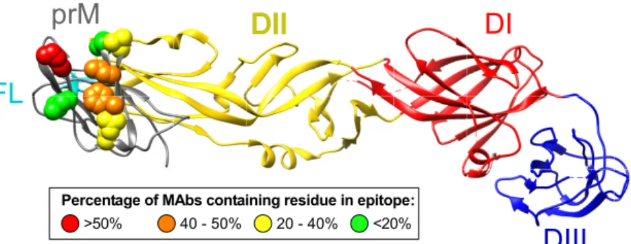
of the human MAbs differ. Seven commonly recognized residues
were identified (F1, L3, S5, E9, E18, L24, and K26), and there
were residues critical for the binding of over 50% of the MAbs in
the panel. Residue K26 was critical for the binding of 71% of the
MAbs. F1 or L3 residues also were found to be necessary for the
binding of 42 or 46% of the MAbs, respectively. We identified critical
residues within both prM and E protein for MAb 1G6.
Figure 3
provides a prM protein map indicating the frequency of residues
participating in MAb epitopes; the reduction of binding for
criti-cal residues is shown in Table S2 in the supplemental material. We
analyzed the distribution of residues recognized by MAbs from
donors with primary versus secondary infection and did not
de-tect major distinguishing patterns. Specifically, F1, L3, S5 E18, and
K26 contributed to the binding for MAbs derived from primary
and secondary cases; E9 contributed to the binding of 3 of 14
(21%) MAbs from primary and 0 of 11 (
⬍
9%) MAbs from
sec-ondary cases.
Genetic features of the antibodies.
Given that each of the
MAbs isolated recognized a single major antigenic site, it was
pos-sible that these diverse individuals were using a single dominant
antibody gene to encode a canonical antibody that is inherently fit
for binding to prM in the germ line configuration. To determine
whether such unique genetic features were characteristic of
hu-man anti-prM protein antibodies, we obtained heavy- and
light-chain-variable gene sequences for 23 clones and analyzed them for
variable gene usage and amino acid motifs (
Table 3
). We noted
that each of the antibodies was encoded by distinct variable gene
sequences using diverse V
H, D, and J
Hgene segments. The V
H1-69
segment was moderately over-represented, being found in 5 of 23
clones (22%). The mean HCDR3 length of 18.3 was about 2 amino
acids longer than the typical mean of 16 in human B cells.
Consis-tent with this, many of the longer HCDR3 regions were encoded
by the longest D gene segments (D2 and D3 family members). We
also determined the light-chain usage and heavy-chain isotype/
subtype by ELISA to determine whether any of these features were
common in the prM panel. We found that all of the clones were of
the IgG1 isotype. Interesting, the lambda light chain was highly
over-represented, being found in 20 of 23 (87%) of clones. The
variable gene sequences did contain somatic mutations consistent
with a memory cell phenotype, but the number of mutations was
not unusual or excessive. Thus, there appears to be a lambda
light-chain bias in the response against prM; otherwise, there are no
distinguishing genetic features in the prM-specific repertoire.
DISCUSSION
We sought to improve our understanding of the molecular
land-scape of the human anti-prM protein antibody response by
deter-mining the number of antigenic sites, the dominant epitope
tar-gets, and how these epitopes relate to the enhancing potencies
exhibited by different MAbs. The results show that human MAbs
target several overlapping epitopes on the surface of prM protein,
but their footprints fall into a single major antigenic site. We did
not identify any particular relationship between the infection
en-hancing properties of the antibodies and the fine specificity of the
epitope. In fact, every prM antibody enhanced infection with
vi-ruses from each of the four serotypes, no matter what the exposure
history of the donor or the fine epitope specificity. The findings
suggest that, as a class, prM antibodies mediate ADE using
essen-tially a single mode of action. All of the MAbs we isolated were
IgG1 subclass. It was also remarkable that this class of antibody
was common to every individual studied, no matter what the type
of DENV exposure history. This panel of human MAbs was
ob-tained from human subjects following diverse types of DENV
in-fection, including DENV serotype 1, 2, or 3 primary or secondary
dengue virus infections, or following DENV1 live attenuated virus
vaccination. We did not detect any major differences in the
bodies or the epitopes recognized by prM-specific MAbs based on
infecting serotype or primary versus secondary infection status.
Despite the common functional profile of these antibodies and
recognition of a single antigenic site, the members of the panel of
human MAbs exhibited a diversity of features that showed that
there are many antibodies and recognition features that target
human MAbs to this site on both the antibody and the virus side of
the interface. We considered whether there was a canonical
anti-body in humans forming a public clonotype across individuals
that accounts for this common recognition pattern. Several
anti-body heavy-chain-variable (V
H) genes have been associated
spe-cifically with recognition of structurally distinct viral epitopes, for
instance the association of V
H1-46 gene use with rotavirus
VP6-specific antibodies (
29–31
), V
H1-69 gene use in influenza
hemag-glutinin stem region antibodies (
32
,
33
), or V
H1-02 or V
H1-46
gene use in human MAbs specific for the CD4 supersite on HIV
envelope protein (
34
). These gene associations generally derive
from optimal interacting amino acids encoded by antibody
heavy-chain complementarity region 2 (HCDR2). Other canonical
amino acid motifs have been identified in the antibody HCDR3,
encoded by the V
H, diversity (D) and joining (J) genes, for
in-stance, the HIV neutralizing antibodies PG9 and PG16 (
35
). We
did not identify any such antibody gene associations in this panel
of antibodies to prM, even though they all recognize a single major
antigenic site. These data suggest that it is likely that most if not all
individuals can make prM-specific ADE antibodies, and they can
do so using a diversity of antibody clones from a wide variety of
naive B cells.
Diversity in mode of recognition by these antibodies also was
suggested by features of their binding characteristics. First,
epitope mapping studies identified seven different residues
neces-sary for binding of antibodies in the panel. There was a clear
hier-archical pattern, with certain prM residues most commonly
in-volved in the interaction. Residue K26 was inin-volved in the binding
of 17 human antibodies, while residues F1 and L3 were critical for
binding of nearly half of our MAbs. These residues, along with E9,
E18, and L24, are highly conserved across the DENV complex.
This finding is the likely explanation for why this class of MAbs is
so universally serotype cross-reactive, since they target areas of the
prM protein that are highly conserved. Interestingly, critical
resi-dues were also identified in the E protein for MAb 1G6. These
residues were focused within the fusion loop and neighboring E
protein domain II. Interpreting the role of critical residues in E
protein for 1G6 antibody binding is complex. The point
muta-tion in the prM/E antigen on the surfaces of cells in the
satura-tion mutagenesis studies could destabilize or alter the fine
structure of prM and cause loss of binding or, alternatively, the
MAb may bind a quaternary epitope with contact residues in
both prM and E. In the ELISA, the prM and E components were
tested separately, and the MAb bound only to prM. Likely 1G6
binds a quaternary prM/E epitope in which most of the contact
residues are in prM, and this component of the epitope is
suf-ficient for binding in ELISA. Clearly, the mode of binding for
antibodies that bind to both prM and E is complex, with
mo-lecular recognition of quaternary epitopes that would be found
only on virus particles. This recognition pattern has been
de-scribed previously for a human anti-prM MAb that was
iso-lated by phage display (
36
). The significance of recognition of a
quaternary epitope involving both E and prM protein residues
for these human anti-prM protein antibodies is unclear, but
FIG 3Percentage of MAbs containing specific prM region residues in the epitope. Colors show the percentage of MAbs for which the indicated residue contributes to the epitope, as determined by the loss of binding in fine epitope mapping studies. Residues are visualized on the DENV2 prM/E protein crystal structure (PDB identifier 3C6E).
TABLE 3Genetic features of prM-reactive human MAbs
MAb IgG subclass
Light chain
Heavy-chain-variable genes
Junctional protein sequence
Heavy-chain CDR3 length (amino acids)
No. of somatic mutations in heavy chains
VH D JH Nucleotides Amino acids
1B22 IgG1 1-69 2-2 6 CATLSLFCDSASCYHDPTSMDVW 21 34 18
1C6 IgG1 1-18 2-2 5 CAKMEDCNSTSCYGGTNWFDPW 20 23 10
1G6 IgG1 1-3 3-22 4 CARVNGDSAYYYGAPDYW 16 30 15
1G10 IgG1 3-15 2-2 6 CTTDLPLEYQLFYYDYMDVW 18 17 7
1H7.2 IgG1 3-23 2-15 4 CAKMGLCSGGSCYTGFIHFW 18 31 17
1H10 IgG1 4-4 3-3 3 CARVEFGFFGVVAKGIDLW 17 27 14
1I12 IgG1 3-74 3-3 5 CARGVNYDLWSAYSTDENWFDPW 21 12 9
1K20 IgG1 4-31 2-15 5 CARGRYCNDDSCYSEESAIWFDPW 22 25 17
1L13 IgG1 4-31 2-21 5 CATESYCRGNNCYPTPVIDPW 19 16 10
1O6 IgG1 1-69 2-2 5 CARATDCSTTSCYSSSWFDPW 19 21 14
2B17 IgG1 3-72 2-2 6 CAREGSCGSSTSCYADHYYGMDVW 22 13 9
2G3 IgG1 1-69 3-22 4 CATDTSGNLDFW 10 24 14
2H12 IgG1 3-49 3-3 4 CTQTPYCSGDKCYPVPFFDSW 19 17 11
2H21 IgG1 3-15 3-9 5 CATVEYCDATSCYNDDEAWFDPW 21 30 19
2J9 IgG1 3-49 2-15 5 CTRVVDCSGVNCYPMGWFDPW 19 15 7
2K2 IgG1 3-21 5-12 6 CARDRDTLPRDYYYHYGMDVW 19 20 9
2M2 IgG1 5-51 3-10 6 CGRHLYYFGSGKALLHGADVW 19 24 15
4E9 IgG1 3-11 2-15 1 CARGPEGYCSGNNCYPAEYFQHW 21 17 9
4F8 IgG1 4-59 2-8 3 CAISLGYCTGGKCHSGLGTFDIW 21 18 14
4G21 IgG1 1-69 5-12 4 CATDSGYVFYFGYW 12 7 4
5E15 IgG1 3-30 2-2 3 CTGGLGYCSSSSCYLGAFDVW 19 28 18
5G22 IgG1 3-53 1-26 4 CARGGSFYDPFDYW 12 8 4
both 2M2 and 5M22 are some of the few neutralizing MAbs
that recognize prM.
Our shotgun mutagenesis approach identifies epitopes
irre-spective of MAb neutralization status or viral fitness and so is not
limited to inhibitory MAbs or to mutations that are compatible
with virus replication. Nonetheless, this strategy cannot detect the
contribution of alpha carbons to an interaction, mutations to
other substitutions could result in different results, and the ability
to differentiate direct from indirect or allosteric effects on MAb
interactions depends on the number and diversity of other
avail-able control MAbs. However, the concordance of mutagenesis,
competition binding, ELISA, and Western blot data provides
con-fidence that the epitope residues identified here are accurate.
Other residues may also be involved in each epitope, but their
mutation may have resulted in misfolding of the protein, or they
did not disrupt the energetics of binding significantly enough to be
identified as critical.
The number of antibodies that can bind one prM protein
mol-ecule is one factor that may contribute to the complexity of
anti-body-dependent enhancement. Only one antibody binding group
was identified by MAb competition-binding assays using whole
virus particles. These data suggest that, despite the overall
com-plexity of the human anti-prM protein antibody response, only a
single antibody molecule is bound to one molecule of the prM
protein in immature particles at a given moment in time.
There-fore, anti-prM antibodies must be in a state of competition with
one another for binding to the single accessible site. This dynamic
is also complicated by the fact that the occupancy of antibodies on
the population of prM molecules on a single particle may not be
complete, and most particles likely contain a mixture of cleaved or
uncleaved prM/E protein. In the case of the polyclonal antibody
response in serum, the antibody concentration, affinity, and
epitope availability are likely to play a critical role in determining
which anti-prM antibodies are most successful in engaging the
virus.
In summary, the human antibody response encoding DENV
prM protein-specific antibodies is complex in terms of their
ge-netic origins and fine specificity, but these antibodies exhibit
con-sistent functional properties. These antibodies are cross-reactive,
possess very limited neutralizing properties, and are potent at
in-fection enhancement
in vitro. The human prM protein-specific
antibody response targets diverse epitopes while competing for
binding to a single antigenic site. Continued discovery of the
structure and nature of the epitopes for this unique group of
MAbs in the future could point the way toward rational design of
DENV vaccine antigens to minimize the cross-reactive
disease-enhancing antibody profiles of those receiving the vaccine.
Devel-opment of vaccine preparations that have reduced tendencies to
induce prM-specific antibodies, such as viruses or particles
lack-ing the immunodominant K26 residue, could be considered.
ACKNOWLEDGMENTS
This work was supported by NIH grants U54 AI057157 (Southeastern Regional Center of Excellence for Emerging Infections and Biode-fense), R01 AI107731 (A.M.D.S.), K08 AI103038 (S.A.S.), and HHSN272200900055C (B.J.D.).
We thank Frances Smith-House (Vanderbilt) for excellent laboratory management support, Edgar Davidson (Integral Molecular) for editorial support, and Bhumi Patel and Sandra Henein (UNC) for technical assis-tance. We thank Eva Harris (University of California, Berkeley), Anna
Durbin (Johns Hopkins University), and Stephen Whitehead (NIH) for providing cryopreserved PBMCs from immune donors. We also thank all dengue-immune travelers who voluntarily donated blood at the UNC and Vanderbilt Schools of Medicine.
FUNDING INFORMATION
HHS | NIH | National Institute of Allergy and Infectious Diseases (NIAID) provided funding to James E. Crowe under grant number U54 AI057157. HHS | NIH | National Institute of Allergy and Infectious Diseases (NIAID) provided funding to Scott A. Smith under grant number K08 AI103038. HHS | NIH | National Institute of Allergy and Infectious Diseases (NIAID) provided funding to Benjamin J. Doranz under grant number HHSN272200900055C. HHS | NIH | National Institute of Allergy and Infectious Diseases (NIAID) provided funding to Aravinda M. de Silva and James E. Crowe under grant number R01 AI107731.
REFERENCES
1.Bhatt S, Gething PW, Brady OJ, Messina JP, Farlow AW, Moyes CL, Drake JM, Brownstein JS, Hoen AG, Sankoh O, Myers MF, George DB, Jaenisch T, Wint GR, Simmons CP, Scott TW, Farrar JJ, Hay SI.2013. The global distribution and burden of dengue. Nature496:504 –507.http: //dx.doi.org/10.1038/nature12060.
2.Gubler DJ.2002. Epidemic dengue/dengue hemorrhagic fever as a public health, social and economic problem in the 21st century. Trends Micro-biol10:100 –103.http://dx.doi.org/10.1016/S0966-842X(01)02288-0. 3.Halstead SB.2003. Neutralization and antibody-dependent enhancement
of dengue viruses. Adv Virus Res60:421– 467.http://dx.doi.org/10.1016 /S0065-3527(03)60011-4.
4.Halstead SB, O’Rourke EJ.1977. Antibody-enhanced dengue virus in-fection in primate leukocytes. Nature265:739 –741.http://dx.doi.org/10 .1038/265739a0.
5.Zellweger RM, Prestwood TR, Shresta S.2010. Enhanced infection of liver sinusoidal endothelial cells in a mouse model of antibody-induced severe dengue disease. Cell Host Microbe7:128 –139.http://dx.doi.org/10 .1016/j.chom.2010.01.004.
6.Balsitis SJ, Williams KL, Lachica R, Flores D, Kyle JL, Mehlhop E, Johnson S, Diamond MS, Beatty PR, Harris E.2010. Lethal antibody enhancement of dengue disease in mice is prevented by Fc modification. PLoS Pathog
6:e1000790.http://dx.doi.org/10.1371/journal.ppat.1000790.
7.Goncalvez AP, Engle RE, St Claire M, Purcell RH, Lai CJ. 2007. Monoclonal antibody-mediated enhancement of dengue virus infection in vitro and in vivo and strategies for prevention. Proc Natl Acad Sci U S A
104:9422–9427.http://dx.doi.org/10.1073/pnas.0703498104.
8.Modis Y, Ogata S, Clements D, Harrison SC. 2005. Variable surface epitopes in the crystal structure of dengue virus type 3 envelope glycopro-tein. J Virol79:1223–1231.http://dx.doi.org/10.1128/JVI.79.2.1223-1231 .2005.
9.Kuhn RJ, Zhang W, Rossmann MG, Pletnev SV, Corver J, Lenches E, Jones CT, Mukhopadhyay S, Chipman PR, Strauss EG, Baker TS, Strauss JH.2002. Structure of dengue virus: implications for flavivirus organization, maturation, and fusion. Cell108:717–725.http://dx.doi.org /10.1016/S0092-8674(02)00660-8.
10. Yu IM, Zhang W, Holdaway HA, Li L, Kostyuchenko VA, Chipman PR, Kuhn RJ, Rossmann MG, Chen J. 2008. Structure of the immature dengue virus at low pH primes proteolytic maturation. Science319:1834 – 1837.http://dx.doi.org/10.1126/science.1153264.
11. Plevka P, Battisti AJ, Junjhon J, Winkler DC, Holdaway HA, Keelapang P, Sittisombut N, Kuhn RJ, Steven AC, Rossmann MG.2011. Matura-tion of flaviviruses starts from one or more icosahedrally independent nucleation centres. EMBO Rep12:602– 606.http://dx.doi.org/10.1038 /embor.2011.75.
12. Rodenhuis-Zybert IA, van der Schaar HM, da Silva Voorham JM, van der Ende-Metselaar H, Lei HY, Wilschut J, Smit JM.2010. Immature dengue virus: a veiled pathogen? PLoS Pathog6:e1000718.http://dx.doi .org/10.1371/journal.ppat.1000718.
13. Cardosa MJ, Wang SM, Sum MS, Tio PH.2002. Antibodies against prM protein distinguish between previous infection with dengue and Japanese encephalitis viruses. BMC Microbiol2:9.http://dx.doi.org/10.1186/1471 -2180-2-9.
322–330. http://dx.doi.org/10.1002/(SICI)1096-9071(199903)57:3⬍322 ::AID-JMV17⬎3.0.CO;2-5.
15. Smith SA, Zhou Y, Olivarez NP, Broadwater AH, de Silva AM, Crowe JE, Jr.2012. Persistence of circulating memory B cell clones with potential for dengue virus disease enhancement for decades following infection. J Virol86:2665–2675.http://dx.doi.org/10.1128/JVI.06335-11.
16. Dejnirattisai W, Jumnainsong A, Onsirisakul N, Fitton P, Va-sanawathana S, Limpitikul W, Puttikhunt C, Edwards C, Duangchinda T, Supasa S, Chawansuntati K, Malasit P, Mongkolsapaya J, Screaton G.
2010. Cross-reacting antibodies enhance dengue virus infection in hu-mans. Science328:745–748.http://dx.doi.org/10.1126/science.1185181. 17. Lai CY, Tsai WY, Lin SR, Kao CL, Hu HP, King CC, Wu HC, Chang GJ,
Wang WK.2008. Antibodies to envelope glycoprotein of dengue virus during the natural course of infection are predominantly cross-reactive and recognize epitopes containing highly conserved residues at the fusion loop of domain II. J Virol82:6631– 6643.http://dx.doi.org/10.1128/JVI .00316-08.
18. Beltramello M, Williams KL, Simmons CP, Macagno A, Simonelli L, Quyen NT, Sukupolvi-Petty S, Navarro-Sanchez E, Young PR, de Silva AM, Rey FA, Varani L, Whitehead SS, Diamond MS, Harris E, Lanza-vecchia A, Sallusto F.2010. The human immune response to dengue virus is dominated by highly cross-reactive antibodies endowed with neu-tralizing and enhancing activity. Cell Host Microbe8:271–283.http://dx .doi.org/10.1016/j.chom.2010.08.007.
19. Smith SA, de Alwis AR, Kose N, Jadi RS, de Silva AM, Crowe JE, Jr.
2014. Isolation of dengue virus-specific memory B cells with live virus antigen from human subjects following natural infection reveals the pres-ence of diverse novel functional groups of antibody clones. J Virol88:
12233–12241.http://dx.doi.org/10.1128/JVI.00247-14.
20. Luo YY, Feng JJ, Zhou JM, Yu ZZ, Fang DY, Yan HJ, Zeng GC, Jiang LF.2013. Identification of a novel infection-enhancing epitope on dengue prM using a dengue cross-reacting monoclonal antibody. BMC Microbiol
13:194.http://dx.doi.org/10.1186/1471-2180-13-194.
21. de Alwis R, Williams KL, Schmid MA, Lai CY, Patel B, Smith SA, Crowe JE, Wang WK, Harris E, de Silva AM.2014. Dengue viruses are enhanced by distinct populations of serotype cross-reactive antibodies in human immune sera. PLoS Pathog10:e1004386.http://dx.doi.org/10.1371/journal.ppat.1004386. 22. Weitkamp JH, Kallewaard N, Kusuhara K, Feigelstock D, Feng N,
Greenberg HB, Crowe JE, Jr.2003. Generation of recombinant human monoclonal antibodies to rotavirus from single antigen-specific B cells selected with fluorescent virus-like particles. J Immunol Methods275:
223–237.http://dx.doi.org/10.1016/S0022-1759(03)00013-9.
23. Ruiz M, Giudicelli V, Ginestoux C, Stoehr P, Robinson J, Bodmer J, Marsh SG, Bontrop R, Lemaitre M, Lefranc G, Chaume D, Lefranc MP.
2000. IMGT, the international ImMunoGeneTics database. Nucleic Acids Res28:219 –221.http://dx.doi.org/10.1093/nar/28.1.219.
24. Smith SA, de Alwis R, Kose N, Durbin AP, Whitehead SS, de Silva AM, Crowe JE, Jr.2013. Human monoclonal antibodies derived from memory B cells following live attenuated dengue virus vaccination or natural infec-tion exhibit similar characteristics. J Infect Dis207:1898 –1908.http://dx .doi.org/10.1093/infdis/jit119.
25. Lambeth CR, White LJ, Johnston RE, de Silva AM.2005. Flow cytom-etry-based assay for titrating dengue virus. J Clin Microbiol43:3267–3272.
http://dx.doi.org/10.1128/JCM.43.7.3267-3272.2005.
26. Kraus AA, Messer W, Haymore LB, de Silva AM.2007. Comparison of plaque- and flow cytometry-based methods for measuring dengue virus neutralization. J Clin Microbiol45:3777–3780.http://dx.doi.org/10.1128 /JCM.00827-07.
27. Li L, Lok SM, Yu IM, Zhang Y, Kuhn RJ, Chen J, Rossmann MG.2008. The flavivirus precursor membrane-envelope protein complex: structure and matu-ration. Science319:830–1834.http://dx.doi.org/10.1126/science.1153263. 28. Smith SA, de Alwis AR, Kose N, Harris E, Ibarra KD, Kahle KM, Pfaff
JM, Xiang X, Doranz BJ, de Silva AM, Austin SK, Sukupolvi-Petty S, Diamond MS, Crowe JE, Jr.2013. The potent and broadly neutralizing human dengue virus-specific monoclonal antibody 1C19 reveals a unique cross-reactive epitope on the bc loop of domain II of the envelope protein. mBio4:e00873-13.http://dx.doi.org/10.1128/mBio.00873-13.
29. Weitkamp JH, Kallewaard N, Kusuhara K, Bures E, Williams JV, LaFleur B, Greenberg HB, Crowe JE, Jr.2003. Infant and adult human B cell responses to rotavirus share common immunodominant variable gene repertoires. J Immunol171:4680 – 4688.http://dx.doi.org/10.4049 /jimmunol.171.9.4680.
30. Weitkamp JH, Kallewaard NL, Bowen AL, LaFleur BJ, Greenberg HB, Crowe JE, Jr.2005. VH1-46 is the dominant immunoglobulin heavy chain gene segment in rotavirus-specific memory B cells expressing the intesti-nal-homing receptor␣47. J Immunol174:3454 –3460.http://dx.doi.org /10.4049/jimmunol.174.6.3454.
31. Tian C, Luskin GK, Dischert KM, Higginbotham JN, Shepherd BE, Crowe JE, Jr.2008. Immunodominance of the VH1-46 antibody gene segment in the primary repertoire of human rotavirus-specific B cells is reduced in the memory compartment through somatic mutation of non-dominant clones. J Immunol180:3279 –3288.http://dx.doi.org/10.4049 /jimmunol.180.5.3279.
32. Sui J, Hwang WC, Perez S, Wei G, Aird D, Chen LM, Santelli E, Stec B, Cadwell G, Ali M, Wan H, Murakami A, Yammanuru A, Han T, Cox NJ, Bankston LA, Donis RO, Liddington RC, Marasco WA. 2009. Structural and functional bases for broad-spectrum neutralization of avian and human influenza A viruses. Nat Struct Mol Biol16:265–273.
http://dx.doi.org/10.1038/nsmb.1566.
33. Ekiert DC, Bhabha G, Elsliger MA, Friesen RH, Jongeneelen M, Thro-sby M, Goudsmit J, Wilson IA.2009. Antibody recognition of a highly conserved influenza virus epitope. Science324:246 –251.http://dx.doi.org /10.1126/science.1171491.
34. Zhou T, Lynch RM, Chen L, Acharya P, Wu X, Doria-Rose NA, Joyce MG, Lingwood D, Soto C, Bailer RT, Ernandes MJ, Kong R, Longo NS, Louder MK, McKee K, O’Dell S, Schmidt SD, Tran L, Yang Z, Druz A, Luongo TS, Moquin S, Srivatsan S, Yang Y, Zhang B, Zheng A, Pancera M, Kirys T, Georgiev IS, Gindin T, Peng HP, Yang AS, Mullikin JC, Gray MD, Stamatatos L, Burton DR, Koff WC, Cohen MS, Haynes BF, Casazza JP, Connors M, Corti D, Lanzavecchia A, Sattentau QJ, Weiss RA, West AP, Jr, Bjorkman PJ, Scheid JF, Nussenzweig MC, Shapiro L, Mascola JR, Kwong PD. 2015. Structural repertoire of HIV-1-neutralizing antibodies targeting the CD4 supersite in 14 donors. Cell
161:1280 –1292.http://dx.doi.org/10.1016/j.cell.2015.05.007.
35. Walker LM, Phogat SK, Chan-Hui PY, Wagner D, Phung P, Goss JL, Wrin T, Simek MD, Fling S, Mitcham JL, Lehrman JK, Priddy FH, Olsen OA, Frey SM, Hammond PW, Kaminsky S, Zamb T, Moyle M, Koff WC, Poignard P, Burton DR.2009. Broad and potent neutralizing antibodies from an African donor reveal a new HIV-1 vaccine target. Sci-ence326:285–289.http://dx.doi.org/10.1126/science.1178746.