doi: http://dx.doi.org/10.18203/2319-2003.ijbcp20150360
IJBCP
International Journal of Basic & Clinical Pharmacology
Review Article
Emerging glaucoma therapeutics
Ranjita Santra (Dhali)*, Shantanu Munshi
CURRENT UNDERSTANDING
Glaucoma is a frequent, pathophysiologically heterogeneous ophthalmologic condition characterized by a typical visual field loss pattern from peripheral to central. As a chronic progressive optic neuropathy, it can lead to severe disability and blindness. Glaucoma is classified according to whether it is congenital or acquired. It is further sub-divided into open-angle or closed-angle, depending on how the aqueous outflow is impaired. Glaucoma is usually associated with an increase in intraocular pressure (IOP) above the normal value, which is usually estimated at 21 mm Hg (mean 15.5, range 10-21). Patients with statistically normal IOP who develop the characteristic changes associated with open-angle glaucoma are said to have low tension or normal pressure glaucoma. Finally, primary or secondary types are identified depending on the presence of underlying contributory factors. The pathophysiology of glaucoma is not fully understood till date. The trabecular outflow pathway is the primary draining tissue for the aqueous
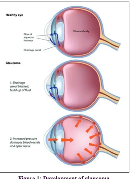
humor in the eye. It consists of 3 structures, the trabecular meshwork (TM), juxtacanalicular tissue, and Schlemm’s canal. In a healthy eye, IOP is maintained within a narrow range through dynamic regulation of trabecular outflow resistance. In a glaucomatous eye, elevated IOP is due to an abnormally high resistance to outflow in the trabecular outflow pathway.1 The causes of increased outflow resistance are not fully understood, but it has been hypothesized to involve an increase in the contractile tone and stiffness of the TM and changes in extracellular matrix composition and/or a change in the conductance of Schlemm’s canal (Figures 1 and 2).2 There is a significant need for glaucoma drugs that specifically target the physiologic cause of elevated IOP and thereby enhance trabecular outflow. Currently, the management of glaucoma is focused on controlling the IOP by pharmacological and surgical measures to avoid these consequences. The tissues of the trabecular outflow pathway are avascular and rely on the aqueous humor to supply nutrients, growth factors, and antioxidants. The most widely prescribed glaucoma drugs,
ABSTRACT
The purpose of this review was to discuss the major recent advances in the field of ophthalmology, particularly as it pertains to glaucoma. We reviewed literature using MEDLINE and PubMed databases with the following search terms: “glaucoma,” “melatonin,” “trabecular outflow pathway,” “adenosine,” “rho kinase,” “norepinephrine,” and “matrix metalloproteinases.” We also reviewed pertinent references from articles found in this search. We looked at various studies concerning the clinical trials of glaucoma therapeutics, and therapeutic potential of putative ocular drug delivery systems in glaucoma. Challenges of assuring safety and efficacy of the newer medicines and techniques are pertinent in this regard. However, more research is needed to better elucidate the mechanism of various investigational drug products and drug delivery devices in glaucoma.
Keywords: Glaucoma, Melatonin, Trabecular outflow pathway, Adenosine, Rho kinase, Norepinephrine, Matrix metalloproteinases
Department of Clinical and Experimental Pharmacology, Calcutta School of Tropical Medicine, Kolkata, West Bengal, India
Received: 15 June 2015
Revised: 02 July 2015
Accepted: 05 July 2015
*Correspondence to:
Dr. Ranjita Santra (Dhali), Email: [email protected]
the prostaglandin analogs (PGAs), lower IOP by increasing the aqueous drainage through the unconventional uveoscleral outflow pathway. The older, non-PGA drug classes, the beta blockers, alpha agonists, and carbonic anhydrase inhibitors, lower IOP by decreasing the production of aqueous humor. By shunting the aqueous humor through the uveoscleral pathway or decreasing the aqueous production, the commonly used glaucoma medications actually decrease the outflow of aqueous humor through the diseased TM. Thus, while protecting the optic nerve from damage by lowering IOP, current medications may be allowing further degradation of the trabecular outflow pathway. Glaucoma has also been reported to be associated with a dysregulation of the circadian system,3 as well as a high incidence of sleep disorders,4,5 depression, and anxiety.6 However, because of the higher incidence of all these conditions in older age, they have not received much clinical or scientific attention and remain often undertreated. Recently, there has been evidence
that glaucoma leads to an impairment of the photo-dependent melatonin production with alterations in circadian rhythms. In the current review, we will present the state of knowledge on the impact of glaucoma on the melatonergic system and possible links to circadian desynchronization, sleep disorders, and depression. The implications indicating that glaucoma may also be a psycho-chronobiological disease and their consequences for research and clinical therapy are discussed. Fortunately, several new drug classes that target the trabecular outflow pathway have entered clinical development. Adenosine A1 receptor agonists represent another new drug class that targets the trabecular outflow pathway. The drug class with the longest history in the clinic is the class of selective rho kinase inhibitors. Rho kinase is a serine/threonine kinase whose activity increases actomyosin contraction in smooth muscle cells, including the smooth muscle-like cells of the TM. Adenosine A1 agonists are thought to increase the trabecular outflow by reducing the cell volume and increasing the expression of matrix metalloproteinases.7 The newest class of trabecular outflow drugs is the dual action rho kinase/norepinephrine transporter (ROCK/NET) class. ROCK/NET inhibitors are single molecules that act through a dual mechanism of action, simultaneously inhibiting rho kinase to increase trabecular outflow and inhibiting the NET to reduce the production of aqueous humor.8,9 In the current review, we will present the state of knowledge on the newer and emerging therapeutic areas of Glaucoma.
METHODS
Medline searches were performed using “glaucoma,” “melatonin,” “trabecular outflow pathway,” “adenosine,” “rho kinase,” “norepinephrine,” and “matrix metalloproteinases.” As search terms, available literature from 1980 onward was screened for relevance, and additional material was added from the bibliography of applicable papers.
RESULTS
[image:2.595.61.277.72.369.2]Glaucoma is a progressive optic neuropathy that is a leading cause of irreversible blindness worldwide.10 Elevated IOP is commonly associated with glaucoma, and multiple longitudinal studies have demonstrated that lowering IOP in patients can slow optic nerve degeneration and preserve vision.11 By shunting the aqueous humor through the uveoscleral pathway or decreasing the aqueous production, the commonly used glaucoma medications actually decrease the outflow of aqueous humor through the diseased TM. Although PGAs often provide adequate efficacy as an initial therapy, nearly half of all patients started on PGA monotherapy ultimately require 1 or more non-PGA drugs to be added to their treatment regimen. This is the primary reason why half of all prescriptions are still written for non-PGA products.12 IOPs continue to rise over time because the function of the trabecular outflow pathway continues to deteriorate over time. Therefore, evolution of newer
Figure 1: Development of glaucoma.
Figure 2: Open angle glaucoma and closed angle
glaucoma. Blue arrow shows the direction of flow of
pharmacotherapeutics has led to the emergence of various classes of anti-glaucoma agents like melatonin receptor agonists, adenosine 1 receptor agonists, rho kinase inhibitors, ROCK/NET inhibitors, newer drug delivery systems, IOP monitoring devices, and neuroprotective drugs such as alpha2 adrenergic agonists, b-adrenergic antagonists, prostaglandin F2alpha agonists, carbonic anhydrase inhibitors, and tumor necrosis factor-alpha (TNF-a) inhibitor etanercept, as well as the investigational drug MRZ-99030.
Melatonin and its implication in glaucoma
The relationship between glaucoma and melatonin in particular seems to be bidirectional, as (i) glaucoma (particularly the death of ipRGC) can affect the rhythm of pineal melatonin production, which in turn, can affect the circadian system activity, and (ii) retinal melatonin could be involved in the pathogenesis of glaucoma. Pharmacologically, there is already an indication for melatonergic treatment in MDD, SAD, and sleep disorders. As we have pointed out, current data implies that glaucoma patients, too, could benefit from melatonergic treatment on multiple levels: (i) there are direct positive effects of melatonin on the core etiopathology of glaucoma: melatonin has been proven to directly reduce IOP significantly through the putative MT3 receptor13 and, thus, may have clinical potential for treating elevated IOP. Furthermore, the antioxidant potency of melatonin in ocular tissue and the neuroprotective role of melatonin in glaucoma could be of use in the management of the disease.14,15 (ii) Indirect positive effects can be achieved by a melatonin-dependent positive influence on systemic blood pressure during the night.16,17 (iii) Melatonin treatment can positively influence comorbid circadian misalignment and sleep disorders. Continuous retinal ganglion cell degeneration in glaucoma supports the hypothesis of this condition being the main ophthalmologic disease affecting the photic input to the circadian system and thereby circadian rhythms. Taking into account that glaucoma is more common in older age individuals and that, in older patients, the ability of the pineal gland to produce melatonin is already decreased and the day–night amplitude in melatonin secretion is physiologically lowered,18 we can assume that melatonin treatment may be of advantage for a high number of glaucoma patients.
Rho kinase inhibitors in glaucoma
Rho kinase is involved in the modulation of aqueous humor outflow facility. Rho kinase, a critical downstream effector of rho GTPase is recognized to control the formation of actin stress fibers, focal adhesions, and cellular contraction in the eye. The therapeutic potential of rho kinase inhibitors was initially demonstrated in preclinical studies using the rho kinase inhibitor Y-27632, which was shown to relax precontracted TM tissue ex vivo,19 increase trabecular outflow in perfused enucleated porcine eyes,20 and lower IOP in live rabbits upon topical ocular application.21 According
to clinical trial registries in the U.S., Europe, and Japan, different selective rho kinase inhibitors have been tested in human clinical trials.22 The most studied compound of this class is Aerie Pharmaceuticals’ AR-12286. One AR-12286 clinical study demonstrated that Rho kinase inhibition can be used successfully in combination with PGAs to achieve a greater IOP lowering than PGA therapy alone.23 In patients with elevated IOP, a fixed-dose combination of 0.5% AR-12286 and 0.004% travoprost achieved IOP reductions of 9-12 mmHg when dosed once daily in the evening and produced a reduction in diurnal IOP that was 2 mmHg greater than travoprost alone. The incidence and severity of hyperemia for the fixed-dose combination was similar to travoprost monotherapy. Given that ophthalmologists often need to prescribe non-PGA drugs as add-on therapy to PGAs, it is important that the Rho kinase mechanism of action is compatible with the PGA mechanism of action. In addition, clinical development has begun for PG324, a fixed-dose combination product that combines AR-13324 with latanoprost. Phase 2 clinical study results have recently been published for another selective Rho kinase inhibitor, K-115, from Kowa.24 In patients with elevated IOP, twice-daily dosing of 0.4% K-115 achieved IOP reductions of 3.1-4.5 mmHg. A 65% incidence of transient, mild hyperemia was reported for this concentration.
Adenosine A1 receptor agonists in glaucoma
but unlike the Inotek compound these drugs are aimed at the adenosine A3 receptor. Targeting this signaling pathway reduces IOP by inhibiting aqueous humor production of the ciliary epithelium. These compounds are also formulated for oral rather than topical dosing, a choice that may provide a greater duration of action and less IOP fluctuation.
ROCK/NET inhibitors in glaucoma
The newest class of trabecular outflow drugs is the dual action ROCK/NET class. ROCK/NET inhibitors are single molecules that act through a dual mechanism of action,
simultaneously inhibiting Rho Kinase to increase trabecular outflow and inhibiting the NET to reduce the production of aqueous humor.8 Aerie Pharmaceuticals’ AR-13324 is the first compound of this class to be tested in the clinic and has replaced AR-12286 as Aerie’s lead trabecular outflow drug. In a 7-day clinical trial in patients with elevated IOP, once-daily A.M. dosing of AR-13324 (0.01%, 0.02%, or 0.04%) produced IOP reductions ranging from 5.6 to 7.2 mmHg.26 The morning dosing regimen produced transient, mild to moderate hyperemia with an incidence of 29% for the 0.02% concentration (the top of the dose-response curve) on day 7. The 0.01% and 0.02% concentrations of AR-13324 have been advanced to a 28-day Phase 2b study to evaluate the efficacy and tolerability of AR-13324 when dosed once-daily in the evening. In addition, clinical development has begun for PG324, a fixed-dose combination product that combines AR-13324 with latanoprost.
Two other additions to the development pipeline are in early-phase clinical trials. One of these is a dose-ranging study of the mixed prostaglandin agonist ONO-9054 (Ono Pharma). Another dose-escalation trial is under way for SYL040012 (Sylentis). The Sylentis compound is an RNAi-based compound designed to target the same b-adrenergic pathway targeted by timolol. Instead of acting as a traditional receptor antagonist, however, SYL040012 blocks the pathway by inhibiting biosynthesis of the receptor protein.
Newer drug delivery systems in glaucoma
One of the greatest hurdles in controlling IOP by means of medical treatment in glaucoma patients is that of compliance: the combination of the chronic nature of treatments, the lack of symptoms, and the age of the affected population is a recipe for poor compliance. Recent estimates suggest that 60% of patients fail to maintain a daily medication regimen.27 One approach to this problem is to take the task out of the hands of the patient by employing sustained-release or other depot forms of existing drugs. A number of strategies employing this approach to drug delivery are currently in development.
The possibilities were wide-ranging, and included:
a. A polymer, like a contact lens that would contain drug and sit under the eyelid and release the medication over several months (from Amorphex Therapeutics, LLC) b. Use of microneedles to inject medication into a specific
spot for it to be most effective (Clearside Biomedical, Inc.)
c. Implantable extended-release devices using engineered highly-precise microparticles and nanoparticles (Envisia Therapeutics)
d. Delivery devices or technology to allow a constant delivery of medication over months or years (pSivida and SKS Ocula, LLC)
[image:4.595.53.284.73.209.2]e. Polymer-based intraocular delivery technologies that would allow customizable sustained release of all therapeutic classes (GrayBug).
Figure 3: Emerging pharmacotherapeutic strategies of glaucoma.
Figure 4: Intraocular pressure (IOP) is kept under control by an matrix metalloproteinases (MMP)-based
feedback mechanism, regulating outflow resistance
of the trabecular meshwork (TM). (a) Homeostatic ECM turnover in the TM is actualized by constitutive
expression of MMP-1, -2, -3, -9, -12, and -14, as well as TIMP-2 (b) Mechanical distortions, provoked by IOP elevations, are sensed by TM cells via ECM-integrin interactions and result in the upregulation of MMP-2, -3, and -14 secretion, while reducing TIMP-2, via an mTOR-mediated intracellular signaling cascade.
Note: Outflow resistance is largely generated in the
juxtacanalicular portion of the TM (JCT); however, MMP production and function are not exclusive to the
JCT portion of the TM, as is depicted in the drawing, but may also occur on the cells of the corneoscleral portion of the meshwork (CSM). SC: Schlemm’s canal.
[image:4.595.52.288.252.391.2]f. Drops that allow medication to get into the eye more easily (Kala Pharmaceuticals)
g. Tear duct plugs that release medication (Ocular Therapeutix).
IOP monitoring devices
Diurnal variations of IOP have been thought to play an important role in progression of the disease for some time.28 Office visit assessments provide a single measure of IOP, so they cannot provide a comprehensive picture of daily IOP fluctuations. The arrival of continuous monitoring devices designed to provide a round-the-clock IOP measurement is a welcome step forward.
A number of different technologies in development can provide continuous IOP monitoring. One approach uses implantable microsensors that transmit pressure data to a handheld external device (Implandata Ophthalmic Products). Another implantable device, the iSense (AcuMEMs), is also in development. Both the Implandata and AcuMEMs systems would be implanted during cataract surgery or another surgery to allow access to the anterior chamber. An alternative technology employs a contact lens with an embedded strain gauge (Triggerfish; Sensimed AG) to record continuous 24-hr changes in ocular surface tension. While this metric is distinct from a true measure of IOP, it provides an indirect means to monitor the fluctuations associated with IOP. Using this device, a recently published study showed a nocturnal peak in tension occurs in about 70% of all patients with diagnosed or suspected POAG.29 These continuous measurement devices will likely have a significant impact on the therapy of POAG going forward.
Targeting neuroprotection
While current therapies, surgeries, and the growing list of devices focus on lowering IOP as the path to successful glaucoma therapy, there have also been some advances in more direct approaches to treatment and prevention of the glaucomatous neuropathology that leads to visual loss. A key point in understanding the pathology underlying POAG is that, while lowering IOP shows a strong correlation with minimizing disease progression, the connection is not absolute: patients with normal pressures can show visual field loss, while the visual fields in others with high IOPs may remain intact. Increasingly, then, the focus of clinicians and researchers has turned to addressing the pathological consequences of the disease.
A growing body of evidence suggests that many of the drugs that are used to treat elevated IOP also have neuroprotective effects.30 For example, there is ample evidence that inflammatory cytokines from neighboring microglia are important contributors to retinal degeneration, and recent studies suggest that a2 adrenergic agonists can minimize cytokine release, perhaps through activation of brain-derived neurotrophic factor, or other neurotrophic pathways.31-33
A clinical study in patients with normal-tension glaucoma has suggested that brimonidine (Alphagan) has an effect beyond lowering IOP.34 Similarly, b-adrenergic antagonists, prostaglandin F2a agonists and carbonic anhydrase inhibitors all have established neuroprotective actions that may provide a therapeutic benefit beyond their IOP-lowering action.35,36
Interest in neuroprotection is evident from clinical studies of the glutamate receptor antagonist memantine, animal studies of other glutamate antagonists, and experimental trials with other drugs with known neuroprotective effects.37,38 Promising animal studies suggest other avenues of addressing the goal of neuroprotection. For instance, the TNF-a inhibitor Etanercept has been shown to effectively prevent RGC loss in a rodent glaucoma model.39 The established association between retinal degeneration seen in POAG, b-amyloid deposition and the more global neurodegeneration of Alzheimer’s disease may provide a route for basic scientific advances as well as new clinical approaches.40 One compound that has shown the ability to reduce b-amyloid aggregation in animal models of glaucoma, MRZ-99030 (Merz Pharmaceuticals GmbH), is currently the subject of clinical trials to assess the safety of a topical formulation (NCT01714960 at clinicaltrials.gov).
DISCUSSION
The past 150 years have produced a plethora of IOP-lowering drugs with a variety of mechanisms of action, most of which remain available and in common usage. Given the accelerated rate of discovery over the past 15 years, the future is likely to produce any number of new drugs to complement our current pharmamentarium. There remain several unmet needs in glaucoma pharmacology. Despite five drug classes, there is no clear choice for adjunctive therapy to PGAs most of the remaining drug classes are relatively inefficacious in combination with the prostaglandins. Adherence is low with glaucoma medications; absolving the patient of the responsibility of daily dosing would be impactful in the management of this chronic disease. Moreover, developing drugs for glaucoma that work by mechanisms other than IOP reduction is an unmet need. Accomplishing this will require that we better understand the pathophysiology of glaucoma on the molecular level. In particular, drugs that protect the optic nerve from damage so-called neuroprotection would be useful in the management of glaucoma. Human trials of memantine for optic neuroprotection have been bitter failures, but other potential neuroprotective drugs are in varying stages of development. These issues are currently being addressed by both industry and academia, and the next 150 years may bring new discoveries that will continue to improve the lives of patients with glaucoma.
CONCLUSION
redundant ocular signaling physiology, but it is likely that future approaches will focus on the mitigation of the retinal pathology that’s the hallmark of open-angle glaucoma. Whatever the route to neuroprotection, this one-two punch of reduced IOP and diminished retinal cell loss is a goal that is in sight, and would be a major step forward in preserving visual function in patients with POAG.
Funding: No funding sources
Conflict of interest: None declared
Ethical approval: Not required
REFERENCES
1. Grant WM. Clinical tonography. Trans Am Acad Ophthalmol
Otolaryngol. 1951;55:774-81.
2. Stamer WD, Acott TS. Current understanding of conventional
outflow dysfunction in glaucoma. Curr Opin Ophthalmol. 2012;23(2):135-43.
3. Jean-Louis G, Zizi F, Lazzaro DR, Wolintz AH. Circadian rhythm dysfunction in glaucoma: a hypothesis. J Circadian
Rhythms. 2008;6:1.
4. Mojon DS, Hess CW, Goldblum D, Boehnke M, Koerner F, Gugger M, et al. Normal-tension glaucoma is associated with sleep apnea syndrome. Ophthalmologica.
2002;216(3):180-4.
5. Mojon DS, Hess CW, Goldblum D, Böhnke M, Körner F, Mathis J. Primary open-angle glaucoma is associated with
sleep apnea syndrome. Ophthalmologica. 2000;214(2):115-8.
6. Mabuchi F, Yoshimura K, Kashiwagi K, Shioe K, Yamagata Z, Kanba S, et al. High prevalence of anxiety and depression in patients with primary open-angle glaucoma.
J Glaucoma. 2008;17(7):552-7.
7. Zhong Y, Yang Z, Huang WC, Luo X. Adenosine, adenosine receptors and glaucoma: an updated overview. Biochim
Biophys Acta. 2013;1830(4):2882-90.
8. deLong MA, Yingling J, Lin CW, Sherman B, Sturdivant J, Heintzelman G, et al. Discovery and SAR of a class of ocularly-active compounds displaying a dual mechanism of activity for the treatment of glaucoma. ARVO Meeting Abstracts.
Invest Ophthalmol Vis Sci 2012;53 E-Abstract:3867.
9. Wang RF, Serle JB, Kopczynski C. Effect of 0.04% AR-13324 on aqueous humor dynamics in normotensive monkey eyes. ARVO Meeting Abstracts. Invest Ophthalmol Vis Sci
2012;53:1994.
10. Quigley HA, Broman AT. The number of people with glaucoma worldwide in 2010 and 2020. Br J Ophthalmol.
2006;90(3):262-7.
11. Sommer A. Intraocular pressure and glaucoma. Am J
Ophthalmol. 1989;107:186-8.
12. IMS Prescription Database, 2012. Available at: http://www. imshealth.com. Accessed 20 October 2014.
13. Serle JB, Wang RF, Peterson WM, Plourde R, Yerxa BR. Effect of 5-MCA-NAT, a putative melatonin MT3 receptor agonist, on intraocular pressure in glaucomatous monkey
eyes. J Glaucoma. 2004;13(5):385-8.
14. Belforte NA, Moreno MC, de Zavalía N, Sande PH, Chianelli MS, Keller Sarmiento MI, et al. Melatonin: a novel neuroprotectant for the treatment of glaucoma. J Pineal Res.
2010;48(4):353-64.
15. Coleman AL, Miglior S. Risk factors for glaucoma onset and
progression. Surv Ophthalmol. 2008;53 Suppl1:S3-10. 16. Jonas M, Garfinkel D, Zisapel N, Laudon M,
Grossman E. Impaired nocturnal melatonin secretion in
non-dipper hypertensive patients. Blood Press. 2003;12(1):19-24.
17. Scheer FA, Van Montfrans GA, van Someren EJ, Mairuhu G, Buijs RM. Daily nighttime melatonin reduces blood pressure in male patients with essential hypertension. Hypertension.
2004;43(2):192-7.
18. Reiter RJ. Pineal function during aging: attenuation of the melatonin rhythm and its neurobiological consequences.
Acta Neurobiol Exp (Wars). 1994;54 Suppl:31-9.
19. Thieme H, Nuskovski M, Nass JU, Pleyer U, Strauss O, Wiederholt M. Mediation of calcium-independent contraction in trabecular meshwork through protein kinase C and rho-A.
Invest Ophthalmol Vis Sci. 2000;41(13):4240-6.
20. Rao PV, Deng PF, Kumar J, Epstein DL. Modulation
of aqueous humor outflow facility by the Rho kinase-specific inhibitor Y-27632. Invest Ophthalmol Vis Sci. 2001;42(5):1029-37.
21. Honjo M, Tanihara H, Inatani M, Kido N, Sawamura T, Yue BY, et al. Effects of rho-associated protein kinase
inhibitor Y-27632 on intraocular pressure and outflow facility. Invest Ophthalmol Vis Sci. 2001;42(1):137-44.
22. Novack GD. Rho kinase inhibitors for the treatment of
glaucoma. Drugs Future 2013;38:107-13.
23. Levy B, Lewis R, Kopczynski C, Van Haarlem T, Novack G.
PG286-CS201 Study Group. Ocular hypotensive efficacy and safety of a fixed dose combination of AR-12286 (a Rho
kinase inhibitor) and travoprost. ARVO Meeting Abstracts.
Invest Ophthalmol Vis Sci. 2013;54 E-Abstract:752.
24. Tanihara H, Inoue T, Yamamoto T, Kuwayama Y, Abe H, Araie M, K-Clinical Study Group. Phase 2 randomized clinical study of a rho kinase inhibitor, K-115, in primary open-angle glaucoma and ocular hypertension. Am J
Ophthalmol. 2013;156(4):731-6.
25. Myers J, Sall K, DuBiner H, Brickman C, Slomowitz N, McVicar W, et al. A randomized, phase II study of trabodenoson (INO-8875) in adults with ocular hypertension (OHT) or primary open-angle glaucoma (POAG). ARVO Meeting Abstracts. Invest Ophthalmol Vis
Sci. 2013;54 E-Abstract:2621.
26. Weiss M, Levy B, Kopczynski C, van Haarlem T, Novack G. AR-13324-CS201 Study Group. Evaluation of AR-13324, a novel dual mechanism agent, in lowering of IOP in glaucoma and ocular hypertension. ARVO Meeting Abstracts. Invest
Ophthalmol Vis Sci. 2013;54 E-Abstract:754.
27. Rossi GC, Pasinetti GM, Scudeller L, Radaelli R, Bianchi PE.
Do adherence rates and glaucomatous visual field progression correlate? Eur J Ophthalmol. 2011;21(4):410-4.
28. Chang EE, Goldberg JL. Glaucoma 2.0: neuroprotection, neuroregeneration, neuroenhancement. Ophthalmology.
2012;119(5):979-86.
29. Mansouri K, Liu JH, Weinreb RN, Tafreshi A, Medeiros FA. Analysis of continuous 24-h intraocular pressure patterns in
glaucoma. Invest Ophthalmol Vis Sci. 2012;53(13):8050-6.
30. Shih GC, Calkins DJ. Secondary neuroprotective effects of hypotensive drugs and potential mechanisms of action.
Expert Rev Ophthalmol. 2012;7(2):161-175.
31. Lambert WS, Ruiz L, Crish SD, Wheeler LA, Calkins DJ. Brimonidine prevents axonal and somatic degeneration of
retinal ganglion cell neurons. Mol Neurodegener. 2011;6(1):4.
Arch Ophthalmol. 2009;127(4):402-6.
33. Gao H, Qiao X, Cantor LB, WuDunn D. Up-regulation of brain-derived neurotrophic factor expression by brimonidine in rat retinal ganglion cells. Arch Ophthalmol.
2002;120(6):797-803.
34. Krupin T, Liebmann JM, Greenfield DS, Ritch R, Gardiner S.
Low-pressure glaucoma study group. A randomized trial of brimonidine versus timolol in preserving visual function: results from the low-pressure glaucoma treatment study. Am
J Ophthalmol. 2011;151(4):671-81.
35. Boltz A, Schmidl D, Weigert G, Lasta M, Pemp B, Resch H, et al. Effect of latanoprost on choroidal blood flow
regulation in healthy subjects. Invest Ophthalmol Vis Sci.
2011;52(7):4410-5.
36. Kniep EM, Roehlecke C, Ozkucur N, Steinberg A, Reber F, Knels L, et al. Inhibition of apoptosis and reduction of intracellular pH decrease in retinal neural cell cultures by a blocker of carbonic
anhydrase. Invest Ophthalmol Vis Sci. 2006;47(3):1185-92. 37. Osborne NN. Recent clinical findings with memantine
should not mean that the idea of neuroprotection in glaucoma
is abandoned. Acta Ophthalmol. 2009;87(4):450-4.
38. Beidoe G, Mousa SA. Current primary open-angle glaucoma treatments and future directions. Clin Ophthalmol.
2012;6:1699-707.
39. Roh M, Zhang Y, Murakami Y, Thanos A, Lee SC, Vavvas DG, et al. Etanercept, a widely used inhibitor of tumor necrosis factor-a (TNF-a), prevents retinal ganglion cell loss in a rat
model of glaucoma. PLoS One. 2012;7(7):e40065.
40. Guo L, Salt TE, Luong V, Wood N, Cheung W, Maass A, et al. Targeting amyloid-beta in glaucoma treatment. Proc
Natl Acad Sci U S A. 2007;104(33):13444-9.
Cite this article as: Santra R, Munshi S. Emerging glaucoma therapeutics. Int J Basic Clin Pharmacol