R E S E A R C H
Open Access
Expression profile analysis of long
non-coding RNA in skeletal muscle of
osteoporosis by microarray and
bioinformatics
Shaojin Liu
1, Hongxing Huang
2, Shuang Chai
1, Hewei Wei
2, Jiachun Huang
1and Lei Wan
2,3*Abstract
Background:Osteoporosis (OP) is a condition featured by bone mass loss and bone tissue microarchitectural alterations due to impaired tissue homeostasis favoring excessive bone resorption versus deposition. The trigger of such an impairment and the downstream molecular pathways involved are yet to be clarified. Long non-coding RNA (lncRNA) plays a role in gene transcription, protein expression and epigenetic regulation; and altered expression results in immune or metabolism related desease development. To determine whether lncRNAs are involved in osteoporosis, we analyzed the expression profile of lncRNAs and mRNAs in osteoporosis.
Method:Three pairs of osteoporosis patients (OP group) and healthy people controls (NC group) were screened by microarray. Quantitative polymerase chain reaction (qRT-PCR) was performed to confirm dysregulated lncRNA expressions in 5 pairs of OP and NC group tissues samples. Gene Ontology (GO) and Kyoto Encyclopedia of Genes and Genomes (KEGG) pathway analyses were performed to construct the lncRNA-mRNA co-expression network. Result:Through co-expression analysis, differently expressed transcripts were divided into modules, and lncRNAs were functionally annotated. We further analyzed the clinical significance of crucial lncRNAs from modules in public data. Finally, the expression of five lncRNAs, CUST_44695_PI430048170-GeneSymbol:CTA-384D8.35;CUST_39447_ PI430048170,CUST_73298_PI430048170,CUST_108340_PI430048170,CUST_118927_PI430048170,this four lncRNAs have not been annotation genes and have not found GeneSymbols, and by quantitative RT-PCR, which may be associated with osteoporosis patients’overall survival.
Conclusion:Analysis of this study revealed that dysregulated lncRNAs and mRNAs in osteoporosis patients and health people controls could affect the immune or metabolism system and musculoskeletal cell differentiation. The biological functions of those lncRNAs need to be further validated.
Keywords:lncRNA, Microarray profile, Musculoskeletal, Osteoporosis, Quantitative real-time PCR
Background
Osteoporosis (OP), is a systemic skeletal disorder, char-acterized by decreased bone mass, deterioration of the microarchitecture of bone tissue and increased fragility as well as consequent increase in risk of bone fracture,
which greatly affects people’s life quality and even gives rise to the increased mortality, arousing extensive con-cerns among the population [1]. Osteopenia is less se-vere and refers to bone density that is below normal peak density but not low enough to be classified as osteoporosis [2].
Our understanding of bone-muscle crosstalk has been historically based on mechanical interactions between the bone and muscle. The bone is shaped by mechanical force applied by muscles, and the bone provides an at-tachment site for the muscle to maintain shape and
© The Author(s). 2019Open AccessThis article is distributed under the terms of the Creative Commons Attribution 4.0 International License (http://creativecommons.org/licenses/by/4.0/), which permits unrestricted use, distribution, and reproduction in any medium, provided you give appropriate credit to the original author(s) and the source, provide a link to the Creative Commons license, and indicate if changes were made. The Creative Commons Public Domain Dedication waiver (http://creativecommons.org/publicdomain/zero/1.0/) applies to the data made available in this article, unless otherwise stated. * Correspondence:[email protected]
2Department of Orthopaedics, The Third Affiliated Hospital, Guangzhou University of Chinese Medicine, Guangzhou 510378, China
3Lingnan Medical Research Center of Guangzhou University of Chinese Medicine, Guangzhou University of Chinese Medicine, Guangzhou 510405, China
drive locomotion [3]. The mechanical aspects of bone-muscle interactions are critical for normal develop-ment and movedevelop-ment and play a large role in changes of these tissues in disease and aging, yet the interactions between the bone and muscle are more complicated [4]. Just as our understanding of other organ system integra-tions has advanced, so too has our understanding of the complex endocrine-based crosstalk between the bone and muscle [3]. Bone and muscle anabolism are tightly coupled during growth and development. Conversely, bone and muscle catabolism occur during aging. Com-promising either the bone or muscle by disease, disuse or aging affects both tissues but the cellular and molecu-lar mechanisms linking these are not well understood [5]. Skeletal muscle and bone mass are influenced by various factors, including diet and nutrition, genetics, hormones, growth factors and mechanical stimuli [6]. The mass of bone and skeletal muscle is maintained in proportion to the mechanical loading experienced by the musculoskeletal system. For example, resistance training leads to an increase in muscle size and bone mineral density, which is associated with improved musculoskel-etal fitness [7].
Long non-coding RNAs (lncRNAs) with length longer than 200 nucleotides are defined as transcripts that are not translated into protein [8]. Long non-coding RNA (lncRNA) regulates gene transcription and protein pressions genetically and epigenetically, and altered ex-pressions result in immune or metabolism related desease development [9]. Among these newly discovered RNA elements, lncRNAs have been identified to have functional roles in a diverse range of cellular functions such as development, differentiation, cell fate, as well as disease pathogenesis [10]. Among the ncRNAs, two of them received lately extensive attention: miRNAs and lncRNAs. Several review the role of miRNAs and/or lncRNAs in adult skeletal myogenesis and muscle dis-eases [5]. LncRNAs can be divided into five broad cat-egories: sense, antisense, bidirectional, intronic, and intergenic [11]. They are involved in diverse biological process and pathogenesis of disorders, such as osteoblast differentiation, osteosarcoma,and cardiovascular [12]. However, to our knowledge, little is known about lncRNAs expression profile in related to skeletal and
muscle, and the potential pathways regulating osteopor-osis remain poorly understood [13].
This pilot study aimed to identify aberrantly expressed lncRNAs and mRNAs profile of muscle disease and ex-plore their potential functions in osteoporosis. In par-ticular, using public databases, we identified the clinical significance of lncRNAs, it may be useful as diagnostic and prognostic biomarkers and provide novel thera-peutic targets in related to skeletal and muscle diseases of the osteoporosis.
Materials and methods Patients and tissue samples
A total of 3 pairs of osteoporosis and health patients skeletal muscle tissues were surgically obtained from adult patients undergoing treatment at The Third Affili-ated Hospital, Guangzhou University of Chinese Medi-cine (Guangzhou,China) between January 2017 and March 2018. The detailed characteristics of the study
subjects are summarizedin Table 1. All the tissues were
collected in anterior cruciate ligament reconstruction with autogenous tendon and fracture surgery patient. The fresh muscle were achieved from operating room and processed immediately in liquid nitrogen within 15 min and then storage in RNA Fixer Reagent (Bioteke,-Beijing,China) at−80∘C prior to total RNA extraction. 3 pairs of tissues underwent microarray analysis and the remaining 10 tissues were used in validation studies by
quantitative real-time polymerase chain reaction
(qRT-PCR).
The Ethics Committee in Clinical Research of The Third Affiliated Hospital, Guangzhou University of Chinese Medical approved this study, and written in-formed consent was provided by all patients.
Transcript analysis
RNA extraction and purification
Total RNA was extracted using TAKARA RNAiso Plus#9109 following the manufacturer’s instructions and checked for a RIN number to inspect RNA integrity by an Agilent Bioanalyzer 2100 (Agilent technologies, Santa Clara, CA, US). Qualified total RNA was further purified by RNeasy micro kit (Cat#74004, QIAGEN, GmBH,
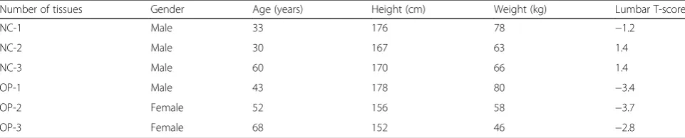
Table 1Characteristics of the study subjects
Number of tissues Gender Age (years) Height (cm) Weight (kg) Lumbar T-score
NC-1 Male 33 176 78 −1.2
NC-2 Male 30 167 63 1.4
NC-3 Male 60 170 66 1.4
OP-1 Male 43 178 80 −3.4
OP-2 Female 52 156 58 −3.7
Germany) and RNase-Free DNase Set (Cat#79254, QIA-GEN, GmBH, Germany).
RNA amplification and labeling
Total RNA was amplified and labeled by Low Input
Quick Amp Labeling Kit, One-Color (Cat.#5190–2305,
Agilent technologies, Santa Clara, CA, US), following
the manufacturer’s instructions. Labeled cRNA were
purified by RNeasy mini kit (Cat.# 74,106, QIAGEN, GmBH, Germany).
Each slide was hybridized with 1.65μg Cy3-labeled
cRNA using Gene Expression Hybridization Kit (Cat.#
5188–5242, Agilent technologies, Santa Clara, CA, US)
in Hybridization Oven (Cat.# G2545A, Agilent technolo-gies, Santa Clara, CA, US), according to the manufac-turer’s instructions. After 17 h hybridization, slides were washed in staining dishes (Cat.# 121, Thermo Shandon, Waltham, MA, US) with Gene Expression Wash Buffer
Kit (Cat.# 5188–5327, Agilent technologies, Santa Clara,
CA, US), followed the manufacturer’s instructions。.
Data acquisition
Slides were scanned by Agilent Microarray Scanner (Cat#G2565CA, Agilent technologies, Santa Clara, CA, US) with default settings, Dye channel: Green, Scan
resolution = 3μm, PMT 100%, 20bit. Data were
ex-tracted with Feature Extraction software 10.7 (Agilent technologies, Santa Clara, CA, US). Raw data were nor-malized by Quantile algorithm, limma packages in R. We slect a standard threshold set for differentially
expressed genes of a fold change≥2.0 and a
value≤0.05.
Gene ontology and Kyoto encyclopedia of genes and genomes pathway analyses
Gene Ontology (GO) analysis was applied to analyze the main function of the differential expression genes ac-cording to the GO database. Pathway analysis was used to find out the significant pathway of the differential genes according to KEGG.
lncRNA-mRNA coexpression networks
R function cor.test (a test for association/correlation
be-tween paired sam-ples) was utilized to compute
Pearson’s correlation coeficient to measure the gene
coexpression. The lncRNAs (fold change≥2.0, value≤0.01
and length 0,10,000),mRNAs (fold change≥4.0 and
value≤0.01) were choosed to draw the network by
Cytoscape.
According to these data, we built lncRNA-mRNA net-work using the correlation coefficients to examine inter-actions between lncRNA and mRNA. The value of “degree” in coexpression network indicated that one
mRNA/lncRNA might be correlated with several
lncRNAs/mRNAs.
qRT-PCR analysis
Total RNA was extracted and purified using standard methods (Life Technologies; RNA Easy, Qiagen, Valen-cia, CA,USA). Bestar qPCR RT Kit reverse transcription (Promega) was utilized to synthesize cDNA. 5 lncRNA
(fold change≥2.0, value≤0.01 and length 0,3000 ChrX)
expressions in sinonasal tissues were measured by qRT-PCR which was performed on the ABI7500 qPCR
system with the primer pairs listed in Table 2. The raw
quantifications were normalized to the beta-actin gene values for each sample and fold changes were shown as mean ± SD in three independent experiments, each in triplicate.
Statistical analysis
All data were expressed as the mean ± SD or proportions where appropriate. Expression levels skeletal muscle tis-sues from osteoporosis patients were analyzed by
paired-sample -tests. values < 0.05(two-tailed)
indi-cated statistical significance. The Statistical Program for Social Sciences (SPSS) 20.0 software was employed to perform all of the statistical analyses.
Results
Differentially expressed lncRNAs and mRNAs in osteoporosis
Volcano plots were used for assessing gene expression variation between osteoporosis and health patient groups. In total, 1594 lncRNAs displayed differential ex-pression in osteoporosis, including 482 upregulated lncRNAs and 1112 down regulated lncRNAs. Of 886 mRNAs that showed differential expression, 436 were upregulated and 450 were downregulated. Among them,
Table 2Real-time quantitative PCR primer sequences used in this study
Probe Name Forward primers (5′-3′) Reverse primers (5′-3′) Amplicon size (bp)
CUST_39447_PI430048170 GTTCCCGAGGTGTCCGA TCGTTAGTCGAGCTGAAACC 148
CUST_44695_PI430048170 CGGTGAGGTAGAAGGAGAAC TCTTCCTCAGCAGCACTTCC 159
CUST_73298_PI430048170 AATCCTTGGCATTGTCTGTAA AGCCAGTAGACGACACCAGC 149
CUST_108340_PI430048170 GAACAAAGGAGCAACAGCCC CCTGATAGGAATCTGCGAAAA 83
94 lncRNAs and 84 mRNAs were significantly upregu-lated, and 140 lncRNAs and 52 mRNAs were
signifi-cantly downregulated> 2-fold in osteoporosis.
Hierarchical clustering analysis showed systematic varia-tions in the expression of lncRNAs and mRNAs among samples. The data suggested that the expression of lncRNAs and mRNAs in osteoporosis differ from those
in health people controls (Fig. 1). As expected, the
lncRNA and mRNA expression proiles were distinguish-ing osteoporosis and normal tissue samples accurately based on the molecular signature.
Hierarchical clustering of the lncRNAs and mRNAs proile was performed using cluster 3.0.2; hierarchical clus-tering of the expression of the dysregulated lncRNAs and mRNAs based on centered Pearson correlation clearly separated osteoporosis tissues from corresponding normal
tissues (Fig. 1). Out of the group of RNAs that were
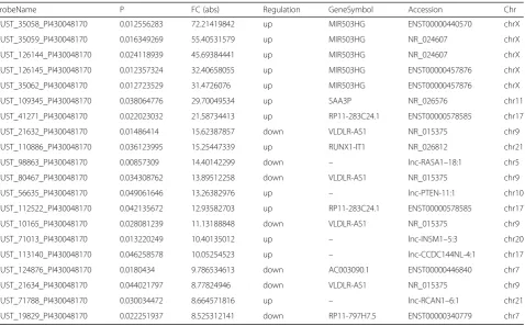
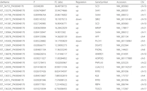
upregulated, lncRNA CUST_35058_PI430048170 and mRNA CUST_56753_PI430048170 showed the greatest
degree of demonstrated upregulation, with 72.214
19842-and 36.48758725-fold increases, respectively; of those that were downregulated, lncRNA CUST_216 32_PI430048170 and mRNA CUST_139172_PI430048170 demonstrated the greatest degree of downregulation, with 15.62387857-and 18.19379213-fold decreases, respectively (Tables3and4).
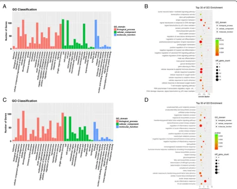
Functional analysis of differentially expressed genes Until now, the functions of most lncRNAs have not been well annotated. Therefore, by analyzing differentially expressed mRNAs, we can forecast the role that lncRNAs play in osteoporosis. The GO and KEGG path-way analyses of differentially expressed lncRNAs and mRNAs could provide a clue about the osteoporosis
disease process. We utilized all differentially expressed mRNAs for the GO analysis and found that the most enriched GO targeted by upregulated and downregulated transcripts were involved in anterior/posterior pattern specification, immune or metabolism system processes,
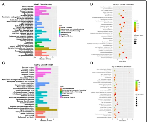
and immune response (Fig. 2). In the KEGG pathway
analysis, the down-and up-regulated mRNAs were found to be mostly enriched in muscle tissues, respectively
(Fig. 3). Many genes involved in muscle tissues
differen-tiation were dysregulated. A pathway network was con-structed using 20 of the most significantly enriched pathways to illustrate the critical pathways in the process of osteoporosis. The bone metabolism pathway were considered to be the most central functions in the net because the exchanges with other pathways strongly
depended on their existence (Fig.3).
Long non-coding RNA/mRNA coexpression network in osteoporosis
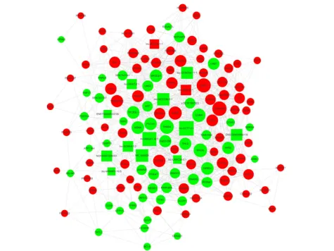
We constructed the lncRNA-mRNA coexpression net-work to identify the interactions between mRNAs and lncRNAs. Transcriptome regulation involves a huge network, among which many transcripts form a com-plex web to function. Coexpression networks facilitate
the intricate network based on gene screening
methods that can be used to identify candidate
biomarkers or therapeutic targets. The most crucial subnetwork was constructed by the transcripts with a
high k-score, which would be the core regulatory
modules of the entire coexpression network (Fig. 4).
What is more, the transcripts in this network are widely distributed in all chromosomes, indicating the widely interconnected regulation network between
lncRNAs and mRNAs (Fig. 4). This subnetwork
in-cludes four lncRNAs,lnc-CCT7–3:1,lnc-RASA1–
18:1,NR_046609,and lnc-SEC23B-2:1, constituting
probably the core of the network. The results implied that mRNA CDKN2B,CIDEC,and THBS4 may play key roles in osteoporosis process and development.
qRT-PCR validation
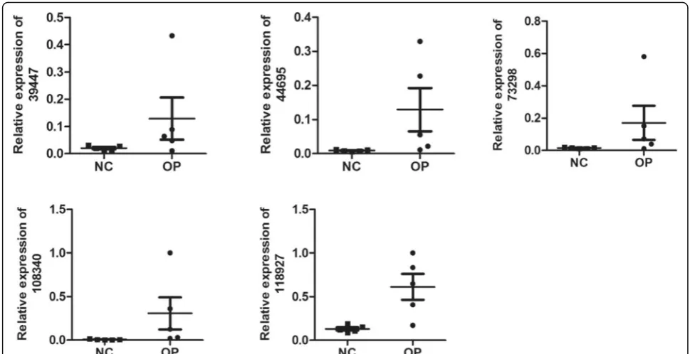
5 differentially expressed lncRNAs were randomly se-lected for validation by means of qRT-PCR according to
the manufacturer’s recommendations. CUST_108
340_PI430048170 was upregulated and CUST_39447_-
PI430048170,CUST_44695_PI430048170,CUST_73298_-PI430048170,and CUST_118927_PI430048170 were
downregulated in osteoporosis. The results of qRT-PCR were consistent with those of the microarray. All of the 5 lncRNAs were differentially expressed with the same
trend (up-or downregulated) (Fig.5).
Table 3Top 20 aberrantly expressed lncRNAs in microarray for 3 pairs of osteoporosis patients and health people skeletal muscle tissues
ProbeName P FC (abs) Regulation GeneSymbol Accession Chr
CUST_35058_PI430048170 0.012556283 72.21419842 up MIR503HG ENST00000440570 chrX
CUST_35059_PI430048170 0.016349269 55.40531579 up MIR503HG NR_024607 chrX
CUST_126144_PI430048170 0.024118939 45.69384441 up MIR503HG NR_024607 chrX
CUST_126145_PI430048170 0.012357324 32.40658055 up MIR503HG ENST00000457876 chrX
CUST_35062_PI430048170 0.012723529 31.4726076 up MIR503HG ENST00000457876 chrX
CUST_109345_PI430048170 0.038064776 29.70049534 up SAA3P NR_026576 chr11
CUST_41271_PI430048170 0.022023032 21.58734413 up RP11-283C24.1 ENST00000578585 chr17
CUST_21632_PI430048170 0.01486414 15.62387857 down VLDLR-AS1 NR_015375 chr9
CUST_110886_PI430048170 0.036123995 15.25447339 up RUNX1-IT1 NR_026812 chr21
CUST_98863_PI430048170 0.00857309 14.40142299 down – lnc-RASA1–18:1 chr5
CUST_80467_PI430048170 0.034308762 13.89512258 down VLDLR-AS1 NR_015375 chr9
CUST_56635_PI430048170 0.049061646 13.26382976 up – lnc-PTEN-11:1 chr10
CUST_112522_PI430048170 0.042135672 12.93582703 up RP11-283C24.1 ENST00000578585 chr17
CUST_10165_PI430048170 0.028081239 11.13188848 down VLDLR-AS1 NR_015375 chr9
CUST_71013_PI430048170 0.013220249 10.40135012 up – lnc-INSM1–5:3 chr20
CUST_113140_PI430048170 0.046258578 10.05254523 up – lnc-CCDC144NL-4:1 chr17
CUST_124876_PI430048170 0.0180434 9.786534613 down AC003090.1 ENST00000446840 chr7
CUST_21634_PI430048170 0.044021797 8.77824946 down VLDLR-AS1 NR_015375 chr9
CUST_71788_PI430048170 0.030034472 8.664571816 up – lnc-RCAN1–6:1 chr21
Discussion
It is well known that the mutation of genes and chromo-somes contribute to the pathogenesis of leukemia. How-ever, lncRNAs, the rising stars in biology, have just begun to be understood, and the majority of them have not yet been researched [14]. To provide some insights into the biological functions of lncRNAs in the patho-genesis of osteoporosis, we undertook a comprehensive analysis of lncRNA and mRNA profiling data from osteoporosis patients and health people, together with data from a public database. We identified the core lncRNAs and their functional annotations, and validated their expression by qRT-PCR. Overall, our work uncov-ered an interlaced transcripts network that is involved in osteoporosis development, in which lncRNAs play an in-dispensable role.
We explored the expression patterns of transcripts
be-tween osteoporosis patients and health people controls’
muscleskeletal. Microarray data identified vast lncRNAs and mRNAs, supporting an extensive involvement of lncRNAs in osteoporosis. There were two important concepts that ran throughout our studies to handle the mass data. First, we simplified the complex transcript network by modularization. The GO and KEGG pathway analyses divided mRNAs into several functional mod-ules, which are related to immunity, metabolism,
osteoclast differentiation, hematopoiesis, and TGF-beta signaling pathway, indicating the validity of the
micro-array [15]. Cytoscape was used to construct
co-expressed networks from the public and our micro-array data, respectively, and then the networks were fa-cilitated into several sub-networks through the k-score method. In the same way, lncRNAs were attributed their correlated functional mRNA modules through the co-expressed network and GO annotations. Overall, modularization contributes to simplifying the intricate
network into modules, which were like“big genes”.
Fur-ther analysis of public data showed that the lncRNA may regulate osteoclast differentiation, leading to the de-crease bone mass, cause to osteoporosis happened [16].
Finally, five lncRNAs,
CUST_39447_PI430048170,- CUST_44695_PI430048170,CUST_73298_PI430048170,-CUST_108340_PI430048170,and
CUST_118927_PI430048170 were confirmed as signifi-cantly differentially expressed in osteoporosis patients and healthy controls by qRT-PCR.
Our work clearly indicated an important role for lncRNAs in OP. However, many lncRNAs were excluded as they failed to be allocated to functional modules and have not been included in public data. It was difficult to originally understand the functions and targets of these lncRNAs, which may also play a key role in osteoporosis Table 4Top 20 aberrantly expressed mRNAs in microarray for 3 pairs of osteoporosis patients and health people skeletal muscle tissues
ProbeName P FC (abs) Regulation GeneSymbol Accession Chr
CUST_56753_PI430048170 0.0348289 36.48758725 up SCD NM_005063 chr10
CUST_133279_PI430048170 0.036740847 33.94274666 up SAA1 NM_000331 chr11
CUST_141790_PI430048170 0.009996764 20.86176002 up SAA4 NM_006512 chr11
CUST_139172_PI430048170 0.005145352 18.19379213 down SBK2 NM_001101401 chr19
CUST_141387_PI430048170 0.027240985 16.90436771 up SCD NM_005063 chr10
CUST_135342_PI430048170 0.003639094 16.28422029 up MRAP NM_178817 chr21
CUST_130964_PI430048170 0.004130947 14.9015582 up SAA4 NM_006512 chr11
CUST_138578_PI430048170 0.004133096 14.36030139 down AFP NM_001134 chr4
CUST_145430_PI430048170 0.005228882 14.17959829 down PLCH1 NM_001130961 chr3
CUST_132601_PI430048170 0.028566771 12.39092273 up DGAT2 NM_032564 chr11
CUST_128647_PI430048170 0.004831104 11.96333249 up PXDNL NM_144651 chr8
CUST_136078_PI430048170 0.046626016 11.41446117 up ANKRD1 NM_014391 chr10
CUST_139601_PI430048170 0.030211027 11.00284852 up ADIPOQ NM_001177800 chr3
CUST_142975_PI430048170 0.017278415 10.02050967 up PNPLA3 NM_025225 chr22
CUST_126930_PI430048170 0.014106518 8.930113104 up LGALS12 NM_001142537 chr11
CUST_142712_PI430048170 0.026107426 8.824194597 up RYR3 NM_001036 chr15
CUST_130312_PI430048170 0.004510607 7.885502819 up KLB NM_175737 chr4
CUST_52015_PI430048170 0.039281946 7.576008123 up RYR3 NM_001036 chr15
CUST_141047_PI430048170 0.009777851 7.231495622 up RBP4 NM_006744 chr10
[17]. In addition, our analysis showed that lncRNA lnc-CCT7–3:1 was highly correlated with osteoporosis, its neighboring gene. The lncRNA may be directly or in-directly correlated with osteoporosis and there may be additional transcripts involved in the lncRNA-associated
biological process [18]. The lncRNA’s biological
func-tions need to be validated further.
The last decades witnessed the discovery of biological functions for non-coding RNA, which triggered the rec-ognition that RNA is not only a simple hinge of the cen-tral dogma but also directly takes part in the regulation of biological networks [19]. With the development of next-generation sequencing, especially in terms of depth and scale, significant data has been accumulated. We have to recognize the system is so complex that it is
beyond the initial recognition. Fortunately, the progress of methodology simplifies the networks. Through modu-larization, thousands of transcripts can be facilitated into
several “big genes,” and then the core lncRNAs of each
module can be researched in details. Moreover, the ac-tive application of accumulated public data will help us to make the functions of lncRNAs more clear. Hopefully, this study can provide a reference for the broad analysis of OP data.
Skeletal muscle possesses a remarkable ability to adapt to various physiologic conditions [20]. AMPK is a sensor of intracellular energy status that maintains energy stores by fine-tuning anabolic and catabolic pathways [21]. At the same time, skeletal muscle has been shown to be important for regulating whole-body metabolism.
Skeletal muscle demonstrates high malleability and can adapt its contractile composition and metabolic proper-ties in response to a number of physiologic conditions [22]. However, bone mass and skeletal muscle mass are controlled by factors such as genetics, diet and nutrition, growth factors and mechanical stimuli. Whereas in-creased mechanical loading of the musculoskeletal sys-tem stimulates an increase in the mass and strength of skeletal muscle and bone, reduced mechanical loading and disuse rapidly promote a decreasein musculoskeletal mass, strength and ultimately performance [26]. More-over, skeletal muscle atrophy often occursinparallel tobo-nelossand activity; musclemassand strength influence bone mass [23]. However, because the mechanisms that
regulate mechanotransduction in bone and muscle are complex, it is not entirely clear which specific mecha-nisms operate in synergy during musculoskeletal disuse [24]. Treatment options for muscle disuse atrophy and osteoporosis are currently limited, with rehabilitative strength training proving to be the most effective method to restore musculoskeletal mass; however, long periods of rehabilitation are necessary to restore muscle, bone and locomotor performance following artificial im-mobilisation in non-hibernating mammals [25]. Mus-cules and bones are the main part of the motor system, regulating processes such as endocrine and metabolism [26]. IGF-1 in muscle tissue promotes bone development by improving muscle mass and strength, and also
Fig. 3Kyoto Encyclopedia of Genes and Genomes (KEGG) pathway analyses. The KEGG pathway is a collection of manually drawn signal pathway maps and provides a valuable tool for mapping a specific gene to its corresponding pathway.aandc, KEGG classification of Cellular Processes, Environmental Information Processing, Genetic Information Processing, Human Diseases, Metabolism and Organismal Systems of lncRNAs,mRNAs.
directly promotes bone development, increasing bone mineral content and bone density [27]. Myostatin is most abundantly expressed in skeletal muscle tissue, in-hibits muscle growth and regeneration, and promotes
osteoclast differentiation, inhibiting TNF-α-induced
osteoclast differentiation [28]. Indian Hedgehog (Ihh), a signaling molecule secreted by chondrocytes and osteo-blasts, promotes muscle growth. Ihh in bone can directly promote muscle growth [29]. Osteocalcin (Glu-OC) par-tially repairs muscles with impaired function. The gap junction protein Connexin43 in bone cells directly regu-lates muscle growth and function [30]. The close con-nection between bone and muscle is not only related to the mechanics of its interaction, but also to the complex and precise endocrine regulation and some common molecular signaling pathways [31]. The new recent study that significant differences in the expression of lncRNAs,
mRNAs, circRNAs, and miRNAs between postmeno-pausal OP patients and healthy controls, and the func-tions of the RNAs were also identified based on our RNA-seq analysis [32]. Taken together, our study indi-cated that lncRNAs and mRNA could associate with the occurrence of OP and may be as possible biomarkers and target genes in OP.
Conclusions
Insummary, to our knowledge, our study analysis of lncRNA expression profile in osteoporosis. The results show that genes regulated by these lncRNAs are in-volved in TGF-beta signaling pathways as a proof of principle, or influence osteoclast differentiation. This may offer new insights into pathogenesis and could be a promising way to dissect the molecular pathogenesis of this refractory bone metabolism desease. Our study lays
the foundation for further investigation of this disease. Further large scale studies are warranted to provide con-vincing evidence for clarifying the functions of lncRNAs in osteoporosis and determining whether these lncRNAs can serve as new diagnostic biomarkers, prognostic fac-tors for survival, and therapeutic targets in osteoporosis.
Abbreviations
GO:Gene Ontology; KEGG: Kyoto Encyclopedia of Genes and Genomes; LncRNA: Long non-coding RNA; OP: Osteoporosis; qRT-PCR: Quantitative polymerase chain reaction
Acknowledgements
Not applicable.
Funding
This study was supported by Grants from the National Natural Science Foundation of China (81674004;81673786).
Availability of data and materials
All data generated or analysed during this study are included in this published article.
Authors’contributions
LW and HH conceived and designed the experiments; SL, SC and JH performed the experiments; HW and SL performed data analysis; SL and HW contributed to sample collection; SL wrote the paper; LW assisted with writing and proofreading. All authors read and approved the final manuscript.
Ethics approval and consent to participate
This study was approved by the Internal Review and the Ethics Boards of Guangzhou University of Chinese Medicine and The Third Affiliated Hospital, Guangzhou University of Chinese Medicine. Informed written consent was obtained from all study subjects.
Consent for publication
Not applicable.
Competing interests
The authors declare that they have no competing interests.
Publisher’s Note
Springer Nature remains neutral with regard to jurisdictional claims in published maps and institutional affiliations.
Author details
1Guangzhou University of Chinese Medicine, Guangzhou 510405, China. 2Department of Orthopaedics, The Third Affiliated Hospital, Guangzhou University of Chinese Medicine, Guangzhou 510378, China.3Lingnan Medical Research Center of Guangzhou University of Chinese Medicine, Guangzhou University of Chinese Medicine, Guangzhou 510405, China.
Received: 31 January 2019 Accepted: 15 May 2019
References
1. Cauley JA. Osteoporosis: fracture epidemiology update 2016. Curr Opin Rheumatol. 2017;29:150–6.
2. Tong X, Gu P-c, Xu S-z, Lin X-j. Long non-coding RNA-DANCR in human circulating monocytes: a potential biomarker associated with
postmenopausal osteoporosis. Biosci Biotechnol Biochem. 2015;79(5):732–7. 3. Picca A, Calvani R, et al. Bone-muscle crosstalk: unraveling new therapeutic
targets for osteoporosis. Curr Pharm Des. 2017;23:1–8.
4. Ferrucci L, Baroni M, Ranchelli A, et al. Interaction between bone and muscle in older persons with mobility limitations. Curr Pharm Des. 2014;20: 3178–97.
5. Goodman CA, Hornberger TA, Robling AG. Bone and skeletal muscle: key players in mechanotransduction and potential overlapping mechanisms. Bone. 2015;80:24–36.
6. Reilly BD, Franklin CE. Prevention of muscle wasting and osteoporosis: the value of examining novel animal models. J Exp Biol. 2016;219:2582–95.
Fig. 5qRT-PCR validation. qRT-PCR verification of 5 candidate lncRNAs in 5 pairs of OP and NC group tissue. Expression of pediatric OP samples vs. control samples was analyzed using qRT-PCR, and summarized as mean average ± standard error (SE).P< 0.05 was considered
7. Brooks NE, Myburgh KH. Skeletal muscle wasting with disuse atrophy is multi-dimensional: the response and interaction of myonuclei, satellite cells and signaling pathways. Front Physiol. 2014;5:99.
8. Cao J. The functional role of long non-coding RNAs and epigenetics. Biol Proced Online. 2014;16:11.
9. Li G, Zhang H, Wan X, Yang X, Zhu C, Wang A, He L, Miao R,Chen S and Zhao H: Long noncoding RNA plays a key role in metastasis and prognosis of hepatocellular carcinoma. Biomed Res Int 2014: 780521, 2014. 10. McMullen JR, Drew BG. Long non-coding RNAs (lncRNAs) in skeletal and
cardiac muscle: potential therapeutic and diagnostic targets? Clin Sci. 2016; 130:2245–56.
11. Kashi K, Henderson L, Bonetti A, Carninci P. Discovery and functional analysis of lncRNAs: methodologies to investigate an uncharacterized transcriptome. Biochim Biophys Acta. 2016;1859:3–15.
12. Beermann J, Piccoli MT, Viereck J, Thum T. Non-coding RNAs in development and disease: background, mechanisms, and therapeutic approaches. Physiol Rev. 2016;96:1297–325.
13. Nelson BR, Makarewich CA, Anderson DM, Winders BR, Troupes CD, Wu F, Reese AL, McAnally JR, Chen X, Kavalali ET, et al. A peptide encoded by a transcript annotated as long noncoding RNA enhances SERCA activity in muscle. Science. 2016;351:271–5.
14. Tong X, Gu PC, Xu SZ, Lin XJ. Long non-coding RNA-DANCR in human circulating monocytes:a potential biomarker associated with post-menopausal osteoporosis. Biosci Biotechnol Biochem. 2015;79:732–7. 15. Mao X, Cai T, Olyarchuk JG, Wei L. Automated genome annotation and
pathway identification using the KEGG Orthology (KO) as a controlled vocabulary. Bioinformatics. 2005;21:3787–93.
16. Jin C, Zheng Y, Huang Y, Liu Y, Jia L, Zhou Y. Long non-coding RNA MIAT knockdown promotes osteogenic differentiation of human adipose-derived stem cells. Cell Biol Int. 2017;41:33–41.
17. Gao KT, Lian D. Long non-coding RNA MALAT1is an independent prognostic factor of osteosarcoma. Eur Rev Med Pharmacol Sci. 2016;20:3561–5. 18. Bhan A, Mandal SS. Long noncoding RNAs: emerging stars in gene regulation,
epigenetics and human disease. ChemMed- Chem. 2014;9:1932–56. 19. Prabhakar B, Zhong XB, Rasmussen TP. Exploiting long noncoding RNAs as
pharmacological targets to modulate epigenetic diseases. Yale J Biol Med. 2017;90:73–86.
20. Egan B, Zierath JR. Exercise metabolism and the molecular regulation of skeletal muscle adaptation. Cell Metab. 2013;17:162–84.
21. Gowans GJ, Hardie DG. AMPK: a cellular energy sensor primarily regulated by AMP. Biochem Soc Trans. 2014;42:71–5.
22. Anderson DM, et al. A micropeptide encoded by a putative long noncoding RNA regulates muscle performance. Cell. 2015;160:595–606.
23. Swift JM, Nilsson MI, Hogan HA, Sumner LR, Bloomfield SA. Simulated resistance training during hindlimb unloading abolishes disuse bone loss and maintains muscle strength. J Bone Miner Res. 2010;25:564–74. 24. Goodman CA. The role of mTORC1 in regulating protein synthesis and
skeletal muscle mass in response to various mechanical stimuli. Rev Physiol Biochem Pharmacol. 2014;166:43–95.
25. Cotton CJ. Skeletal muscle mass and composition during mammalian hibernation. J Exp Biol. 2016;219:226–34.
26. Dallas SL, Prideaux M, Bonewald LF. The osteocyte:an endocrine cell and more. Endocr Rev. 2013;34:658–90.
27. Banu J, Wang L, Kalu DN. Effects of increased muscle mass on bone in male mice overexpressing IGFI in skeletal muscles. In: Calcif tissue Int, 2008, 73. p. 196–201.
28. Dankbar B, Fennen M, Brunert D, et al. Myostatin is a direct regulator of osteoclast differentiation and its inhibition reduces inflammatory joint destruction in mice. Nat Med. 2015;21:1085–90.
29. Bren-Mattison Y, Hausburg M, Olwin BB. Growth of limb muscle is dependent on skeletal-derived Indian hedgehog. Dev Biol. 2011;356:486–95. 30. Shen H, Grimston S, Civitelli R, et al. Deletion of connexin43 in osteoblasts
/osteocytes leads to impaired muscle formation in mice. J Bone Miner Res. 2015;30:596–605.
31. Borsheim E, Herndon DN, Hawkins HK, et al. Pamidronate attenuates muscle loss after pediatric burn injury. J Bone Miner Res. 2014;29:1369–72. 32. Jin D, Wu X, Yu H, et al. Systematic analysis of lncRNAs, mRNAs, circRNAs