RESEARCH
Production of fluorescent and cytotoxic
K28 killer toxin variants through high cell
density fermentation of recombinant
Pichia
pastoris
Esther Giesselmann, Björn Becker and Manfred J. Schmitt
*Abstract
Background: Virus infected killer strains of the baker’s yeast Saccharomyces cerevisiae secrete protein toxins such as K28, K1, K2 and Klus which are lethal to sensitive yeast strains of the same or related species. K28 is somewhat unique as it represents an α/β heterodimeric protein of the A/B toxin family which, after having bound to the surface of sensi-tive target cells, is taken up by receptor-mediated endocytosis and transported through the secretory pathway in a retrograde manner. While the current knowledge on yeast killer toxins is largely based on genetic screens for yeast mutants with altered toxin sensitivity, in vivo imaging of cell surface binding and intracellular toxin transport is still largely hampered by a lack of fluorescently labelled and biologically active killer toxin variants.
Results: In this study, we succeeded for the first time in the heterologous K28 preprotoxin expression and produc-tion of fluorescent K28 variants in Pichia pastoris. Recombinant P. pastoris GS115 cells were shown to successfully process and secrete K28 variants fused to mCherry or mTFP by high cell density fermentation. The fluorescent K28 derivatives were obtained in high yield and possessed in vivo toxicity and specificity against sensitive yeast cells. In cell binding studies the resulting K28 variants caused strong fluorescence signals at the cell periphery due to toxin binding to primary K28 receptors within the yeast cell wall. Thereby, the β-subunit of K28 was confirmed to be the sole component required and sufficient for K28 cell wall binding.
Conclusion: Successful production of fluorescent killer toxin variants of S. cerevisiae by high cell density fermentation of recombinant, K28 expressing strains of P. pastoris now opens the possibility to study and monitor killer toxin cell surface binding, in particular in toxin resistant yeast mutants in which toxin resistance is caused by defects in toxin binding due to alterations in cell wall structure and composition. This novel approach might be easily transferable to other killer toxins from different yeast species and genera. Furthermore, the fluorescent toxin variants described here might likewise represent a powerful tool in future studies to visualize intracellular A/B toxin trafficking with the help of high resolution single molecule imaging techniques.
Keywords: High cell density fermentation, Fluorescence labelling, Heterologous protein expression, Killer toxin, Pichia pastoris, Saccharomyces cerevisiae, A/B toxins
© The Author(s) 2017. This article is distributed under the terms of the Creative Commons Attribution 4.0 International License (http://creativecommons.org/licenses/by/4.0/), which permits unrestricted use, distribution, and reproduction in any medium, provided you give appropriate credit to the original author(s) and the source, provide a link to the Creative Commons license, and indicate if changes were made. The Creative Commons Public Domain Dedication waiver (http://creativecommons.org/ publicdomain/zero/1.0/) applies to the data made available in this article, unless otherwise stated.
Background
The killer phenotype of virus infected Saccharomyces cerevisiae strains is elicited by the secretion of antifungal
killer toxins which are able to kill sensitive strains of various yeast and fungal species [1]. Due to an intrinsic mechanism of toxin immunity, killer strains are effec-tively protected against their own toxin and, thereby, possess a growth advantage towards non-killer strains [2, 3]. The vast majority of killer toxins in S. cerevisiae
is encoded by cytoplasmic dsRNA viruses [3, 4]. In case
Open Access
*Correspondence: [email protected]
of K28, the primary gene product of the K28 encoding dsRNA is a preprotoxin whose intracellular processing and maturation within the secretory pathway is mecha-nistically similar to prepro-α-factor processing in yeast and pro-hormone conversion in higher eukaryotes [3,
5–8].
Maturation of K28 from its precursor resembles a multi-step process initiated by posttranslational import into the lumen of the endoplasmic reticulum (ER) and subsequent removal of the N-terminal signal peptide by signal peptidase cleavage at the ER membrane. Fur-ther proteolytic preprotoxin processing in the late Golgi catalysed by the activities of Kex2p and Kex1p results in the formation and final secretion of a disulphide-bonded α/β heterodimeric protein toxin whose β-subunit carries a carboxyterminal ER retention motif (HDEL) which is essential for host cell intoxication and intracellular toxin transport [5, 8, 9].
Internalization of K28 by sensitive yeast cells is real-ized in a two step mechanism: while α-1,3-linked cell wall mannoproteins are used as primary K28 binding sites at the outer yeast cell surface, the secondary plasma mem-brane receptor of K28 has recently been identified as the HDEL-receptor Erd2p [10, 11] which ensures endo-cytotic toxin uptake and retrograde transport through the secretory pathway [9]. After toxin retro-transloca-tion from the ER into the cytosol, the β-subunit of K28 becomes ubiquitylated and proteasomally degraded while α enters the nucleus and causes final cell death [12–15]. Since the dimeric α/β structure is characteristic for A/B toxin family members including clinically relevant repre-sentatives like cholera, anthrax and Shiga toxin, K28 rep-resents an attractive model to study A/B toxin trafficking in yeast [16, 17].
In the last decades, mammalian cells have been inten-sively used to study the mode of intoxification, uptake and intracellular transport of A/B toxins through live cell imaging techniques. To avoid procedures requiring cell fixation and permeabilization (e.g. immunostaining), fluorescently labelled toxins were used to analyze the dynamics of toxin transport in living cells. In this respect, the toxin subunit of interest is either coupled with a fluo-rophore or fused to a fluorescent protein to microscopi-cally track toxin uptake and intracellular trafficking in real-time [18–20]. In contrast to A/B toxins that pene-trate and kill mammalian cells, the respective knowledge on yeast killer toxins is mostly based on genetic screens for mutants with altered toxin sensitivity [21] since fluo-rescent killer toxin variants for live cell imaging are still lacking due to the pronounced sensitivity of yeast killer toxins to pH changes or fusions to its cytotoxic subu-nits. In the present study, we used the methylotrophic yeast Pichia pastoris as platform for the expression and
production of fluorescent variants of killer toxin K28. Tightly controlled cultivation conditions during P. pas-toris fermentation in a bioreactor led to the secretion of high yields of fluorescent and toxic K28 variants. Further toxin binding studies on sensitive yeast cells likewise con-strained that the β-subunit is exclusively responsible for cell wall binding of the fluorescent K28 variants.
Results and discussion
Construction and expression of fluorescent K28 variants in P. pastoris
Fluorescence labelling of proteins and protein toxins is usually achieved by chemically coupling with organic fluorophores or quantum dots [22]. Although the chol-era toxin B-subunit was successfully labelled with Cy3 and used for in vivo tracking of its retrograde trans-port in Vero cells [23], this technique is inappropriate for the viral toxin K28 since the low pH and/or reduc-ing redox milieu durreduc-ing the chemical couplreduc-ing reaction would immediately inactivate the pH and redox sensi-tive K28 toxin (Suzuki et al. [32]). Furthermore, since the α-subunit of K28 is highly sensitive to any addition of a tag, we followed another approach and inserted the DNA sequence of the fluorescent protein mCherry between the β-subunit of K28 and its C-terminal HDELR motif. In addition, the native K28 signal sequence was replaced by the α mating factor signal peptide encoded by the expression vector pPIC9 to ensure efficient ER import of the toxin precursor in P. pastoris [24, 25]. As illustrated in Fig. 1a, the resulting toxin chimera is predicted to be composed of K28α covalently coupled to β-mCherryHDEL
via a single inter-chain disulphide bond.
For in vivo expression of the chimeric K28 variant, the methylotropic yeast P. pastoris was used as host to ensure proper posttranslational toxin processing and folding in the eukaryotic secretory pathway, reflecting the natural situation of toxin secretion in K28 killer strains of S. cer-evisiae [26]. Since previous studies already demonstrated that P. pastoris is a suitable host for the expression of biologically active wild-type K28 toxin [27], we checked the in vivo processing of the chimeric toxin by analysing the cell-free culture supernatant of recombinant P. pas-toris GS115 [pPIC9 K28-mCherryHDEL] by SDS-PAGE
and Western blotting (Fig. 1b). Electrophoretic mobility of K28-mCherryHDEL is in agreement with the calculated
size of the K28 chimera. The observed mobility down-shift to approximately 11 kDa under reducing conditions is due to a loss of the α-subunit caused by reduction of the disulphide bond which joins α and β-mCherryHDEL.
of a concentrated culture supernatant sample under non-reducing conditions (Fig. 1c). In this study, mCherry was selected because of its insensitivity towards C- and N-terminal fusions and its high acid-stability [28]. In par-ticular, this latter property is of special importance due to the narrow intrinsic pH-optimum of K28 (pH 4.7). Interaction between the secondary toxin receptor Erd2p and the C-terminal HDEL retention motif of K28 only occurs in a mildly acidic pH milieu [11, 29, 30]. The pro-nounced pH-sensitivity of K28 is probably responsible for the observed lack of killer activity after cultivation of
P. pastoris in shake flasks for toxin production (data not shown) as no buffer system was capable to stabilize an appropriate pH during longer fermentation periods.
Fermentation of P. pastoris yields in fluorescent and toxic K28 variants
High yields in recombinant protein production after cultivation of P. pastoris in shake flasks proved difficult due to limitations in culture volume, oxygen transfer, substrate addition and monitoring [31]. Especially to maintain a constant acidic pH to prevent K28 oligomer formation and inactivation [32], fermentation is the bet-ter choice for killer toxin production. Due to the dra-matic reduction of K28 killer activity at pH 7.0 and the inadequate fluorescence intensity of mCherry at pH 4.7 (data not shown), medium and culture pH was adjusted to pH 5.3 during the fermentation process. This pH value represents a suitable compromise between efficient
a
b c
Fig. 1 a Schematic structure of K28-mCherryHDEL. The K28 chimera contains an N-terminal signal peptide derived from the α-mating factor which is
removed by signal peptidase (SP) cleavage after ER import. In the cis-Golgi, endopeptidase Kex2p cleavage removes the pro- and γ-sequence which leads to the separation of α and β. Both subunits remain connected by a single disulphide which was previously formed in the ER. Carboxypepti-dase Kex1p removes the C-terminal arginine residue in a trans-Golgi compartment and, thereby, unmasks the HDEL motif. b Western blot analysis of secreted K28-mCherryHDEL from P. pastoris. Cell-free culture supernatants of the recombinant strain GS115 [pPIC9 K28-mCherryHDEL] and the negative
control strain GS115 [pPIC9] grown for 120 h in BMM and ×125 concentrated by centrifugal concentrators were separated under non-reducing (left) and reducing (right) conditions by SDS-PAGE and probed with an anti-DsRed antibody. Position and size of the correctly processed toxin is indicated. c Detection of fluorescent K28-mCherryHDEL. GS115 [pPIC9 K28-mCherryHDEL] was grown for 120 h in BMM medium (pH 7) in the presence
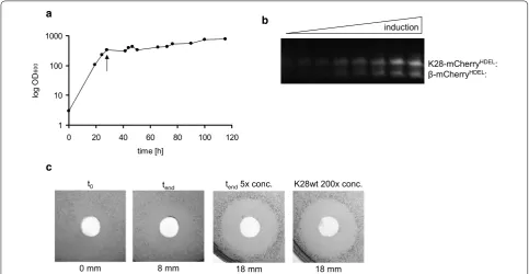
in vivo K28 killing activity and sufficient mCherry fluo-rescence. As soon as the methanol-induced toxin expres-sion started, cultivation temperature was lowered to 20 °C, in turn resembling the temperature optimum for killer toxin production. Usually, a moderate decrease in cultivation temperature can improve heterologous pro-tein production, secretion and folding of fluorescent proteins [33, 34]. Yeast cell growth was controlled by measuring the optical density which achieved values of OD600 > 700 (Fig. 2a). To avoid high oxygen
concentra-tions harming K28 production, methanol feedings were reduced as far as possible. As methanol is required to induce protein expression, culture pO2 could only be
maintained without supplying extra oxygen by regulating the stirring rate during methanol feeding. High oxygen concentrations have been described to induce cell stress and, thereby, to cause the release of proteases, whereas hypoxic conditions positively affect recombinant protein secretion [35–37]. In addition, precisely adjusted metha-nol feedings during fermentation, which is not possible in shake flask cultures, prevent fluctuations in methanol concentrations [38].
As shown in Fig. 2b, fluorescence of the fermen-tation product K28-mCherryHDEL was immediately
detectable after SDS-PAGE of the cell-free culture super-natant. Again, the heterodimeric K28-mCherryHDEL
fusion and the β-mCherryHDEL variant were detectable
during fermentation as fluorescent bands under UV illu-mination, resulting from reductive cleavage of the inter-chain connecting disulphide. In contrast to shaking flask experiments (data not shown), the optimized culture conditions for killer toxin production led to K28 variants with in vivo killing activity (Fig. 2c). In the time course of induction of K28 expression, the culture supernatant showed increasing killer activity against a sensitive yeast tester strain in an agar diffusion assay. The fivefold fer-menter concentrate caused the same growth inhibition zone as a 200× concentrated cell-free culture superna-tant of a K28 wild-type killer strain of S. cerevisiae [39], emphasizing the efficiency of bioactive killer toxin pro-duction by P. pastoris fermentation. Furthermore, this is the first time that a biologically active and fluorescently labelled killer toxin has been expressed and secreted.
Besides mCherry, we also introduced an alternative fluorescent protein that can be easily quenched for future studies. mTFP (monomeric teal fluorescent protein) rep-resents one of the brightest and most photo-stable fluo-rescent proteins which is likewise less pH sensitive [40].
a b
c
Fig. 2 a Growth curve of the recombinant P. pastoris strain GS115 [pPIC9 K28-mCherryHDEL] during fermentation. The starting point of methanol
feeding (K28 expression induction) is marked by an arrow. b Fluorescence development of mCherry-tagged K28. Cell-free culture supernatant samples from different fermentation time points were separated by SDS-PAGE under non-reducing conditions. Fusion proteins were detected by UV illumination. Position and size of the fusion proteins are indicated. c Killer activity of the fermentation product K28-mCherryHDEL. Cell-free
culture supernatants were tested against the sensitive S.cerevisiae strain 192.2d in an agar diffusion assay (t0= starting point of methanol feeding;
tend= 120 h post methanol induction). Cell-free zones of growth inhibition were determined and killer activity was compared to a ×200
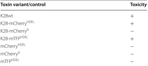
By using identical fermentation conditions we were able to successfully express a biologically active and fluo-rescent K28-mTFPHDEL fusion as well as mTFPHDEL in P. pastoris. To elucidate the influence of the fluorescent proteins and the HDEL-signal on killer activity, addi-tional controls including K28-mCherryΔ, mCherryHDEL
as well as mCherryΔ were likewise expressed and their
in vivo killing ability was tested in toxin activity assays (Table 1). As expected, only wild-type K28 as well as toxin chimera composed of the K28 α/β heterodimer with its C-terminal HDEL-motif showed killer activity. These results also nicely confirm that the α moiety rep-resents the cytotoxic polypeptide of K28 responsible for killing a sensitive target cell [12]. In contrast to α, the C-terminal HDEL-signal is required for toxin interaction with the HDEL-receptor Erd2p at the plasma membrane level, subsequently ensuring endocytotic uptake and ret-rograde transport of the toxin to the ER [9, 11]. There-fore, K28 lacking a HDEL sequence is neither toxic nor capable to enter sensitive target cells [9]. As summarized in Table 1, the absence of either K28α or the ER targeting motif HDEL prevents efficient killing.
β‑subunit of K28 mediates efficient toxin binding to sensitive yeast cells
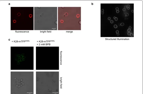
As fluorescently labelled K28 variants possess killer activity (Table 1), the toxin chimera should be taken up by yeast cells and reach the nucleus to finally kill. Con-sequently, incubation of intact S. cerevisiae cells with K28-mCherryHDEL led to an intensive fluorescence of
the cell periphery (Fig. 3a). Despite extensive variations of the experimental conditions, including changes of the duration, temperature and buffer composition during cell treatment, no intracellular signals were detectable (data not shown). Even by using high-resolution structured illumination microscopy [41] to confirm the fluorescence pattern of K28-mCherryHDEL treated cells of the
hyper-sensitive strain 192.2d [21, 42], only showed strong fluo-rescence signals around the outer cell periphery (Fig. 3b).
To remove this intensive cell surface signal, we tried to quench the extracellular K28-mTFPHDEL fluorescence
by using the plasma membrane impermeable quencher bromophenol blue (BPB). BPB is known to efficiently quench the emission of EGFP-fusions. The emission maxima of EGFP and mTFP rarely diverge so that mTFP fluorescence can likewise be quenched by BPB. Conse-quently, the intracellular killer toxin fraction should be visualized under these conditions. However, as illustrated in Fig. 3a, c, the fluorescence pattern of K28-mTFPHDEL
or K28-mCherryHDEL treated cells was not significantly
different. BPB addition to cells pre-incubated with K28-mTFPHDEL caused a complete quenching of mTFP
fluorescence, indicating that the strong fluorescence observed at the cell periphery is due to K28 binding to the cell wall and not derived from endocytosed intracel-lular K28 molecules.
The lack of intracellular toxin signals after incubation of sensitive yeast cells with fluorescent K28 variants can somehow be explained by the extremely low amount of toxin molecules that are internalized in vivo. Visu-alization of compartmental A/B toxin trafficking is even complicated by the low toxin quantity in certain com-partments such that toxin visualization by microscopy is often misleading [18]. Based on the high biological activ-ity of A/B toxins, only a few molecules of protein tox-ins such as ricin or diphtheria toxin are required to kill a cell [43, 44]. A crude estimation for the in vivo toxic-ity of yeast killer toxin K1 is based on experiments with radioactive labelled K1 in which it was determined that approximately 3 × 10⁴ molecules are required to kill
a single cell [45]. However, no information exists about the number of K28 molecules that is needed to reach the nucleus to mediate cell death.
Interestingly, even treatment with the metabolic inhibi-tor azide, which is known to prevent in vivo cell killing by blocking the energy-dependent K28 internalization step (unpublished data and [39]), did not alter the fluores-cence pattern of K28-mCherryHDEL treated cells (data not
shown). It, therefore, can be concluded that the observed fluorescence is caused by an energy-independent cell wall binding process of K28. This assumption is further fortified by comparison of different fluorescent K28 derivatives (Table 2). A non-toxic fluorescent K28 vari-ant without a C-terminal HDEL-motif (K28-mCherryΔ)
caused the same fluorescence pattern as
K28-mCherry-HDEL. As the HDEL-peptide is described to be only
cru-cial for Erd2p-mediated endocytosis and retrograde toxin trafficking [3, 5, 9, 11], the observed K28-mCherryΔ
fluo-rescence in the cell periphery simply arises from energy-independent binding to the outer yeast cell wall.
To exclude that fluorescent proteins somehow alter the cell wall binding behaviour of K28, yeast cells were
Table 1 Killer activity of K28 variants produced by P.
pas-toris fermentation
K28 variants which show killing activity in an agar diffusion assay on methylene blue agar plates in form of a zone of growth inhibition (mm) are marked with “+”, whereas the absence of a killing zone is displayed by “−”
Toxin variant/control Toxicity
K28wt +
K28-mCherryHDEL +
K28-mCherryΔ −
K28-mTFPHDEL +
mCherryHDEL −
mCherryΔ −
analyzed after treatment with fluorescent proteins either containing or lacking the motif. Again, the HDEL-motif had no effect on cell binding. Furthermore, fluo-rescent proteins without any K28 moiety were not able to evoke fluorescence signals at the yeast cell periphery (Table 2). To further substantiate that the β-subunit of K28 is responsible for cell binding, yeast cells were incu-bated with the β-subunit of K28 fused to mCherryHDEL.
Incubation with β-mCherryHDEL indeed caused a strong
peripheric fluorescence comparable to the fluorescence pattern of K28α/β variants, confirming the β subunit as sole cell binding component of K28.
Conclusions
Taken together, our data demonstrate that the viral killer toxin K28 can be fluorescently labelled by fusion with flu-orescent proteins such as mCherry or mTFP and heterol-ogously expressed in the methylotrophic yeast P. pastoris
resulting in high-level production and secretion of fluo-rescent and bioactive toxin chimera which show strong killing activity against sensitive yeast cells. This strategy might be transferable to other yeast killer toxins and used as tool for live cell imaging and high resolution micros-copy to analyse cell surface binding. Although no intra-cellular K28 signals were visible in this study, it might be possible to visualize intracellular toxin trafficking in the future through high resolution single molecule fluores-cence microscopy.
a b
c
Fig. 3 a Fluorescence distribution of S. cerevisiae cells after incubation with K28-mCherryHDEL. S. cerevisiae BY4742 was washed twice after 2 h
incu-bation with K28-mCherryHDEL and analyzed by confocal laser scanning microscopy. b Same cells analysed by structured illumination microscopy
(SIM). The hypersensitive yeast strain 192.2d was treated with K28-mCherryHDEL as described in a and subjected to SIM. (c) Fluorescence quenching
was performed on cells of S. cerevisiae BY4742 after incubation with K28-mTFPHDEL as described in a, however extracellular mTFP fluorescence was
subsequently quenched by the addition of 2 mM bromophenol blue (BPB)
Table 2 Cell wall binding properties of various fluorescent K28 variants
FP fluorescent protein, mCherry or mTFP
K28 derivates and
control proteins Cell binding/ periphery fluorescence
α/β-FPHDEL +
α/β-FPΔ +
β-FPHDEL +
FPHDEL −
Methods
Yeast and E. coli strains, culture media and conditions
Pichia pastoris GS115 (his4, ThermoScientific, Waltham, Massachusetts, USA) was selected for heterol-ogous protein expression. For selection of positive trans-formed clones, cells were cultured in glucose containing histidine d/o medium [27]. For protein expression, cells were initially grown in buffered minimal glycerol medium for biomass production (BMG, 100 mM potas-sium phosphate (pH 6.0), 0.34% YNB, 1% (NH4)2SO4,
0.00004% biotin, 1% glycerol), followed by minimal meth-anol medium (BMM, 100 mM potassium phosphate (pH 6.0 or pH 7), 0.34% YNB, 1% (NH4)2SO4, 0.00004% biotin,
0.5% methanol) for induction of gene expression. Cul-tures were grown at 30 °C for biomass production or at 20 °C for recombinant protein production. The fermenta-tion medium was described previously [35]. Saccharomy-ces cerevisiae 192.2 (MATα ura3 leu2) [42] and BY4742 (MATα his3Δ1 leu2Δ0 lys2Δ0 ura3Δ0) were grown at 30 °C in complex yeast extract-peptone-dextrose medium. Escherichia coli strain TOP 10F′ (lacIq Tn10
(TetR) mcrA Δ(mrr-hsdRMSmcrBC) Φ80lacZΔM15 ΔlacX74 recA1araD139 Δ(ara-leu)7697 galU galK rpsL
endA1 nupG) was used for all cloning purposes. Bacte-rial strains were grown in LB media complemented with 100 mg/ml ampicillin.
Vector construction and transformation
DNA sequences encoding mTFPHDEL, mCherryHDEL and
mCherry without HDEL motif (mCherry∆) were
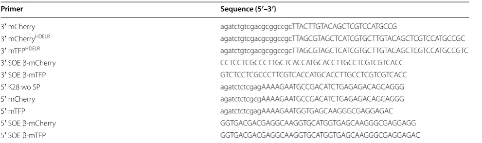
ampli-fied by PCR, for K28-mTFPHDEL, K28-mCherryHDEL
and K28-mCherry∆ by SOE-PCR (Table 3) [46]. Correct
sequenced constructs were cloned into pPIC9 (Thermo-Scientific, Waltham, Massachusetts, USA) via XhoI/NotI restriction. For P. pastoris transformation, plasmids were linearized with SalI to allow integration into the
chromosomal HIS4 locus. P. pastoris GS115 transfor-mation was performed as described previously [27]. Genomic integration was subsequently confirmed by PCR with appropriate primers.
Expression, fermentation and purification of K28 variants Recombinant protein production in P. pastoris cultivated in shaking flasks was performed as described previously [27]. After 120 h induction, the culture supernatant was concentrated through 10 kDa cut-off spin columns (Sar-torius, Vivaspin 20, Göttingen, Germany).
Fermentation of P. pastoris was conducted as described [35] in a Labfors3 bioreactor (Infors, Bottmingen, Swit-zerland) combined with a MultiTemp III cryostat (Pharmacia, Uppsala, Sweden). Pre-cultures (150 ml fermentation medium) were grown at 30 °C to an opti-cal density (OD600) of 10 to 12 and used to inoculate the
bioreactor. At the beginning of the fermentation, the bio-reactor contained a total volume of 900 ml fermentation medium already completed with hexametaphosphate, trace elements and 0.5 ml anti foam (Breox). 25% ammo-nia and 20% phosphoric acid were used to maintain the pH at 5.3. The ventilation was adjusted to 2 vvm and the pO2 to 25%, regulated by a two stage sequential
cas-cade control (1. stirrer, maximum 1250 rpm; 2. addition of O2). After complete glycerol consumption (~ 24 h),
indicated by increasing pO2, 50% glycerol with trace
ele-ments was added (9 ml/h) to increase biomass. After additional 24 h, an optical density at 600 nm of 350–400 was achieved and glycerol feeding terminated. The tem-perature was decreased to 20 °C for induction because of the temperature sensitivity of K28. Addition of methanol solution with trace elements (1.7 ml/h) led to induction of protein expression. For methanol adaptation, feed-ing was stopped several times until a limitfeed-ing growth could be recognized (manual disruption of methanol
Table 3 PCR primers used in this study
Restriction endonuclease cleavage sites used for cloning are shown in small form letters
Primer Sequence (5′–3′)
3′ mCherry agatctgtcgacgcggccgcTTACTTGTACAGCTCGTCCATGCCG
3′ mCherryHDELR agatctgtcgacgcggccgcTTAGCGTAGCTCATCGTGCTTGTACAGCTCGTCCATGCCGC
3′ mTFPHDELR agatctgtcgacgcggccgcTTAGCGTAGCTCATCGTGCTTGTACAGCTCGTCCATGCCGTC
3′ SOE β-mCherry CCTCCTCGCCCTTGCTCACCATGCACCTTGCCTCGTCGTCACC
3′ SOE β-mTFP GTCTCCTCGCCCTTCGTCACCATGCACCTTGCCTCGTCGTCACC
5′ K28 wo SP agatctctcgagAAAAGAATGCCGACATCTGAGAGACAGCAGGG
5′ mCherry agatctctcgcgAAAAGAATGCCGACATCTGAGAGACAGCAGGG
5′ mTFP agatctctcgagAAAAGAATGGTGAGCAAGGGCGAGGAGAC
5′ SOE β-mCherry GGTGACGACGAGGCAAGGTGCATGGTGAGCAAGGGCGAGGAGG
addition causes a pO2 increase). Fermentation process
was finished after 100–120 h and culture supernatant was gained by centrifugation (20 min, 10,000 rpm, 4 °C) and stored on ice. Amicon Stirred Cell 8400 (Millipore, Billerica, Massachusetts, USA) was used to concentrate the supernatant (4 °C, 2.5 bar N2). Gel filtration
chroma-tography (10% McIlvaine buffer pH 4.8 or 5.3; column material Sephadex G-25 fine (Pharmacia, Uppsala, Swe-den)) was performed to desalt the concentrate, followed by sterile filtration allowing long-term storage of K28 variants at − 20 °C or lyophilised and stored at 4 °C in the dark.
Toxin activity assays
Killer activity of toxin containing culture supernatants was determined in a standard agar diffusion assay on methylene blue agar (MBA) plates (pH 4.7) as previously described [12]. In brief, 1 × 10⁶ sensitive cells of S. cer-evisiae 192.2d were embedded into 15 ml MBA. 100 µl of culture supernatant or desalted toxin concentrate was pipetted into wells (9 mm in diameter) which were cut into the agar. For comparison, culture supernatant of
S. cerevisiae MS300b [47] was concentrated 200× and filled into the well. After 4 days at 20 °C, the diameter of the resulting cell-free zone of growth inhibition sur-rounding the well was measured, which is proportional to the logarithm of killer toxin activity.
Fluorescence microscopy
Exponentially growing S. cerevisiae cells were harvested, washed with incubation buffer (10% McIlvaine buffer pH 4.7, 10 mM glucose, 10 mM CaCl2) and resuspended
in 250 µl incubation buffer containing fluorescent toxin variants (1.4 µg/ml). After 2 h incubation (20 °C, 60 rpm) and three washing steps with incubation buffer, cells were immobilized on concanavalin A coated slides. Zeiss LSM 510 META (mCherry: 543 nm excitation, HFT 514, NFT 545, LP 560 filter, mTFP: 488 nm excitation, HFT 488, NFT 490, BP 500–530 filter) was used for obtaining confocal images. For image acquisition and processing, Zeiss LSM Image Examiner software was employed. The setup for Structured Illumination Microscopy was a pro-totype from Zeiss. Images were taken with a 63× Plan-Apochromat (NA 1.4) with excitation light of 561 nm wavelength and then processed for SIM to obtain higher resolutions. Zen2009 software was used for acquisition and processing of the images for higher resolution (Zeiss, Oberkochen, Germany).
Western analysis
SDS-PAGE was performed under non-reducing condi-tions in 10% Tris-Tricine gels unless otherwise indicated. Semi-dry blotting onto PVDF membranes was carried
out in transfer buffer (25 mM Tris, 190 mM glycin, 0.1% SDS, 20% methanol). To allow mCherry detection by immunoblotting and UV illumination (ChemiDoc XRS system, Biorad, Hercules, California, USA) samples were not heated before application. For toxin detection, blots were incubated with primary anti-DsRed antibodies (Clontech, Kyoto, Japan) and secondary HRP-conjugated goat anti-rabbit antibodies (Sigma, St. Louis, Missouri, USA). Chemiluminescent detection was obtained by the addition of “Western Lightning Plus-ECL” (PerkinElmer, Waltham, Massachusetts, USA).
Abbreviations
ER: endoplasmic reticulum; SP: signal peptidase; BPB: bromophenol blue; SIM: structured illumination microscopy; Vvm: vessel volumes per minute.
Authors’ contributions
EG, BB and MJS conceived and designed the experiments; EG performed the experiments; EG, BB and MJS wrote the paper. All authors read and approved the final manuscript.
Acknowledgements
We thank Peter Lipp for kindly providing the mCherry plasmid pmCherry-N1, Klaus Witte, Roswitha Schepp and Leon Muijs for technical assistance.
Competing interests
The authors declare that they have no competing interests.
Availability of data and materials
The constructed P. pastoris strains and plasmids are available at the Depart-ment of Molecular and Cell Biology of Saarland University, Saarbrücken, Germany.
Consent for publication
Not applicable.
Ethics approval and consent to participate
Not applicable.
Funding
This study was in part supported by the Deutsche Forschungsgemeinschaft (GRK845 and SFB1027).
Publisher’s Note
Springer Nature remains neutral with regard to jurisdictional claims in pub-lished maps and institutional affiliations.
Received: 11 September 2017 Accepted: 13 December 2017
References
1. Bevan EA, Makower M. The physiological basis of the killer character in yeast. Proc Xlth Int Congr Genet. 1963;1:202–3.
2. Starmer WT, Ganter PF, Aberdeen V, Lachance MA, Phaff HJ. The ecologi-cal role of killer yeasts in natural communities of yeasts. Can J Microbiol. 1987;33:783–96.
3. Schmitt MJ, Breinig F. Yeast viral killer toxins: lethality and self-protection. Nat Rev Microbiol. 2006;4(3):212–21.
evolutionarily related to a chromosomal host gene. Appl Environ Micro-biol. 2011;77(5):1822–32.
5. Riffer F, Eisfeld K, Breinig F, Schmitt MJ. Mutational analysis of K28 pre-protoxin processing in the yeast Saccharomyces cerevisiae. Microbiology. 2002;148:1317–28.
6. Dmochowska A, Dignard D, Henning D, Thomas DY, Bussey H. Yeast KEX1 gene encodes a putative protease with a carboxypeptidase B-like func-tion involved in killer toxin and alpha-factor precursor processing. Cell. 1987;50(4):573–84.
7. Steiner DF, Smeekens SP, Ohagi S, Chan SJ. The new enzymology of precursor processing endoproteases. J Biol Chem. 1992;267(33):23435–8. 8. Schmitt MJ, Tipper DJ. Sequence of the M28 dsRNA: preprotoxin is
processed to an alpha/beta heterodimeric protein toxin. Virology. 1995;213(2):341–51.
9. Eisfeld K, Riffer F, Mentges J, Schmitt MJ. Endocytotic uptake and retro-grade transport of a virally encoded killer toxin in yeast. Mol Microbiol. 2000;37(4):926–40.
10. Schmitt M, Radler F. Molecular structure of the cell wall receptor for killer toxin KT28 in Saccharomyces cerevisiae. J Bacteriol. 1988;170(5):2192–6. 11. Becker B, Blum A, Giesselmann E, Dausend J, Rammo D, Muller NC,
Tschacksch E, Steimer M, Spindler J, Becherer U, Rettig J, Breinig F, Schmitt MJ. H/KDEL receptors mediate host cell intoxication by a viral A/B toxin in yeast. Sci Rep. 2016;6:31105.
12. Heiligenstein S, Eisfeld K, Sendzik T, Jimenez-Becker N, Breinig F, Schmitt MJ. Retrotranslocation of a viral A/B toxin from the yeast endoplas-mic reticulum is independent of ubiquitination and ERAD. EMBO J. 2006;25(20):4717–27.
13. Pelham HR, Hardwick KG, Lewis MJ. Sorting of soluble ER proteins in yeast. EMBO J. 1988;7(6):1757–62.
14. Reiter J, Herker E, Madeo F, Schmitt MJ. Viral killer toxins induce caspase-mediated apoptosis in yeast. J Cell Biol. 2005;168(3):353–8.
15. Schmitt MJ, Reiter J. Viral induced yeast apoptosis. Biochim Biophys Acta. 2008;1783(7):1413–7.
16. Botstein D, Chervitz SA, Cherry JM. Yeast as a model organism. Science. 1997;277(5330):1259–60.
17. Odumosu O, Nicholas D, Yano H, Langridge W. AB toxins: a paradigm switch from deadly to desirable. Toxins (Basel). 2010;2(7):1612–45. 18. Sandvig K, van Deurs B. Membrane traffic exploited by protein toxins.
Annu Rev Cell Dev Biol. 2002;18:1–24.
19. Mallard F, Antony C, Tenza D, Salamero J, Goud B, Johannes L. Direct pathway from early/recycling endosomes to the Golgi apparatus revealed through the study of shiga toxin B-fragment transport. J Cell Biol. 1998;143(4):973–90.
20. Zornetta I, Brandi L, Janowiak B, Dal Molin F, Tonello F, Collier RJ, Monte-cucco C. Imaging the cell entry of the anthrax oedema and lethal toxins with fluorescent protein chimeras. Cell Microbiol. 2010;12(10):1435–45. 21. Carroll SY, Stirling PC, Stimpson HE, Giesselmann E, Schmitt MJ, Drubin DG. A yeast killer toxin screen provides insights into a/b toxin entry, traf-ficking, and killing mechanisms. Dev Cell. 2009;17(4):552–60.
22. Giepmans BN, Adams SR, Ellisman MH, Tsien RY. The fluorescent toolbox for assessing protein location and function. Science. 2006;312(5771):217–24.
23. Majoul IV, Bastiaens PI, Soling HD. Transport of an external Lys-Asp-Glu-Leu (KDEL) protein from the plasma membrane to the endo-plasmic reticulum: studies with cholera toxin in Vero cells. J Cell Biol. 1996;133(4):777–89.
24. Murasugi A, Tohma-Aiba Y. Comparison of three signals for secretory expression of recombinant human midkine in Pichia pastoris. Biosci Biotechnol Biochem. 2001;65(10):2291–3.
25. Cereghino GP, Cregg JM. Applications of yeast in biotechnology: protein production and genetic analysis. Curr Opin Biotechnol. 1999;10(5):422–7. 26. Bollok M, Resina D, Valero F, Ferrer P. Recent patents on the Pichia pastoris
expression system: expanding the toolbox for recombinant protein production. Recent Pat Biotechnol. 2009;3(3):192–201.
27. Eiden-Plach A, Zagorc T, Heintel T, Carius Y, Breinig F, Schmitt MJ. Viral pre-protoxin signal sequence allows efficient secretion of green fluorescent protein by Candida glabrata, Pichia pastoris, Saccharomyces cerevisiae, and Schizosaccharomyces pombe. Appl Environ Microbiol. 2004;70(2):961–6. 28. Shaner NC, Steinbach PA, Tsien RY. A guide to choosing fluorescent
proteins. Nat Methods. 2005;2(12):905–9.
29. Semenza JC, Hardwick KG, Dean N, Pelham HR. ERD2, a yeast gene required for the receptor-mediated retrieval of luminal ER proteins from the secretory pathway. Cell. 1990;61(7):1349–57.
30. Wilson DW, Lewis MJ, Pelham HR. pH-dependent binding of KDEL to its receptor in vitro. J Biol Chem. 1993;268(10):7465–8.
31. Macauley-Patrick S, Fazenda ML, McNeil B, Harvey LM. Heterologous protein production using the Pichia pastoris expression system. Yeast. 2005;22(4):249–70.
32. Suzuki Y, Schwartz SL, Mueller NC, Schmitt MJ. Cysteine residues in a yeast viral A/B toxin crucially control host cell killing via pH-triggered disulfide rearrangements. Mol Biol Cell. 2017;28(8):1123–31.
33. Lim CR, Kimata Y, Oka M, Nomaguchi K, Kohno K. Thermosensitiv-ity of green fluorescent protein fluorescence utilized to reveal novel nuclear-like compartments in a mutant nucleoporin NSP1. J Biochem. 1995;118(1):13–7.
34. Li Z, Xiong F, Lin Q, d’Anjou M, Daugulis AJ, Yang DS, Hew CL. Low-temperature increases the yield of biologically active herring antifreeze protein in Pichia pastoris. Protein Expr Purif. 2001;21(3):438–45. 35. Tolner B, Smith L, Begent RH, Chester KA. Production of recombinant
protein in Pichia pastoris by fermentation. Nat Protoc. 2006;1(2):1006–21. 36. Baumann K, Maurer M, Dragosits M, Cos O, Ferrer P, Mattanovich D.
Hypoxic fed-batch cultivation of Pichia pastoris increases specific and volumetric productivity of recombinant proteins. Biotechnol Bioeng. 2008;100(1):177–83.
37. Baumann K, Carnicer M, Dragosits M, Graf AB, Stadlmann J, Jouhten P, Maaheimo H, Gasser B, Albiol J, Mattanovich D, Ferrer P. A multi-level study of recombinant Pichia pastoris in different oxygen conditions. BMC Syst Biol. 2010;4:141.
38. Mayson BE, Kilburn DG, Zamost BL, Raymond CK, Lesnicki GJ. Effects of methanol concentration on expression levels of recombinant pro-tein in fed-batch cultures of Pichia methanolica. Biotechnol Bioeng. 2003;81(3):291–8.
39. Schmitt MJ, Brendel M, Schwarz R, Radler F. Inhibition of DNA synthesis in S. cerevisiae by yeast killer toxin KT28. J Gen Microbiol. 1989;135:1529–35. 40. Ai HW, Henderson JN, Remington SJ, Campbell RE. Directed evolution of a monomeric, bright and photostable version of Clavularia cyan fluores-cent protein: structural characterization and applications in fluorescence imaging. Biochem J. 2006;400(3):531–40.
41. Schermelleh L, Carlton PM, Haase S, Shao L, Winoto L, Kner P, Burke B, Cardoso MC, Agard DA, Gustafsson MG, Leonhardt H, Sedat JW. Subdif-fraction multicolor imaging of the nuclear periphery with 3D structured illumination microscopy. Science. 2008;320(5881):1332–6.
42. Schmitt MJ, Klavehn P, Wang J, Schonig I, Tipper DJ. Cell cycle studies on the mode of action of yeast K28 killer toxin. Microbiology. 1996;142(Pt 9):2655–62.
43. Eiklid K, Olsnes S, Pihl A. Entry of lethal doses of abrin, ricin and modeccin into the cytosol of HeLa cells. Exp Cell Res. 1980;126(2):321–6.
44. Yamaizumi M, Mekada E, Uchida T, Okada Y. One molecule of diph-theria toxin fragment A introduced into a cell can kill the cell. Cell. 1978;15(1):245–50.
45. Bussey H, Saville D, Hutchins K, Palfree RG. Binding of yeast killer toxin to a cell wall receptor on sensitive Saccharomyces cerevisiae. J Bacteriol. 1979;140(3):888–92.
46. Horton RM, Hunt HD, Ho SN, Pullen JK, Pease LR. Engineering hybrid genes without the use of restriction enzymes: gene splicing by overlap extension. Gene. 1989;77(1):61–8.
![Fig. 1 of protease inhibitors. SDS-PAGE of the control strain GS115 [pPIC9] grown for 120 h in BMM and (left) and reducing (right) conditions by SDS-PAGE and probed with an anti-DsRed antibody](https://thumb-us.123doks.com/thumbv2/123dok_us/9110434.1903689/3.595.62.537.85.410/protease-inhibitors-control-strain-reducing-conditions-dsred-antibody.webp)