STATISTICAL DEVELOPMENT AND OPTIMIZATION OF SOLID DISPERSION SELF EMULSIFYING PELLETS
Jessy Shaji *, Digambar Jadhav.
Prin. K. M. Kundnani College of Pharmacy, Rambhau Salgaokar Marg. Cuff Parade, Colaba. Mumbai -400005, India. *Email: [email protected]
ABSTRACT
The aim of this study is to developed solid dispersion self emulsification pellets (SDSEP). This is a recent technique for poorly water soluble drugs, where it increase water solubility and enhances the bioavailability. A 32 full factorial design approach was used for the optimization of MCC: Lactose (X1) and % of croscarmellose sodium (X2) were taken as an independent variable. Cumulative % drug release (Y1), Disintegration Time (Y2) and Friability (Y3) were studied as response variables. The differential scanning calorimetry and x-ray diffraction studies demonstrated that enhanced dissolution of Ibuprofen from SDSEP might be due to a decrease in the crystallinity of IBU. In conclusion, dissolution enhancement of Ibuprofen was obtained by preparing a Solid Dispersion Self Emulsion using melt technique. The use of a factorial design approach helped in identifying the critical factors in the preparation and formulation of SDSE.
Keywords: Solid Dispersion, Self emulsification, Extrusion speronization, response variables etc.
1. INTRODUCTION
The improvement of oral bioavailability of poorly water soluble drugs remains one of the most challenging aspect of formulation development. Amidon et al. defined ‘low solubility /high permeability class’, dissolution in the environmental lumen as the rate-controlling step in the absorption process1. To enhance the oral bioavailability of lipophilic drugs in order to increase their clinical efficacy, the most popular approach would be the incorporation of the active lipophilic component into lipid vehicle2, oils3, Salt Formation, Cyclodextrin complexation, Solid dispersion4-6 self emulsifying formulation6-9 Micronization, Liposomes10, Permeation enhancers, lipid solution and microemulsion11-15. Of this Solid dispersion self emulsification technique can be used to increase the solubility and bioavailability.
Soild dispersion self emulsifying Pellets were formulated as a lipid carrier in which the drug is solublized and then blended with spheronizing agent as Microcrystalline Cellulose and Lactose. From this system drug releases by self emulsification mechanism when it comes in contact with the gastric fluid and the gentle agitation due to the natural peristaltic movement of the GIT16-19.
Experimental design techniques such as factorial design and optimization are useful tools in the characterization of the pharmaceutical processes by studying the effects of variables affecting them and their possible interactions20.
The objective of this study were to determine the effects of the % w/w content of Microcrystalline cellulose and Lactose monohydrate and the concentration of super disintegrant as Croscarmellose sodium using a 32 factorial design.
2. MATERIALS
Ibuprofen (Cipla Pvt Ltd Mumbai), Cithrol GMS 0402 (Croda Chemicals, Singapore) Tween 80, Microcrystalline
Cellulose PH101, Lactose Anhydride Granule and Crosscarmellose Sodium.
3. METHOD
3.1 Experimental Design
In experimental design we used full factorial 32 method for the optimization procedure. Factors studied were (independent variables) MCC: Lactose monohydrate (X1) and concentration of superdisintegrant (X2). The dependent variables were drug release studied at pH 6.8 buffer (Y1), Disintegration time of pellets (Y2) and Friability rate (Y3).
Table 1 summarizes the independent and dependent variables. Tables 2 shows resulted formulation. Table 3 Optimization parameters for pellet formation.
Tables 1: Experimental design: factors and response
Factors (independent
variables)
Levels used Response (dependent
variables)
-1 0 1
X1 = ratio of Microcrystalline Cellulose: Lactose
100:0 75:25 50:50 Y1 =
cumulative % drug release within 30 min
X2 = %
Concentration of Croscarmellose Na
1 2 3 Y2 =
disintegration time (min)
Y3 = %
Friability
3.2 Preparation of Self Emulsifying System
Table 2: Composition of experimental formulation Standard Run Factor-1 MCC: Lactose Factor-2 Croscarmellose Na
F1 -1 -1
F 2 1 -1
F 3 -1 1
F 4 1 1
F 5 -1.41 0.00
F 6 1.41 0.00
F 7 0.00 -1.41
F 8 0.00 1.41
F 9 0.00 0.00
F 10 0.00 0.00
F 11 0.00 0.00
F12 0.00 0.00
F13 0.00 0.00
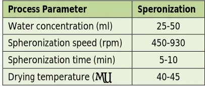
Table 3: Optimization parameters for pellet formation
Process Parameter Speronization
Water concentration (ml) 25-50 Spheronization speed (rpm) 450-930 Spheronization time (min) 5-10 Drying temperature (˚C) 40-45
3.3 Extrusion and Speronization Process:
This cream paste was homogeneously blended with Microcrystalline cellulose, lactose anhydride and croscarmellose sodium. Added sufficient quantity of water to make a dough for extrusion and speronization21. 3.4 Solubility Studies
3.4.1 Microscopy
Solubilized of ibuprofen in Cithrol, Tween 80 was added to water to produce a cream. Ibuprofen at concentrations of 16%, 25%, 33.33% and 50% in the mixture were examined under a compound microscope with 10X objective lens for the presence of ibuprofen crystals and photographs were taken with a high resolution digital camera. Solubility studies of Ibuprofen in the matrix was measured by microscopy, as reported by Gbolahan Samuel Oladiran22.
3.4.2 Differential Scanning Calorimetry (DSC):
Physical state of the drug in lipid carrier was analyze by DSC studies. Thermal analysis of ibuprofen, the physical mixture of ibuprofen and Lipid carrier, and the solid dispersions were carried out using differential scanning calorimetry method. Sample were examined using a Shimadzu TGA-50DSC instrument (Shimadzu Corporation, Japan). Sample equivalent to approximately 8mg ibuprofen were placed in aluminum pans and heated from 25 to 200ºC at heating rate of 10ºC/min.
3.4.3 X-ray diffraction studies
X-ray diffraction studies analyze the crystalline nature of the drug in the mixture. X-ray diffraction was performed using a Philiphs PW 1830 diffractometer (Philiphs,
Netherland), with a monochromatics Cu Kα radiation (λ =
1.54056), having voltage of 50kV, current of 100mA at an
angle of 2θ over a range of 5º40º. Diffraction patterns of
pure ibuprofen, Lipid carrier and solid dispersion were prepared at the drug-to-lipid ratio of 1:3 and 1:4 for the determination.
3.5 Assessment of self emulsification: The self emulsifying properties were tested by preparing different pellets containing oil soluble dye without the drug. 1g pellets were added to 50 ml of distilled water and gently agitated for 30min, 1-2 drops of this solution was examined under light microscope21.
3.6 Pellets Analysis
3.6.1 Pellets size: Particle size analysis was performed using a set of sieves in increasing order placed vertically. 100gm pellets of the were place on the top sieve and agitated on a sieve shaker for 20 min.
3.6.2 Scanning Electron Microscopy
The shape and surface characteristics of Pellets were observed by a scanning electron microscope (JEOL JSM-840A). The samples were dusted onto double sided tape on an aluminum stub and sputter- coated with gold particles in an argon atmosphere.
3.6.3 3.6.3 Friability: Friability testing was done using friability tester. 10g pellet sample was placed in the drum and rotated for 10 min at 25 rpm.
3.6.4 Disintegration testing of pellets: Modified disintegration tester testing 10 pellets were done using distilled water as the medium at 37° C, and the end point was taken as the point at which no particle were present on the sieve.
3.6.5 In-vitro Drug Release Studies: The in vitro dissolution testing was done using USP method II (paddle method) in 900ml of pH 6.8 phosphate buffer, at 37° C and 70 rpm. Accurately weighted pellets containing the equivalent of 50mg of Ibuprofen were transferred to the dissolution medium. At predetermined intervals of 5, 10, 15, 30, 60 and 120 min., the samples were withdrawn for
UV spectrophotometer at a λmax of 221nm.
4 RESULTS AND DISCUSSION
4.1 Microscopic Solubility Studies:
Ibuprofen concentration in the carrier above 33.33 % needle shaped crystal were seen.
4.2 DSC studies of Lipid Carrier with Ibuprofen:
Thermal Analysis of Lipid carrier, a mixture of Cithrol, Tween 80 and Water is shown in fig.2 A. Physical mixture of Ibuprofen and Lipid carrier is shown in fig. 2 B and Ibuprofen-to-lipid carrier composition ratio (w/w) of 1:1, 1:2, 1:3 and 1:4 were carried out using DSC method as shows in Fig 2 (C to F). Fig 2 A shows DSC results of the lipid carrier with endothermic peak at 56.2ºC. The physical mixture of Ibuprofen and lipid carrier at the ratio of 1:1 showed an endothermic peak for ibuprofen at 75.4ºC and Carrier peak at 56.4ºC, which indicated that ibuprofen was still present in crystalline state. On the other hand, as the amount of Lipid carrier were increased, the endothermic peak of ibuprofen disappeared gradually. At the ratio of 1:2 and above, the DSC curve without endothermic peak of ibuprofen indicate the absence of crystalline ibuprofen and the formation of a solid dispersions. However, an X-ray diffraction study would be needed to confirm this. 4.3 X-rays Studies of carrier and Ibuprofen
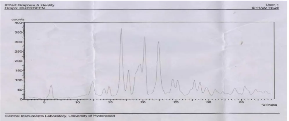
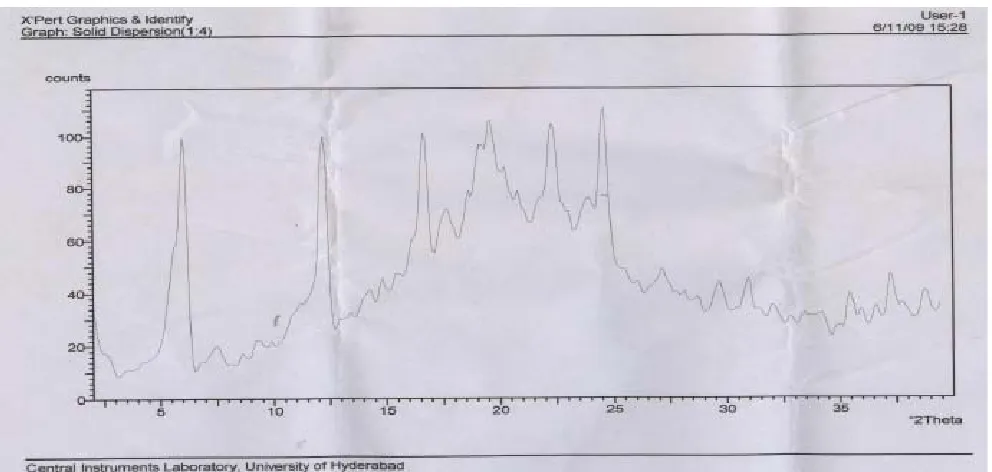
X rays powder diffractometery studies for the pure Lipid Carrier (Cithrol+Tween 80+Water), Ibuprofen, Solid Dispersion (1:3) and (1:4) are depicted in fig 3 (A to D) respectively. Pure Ibuprofen gave a diffraction peak corresponding to a separate crystalline drug phage. Soild Dispersion prepared by fusion method showed less intensity of diffraction peak of Ibuprofen pointing to a transition of Ibuprofen from a crystalline to an amorphous state as a consequence of the preparation procedure.
The Microscopic, DSC and X-ray results confirmed that Ibuprofen is present in an amorphous state in the lipid carrier as solid dispersion.
4.4 Assessment of Self emulsification:
The successively self emulsification was evaluated with increasing time and compared with one without a surfactant as it shows in Fig 4 (B to E). In non surfactant batch no oil droplets were seen as show in Fig 4 A and with increasing time increasing oil droplets were seen surfactant in the batches.
4.5 Statistical Design
32 factorial designs were selected for the final optimization of the formulation. 13 experiments were required for the complete design shown in table 4. Formulations were prepared by the varying composition of the two variables viz. quantity of Microcrystalline cellulose: Lactose monohydrate and quantity of percentage of croscarmellose sodium at three different levels i.e. high, medium and low responses (cumulative drug release within 30 min, disintegration time and friability) were measured after each run and the mathematical polynomial equation for the measured response were obtained with statistical package Design expert® version 6 software The polynomial equation relating the responses Y1,Y2 and Y3 and variable was: Cumulative drug release within 30 min (Y1) = +92.77-4.00 * A +9.19 * B -0.33 * A * -+92.77-4.00 * A2 -14.07 * B2 Disintegration time (Y2) = +7.40 +1.71*A-10.42*B +0.50*A*B +2.11 * A2 +8.36* B2
Friability (Y3) = +0.58 -0.34 * A +7.322E-003 * B +0.000 * A * B +0.091 * A2 +0.12 * B2
where A= ratio of Microcrystalline cellulose:Lactose monohydrate and B= Percentage croscarmellose sodium.
Table 4: Design of the experiment showing different batches responses Normal
Run Standard Run
Response Y1 Cumulative % Drug release within 30 min
Response Y2 Disintegration time (min)
ResponceY3 Friability %
1 F 13 56.42 32 0.3
2 F 2 62.79 35 1.2
3 F 8 75.72 7 0.5
4 F 5 83.41 12 1.3
5 F 1 83.56 6 0.4
6 F 7 96.25 10 1.1
7 F 4 57.89 33 0.8
8 F 11 81.65 8 0.7
9 F 10 92.42 7 0.6
10 F 3 91.88 8 0.6
11 F 6 93.58 7 0.5
12 F 9 92.84 7 0.6
Table 5: One solution found from numerical Optimization
Number MCC:Lactose Croscarmellose Na
Cumulative % Drug release within 30 min
Disintegration
Time Friability Desirability
F14 -0.72 0.17 88.9124 5.64543 0.400658 0.861
Table 6: Quantity of excipient used in SDSEP and their predicted and observed response.
Variable Quantity (mg)
Expected Observed
Cumulative % drug release with
in 30 min
Disintegraton time (min)
Friability %
Cumulative % drug release with
in 30 min
Disintegraton time
Friability %
A-MCC: Lactose 93.75:6.25 (312:20)
88.92 5.6 0.4 92.42 7 0.6
B-Croscarmellose
Na 1
Figure 2: DSC Studies of Ibuprofen and Lipid carrier
A: DSC scan of Lipid Carrier
B: DSC scan of Physical Mixture of Ibuprofen and lipid carrier (1:1)
D: DSC scan of Mixture of Ibuprofen: Lipid carrier (1:2)
E: DSC scan of Mixture of Ibuprofen: Lipid Carrier (1:3)
Figure 3: X rays Diffractometery Studies of Ibuprofen and Lipid at 1:3 and 1:4 ratio A: X ray diffractometery of Lipid Carrier
B: X ray diffractometery Scan of Ibuprofen
D: X ray Diffractometery Scan of Mixture of Ibuprofen: Lipid Carrier (1:4)
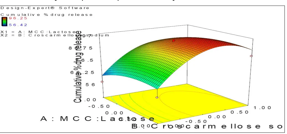
Figure 5: Response surface plot of cumulative drug release within 30 min.
Figure 6: Response surface plot of disintegration time
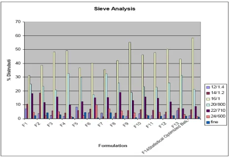
Figure 8: Pellets size analysis
The above equations represent the quantitative effect of process variables (A, B) and their interaction on the responses (Y1, Y2, Y3). The values of the coefficient A and B are related to the effect of these variables on the responses Y1, Y2 and Y3. Coefficient with more than one factor term and those with higher order terms represent interaction term.
Cumulative percent drug release within 30 min of various batches was in the range of 56.42 to 96.25 as shown in fig 5. The cumulative percent drug release within 30 min of ibuprofen was governed mainly by the % of croscarmellose sodium. It was found that as the concentration of croscarmellose sodium increased, there was an increase in the cumulative drug release within 30 min. Croscarmellose sodium was used as super disintegrant, increasing disintegration and cumulative drug release within 30 min. The second factor MCC: Lactose had less effect on cumulative drug release. Lactose concentration was increased showing increase in cumulative drug release within 30 min. These results are comparable to the results obtained by Blanque et al., 1995, who found that the high solubility of lactose provides a faster drug release23.
Disintegration time of the various batches was in the range of 5 to 35 min as shown in fig 6. Disintgration time of pellets was governed mainly by % of croscarmellose sodium. It was found that as the concentration of croscarmellose sodium increases, there was a decrease in disintegration time. Croscarmellose sodium was used as super disintegrant, it increases disintegration of pellets and decrease disintegration time. The other factor MCC: Lactose showed less effect on disintegration time. Lactose concentration increases disintegration time.
This result is similar with Blanque et al, who reported that highly porous pellets were formed during disintegration and dissolution24.
Friability of the various batches was in the range of 0.3 to 1.3 as shown in fig.7. Friability of pellets were governed mainly by the rate of MCC: Lactose. It was found that as the concentration of lactose increased in the ratio of MCC: Lactose, there was an increase in friability and vice versa. But high concentration of MCC decreased friability of pellets and decrease drug release. Therefore the optimum concentration of lactose was required to get less friability and optimum drug release. The other factor namely % of Croscarmellose sodium had less effect on friability. This result is similar to the results obtained by Sousa et al., 2002 who found that the higher the proportion of MCC in the pellets, the greater was the mechanical strength of the pellets23. The other factor namely % Croscarmellose sodium had less effect on friability
Statistical optimization
The experimental runs with the predicted solution are shown in Table No 5. Formulated the solution Batch F14 as shown in Table no 5, then observed the results and
compared the results with the predicated results as shown in table no 6 This demonstrates the reliability of the optimization procedure in predicting the Cumulative percent drug release within 30 min, Disintegration time (min) and Friability (%).
4.6 Pellets Size analysis: Sieve analysis was done and its value of geometric mean diameter (dg) was 1mm. All batches showed pellets size in the optimized range of
800µm to 1000µm as shown in fig 8.
4.7 In-vitro Drug Release Studies:
In vitro dissolution tests of F14 showed 92.42 % drug
release within 30 min (fig 9). Ibuprofen release was optimized from different ratios of MCC: Lactose monohydrate and croscarmellose sodium.
CONCLUSION
The immediate release of water insoluble Ibuprofen from pellet formulation was possible by incorporating them into a Solid Dispersion Self Emulsifying system with croscarmellose Na, which enhanced their rate of release. Increasing Lactose % showed fast release but friability increased and high MCC level was required to achieve low level of friability. 1% Croscarmalose Na was sufficient to get 92.72% drug release within 30 min.
The SEDDS pellets were successfully characterized for its physical parameters such as size analysis, friability, disintegration and invitro dissolution.
Acknowledgement: The authors are grateful to Cipla Pharmaceutical Pvt.Ltd. and Croda Chemicals Mumbai for providing gift sample of Ibuprofen and Cithrol GMS 402 respectively.
REFERENCES
1. Amidon GL, Lennernas VP, Shah VP, Crison JR, A theoretical basis for a biopharmaceutic drug classification: the correlation of in vitro drug product dissolution and in vivo bioavailability, Pharm. Res. 12, 1995, 413-420
2. Aungst BJ, Novel formulation strategies for improving oral bioavailability of drugs with poor membrane permeation or presystemic metabolism, J. Pharma.Sci. 82, 1993, 979-986.
3. Burcham DL, Maurin MB, Hausner EA, Huang SM, Improved oral bioavailability of the hypocholesterolemic DMP 565 in dogs following oral dosing in oil and glycol solutions Biopharm.Drug Dispos. 18, 1997, 737-742.
4. Serajuddin ATM, Sheen PC, Musfson D, Bernstein DF, Augustine MA, Effect of vehicle amphilicity on the dissolution and bioavailability of a poorly water – soluble drug from solid dispersion. J.Pharm.Sci. 77, 1988, 414-417.
HIVprotease inhibitor using Gelucire 44/14 and Labrasol vehicles, B.T. Gattefosse. 87, 1994, 49-54. 6. Nazzal S, Guven N, Reddy IK, Khan MA, Preparation
and characterization of Coenzyme Q10 – Eudragit solid dispersation. Drug Dev. Ind. Pharm. 28, 2002, 49-57.
7. Wakerly MG, Pouton CW, Meakin BJ, Morton FS, Self emulsification of vegetable oil-non-ionic surfactant mixture. ACS Sympser. 311, 1986, 242-255.
8. Charman SA, Charman WN, Rogge MC, Wilson TD, Dutko FJ, Pouton CW, Self emulsifying drug delivery systems: formulation and biopharmaceutic evaluation of an investigational lipophilic compound, Pharm. Res. 9, 1992, 87-93.
9. Shah NH, Carvajal MT, Patel CI, Infeld MH, Malick AW, Self emulsifying Drug Delivery Systems (SEDDS) with polyglycolyzed glycerides for improving in vitro dissolution and oral absorption of lipophilic drugs. Int.J.Pharm. 106, 1994, 15-23.
10. Craig DQM, Lievens HSR, Pitt KG, Storey DE, An investiationinto the physico- chemical properties of self emulsifying systems using low frequency dielectric spectroscopy, surface tension measurements and particle size analysis. Int. J .Pharm. 96, 1993, 147-155.
11. Toguchi H, Ogawa Y, Iga K, Yashiki T, Shimamoto T, Gastrointestinal absorption of ethyl 2-chloro-3- (4-(2-methyl-2-phenylpropyloxy) propionate from different dosage forms in rats and dogs. Chem. Pharm. Bull. 38, 1990, 2792-2796.
12. Palin KJ, Phillips AJ, Ning A, The oral absorption of cefoxitin from oil and emulsion vehicles in rats Int. J.Pharm. 33, 1986, 99-104.
13. Myers RA, Stella VJ, Systemic bioavailability of penclomedine (NSC-338720) from oil-in-water emulsion administered intraduodenally to rats. Int.J. Pharm. 78, 1992, 217-226.
14. Stella V, Haslam J, Yata N, Okada H, Lindenbaum S, Higuchi T, Enhancement of bioavailability of a hydrophobic amine antimalarial by formulation with oleic acid in a soft gelatin capsule. J. Pharm. Sci. 67, 1978, 1375-1377.
15. Kararli TT, Needham TE, Griffin M, Schoenhard G, Ferro LJ, Alcorn L, Oral delivery of a rennin inhibitor compound using emulsion formulations. Pharm. Res. 9, 1992, 888-893.
16. Schwendener RA, Schott H, Lipophilic 1-beta-D-arabinofuranosyl cytosine derivatives in liposomal formulations for oral and parentral antileukemic therapy in the murine L 1210 leukemia model. J.Cancer Res. Clin. Oncol. 122, 1996, 723-726.
17. Constantinides PP, Lipid microemulsion for improving drug dissolution and oral absorption: physical and iopharmaceutical aspects. Pharm.Res.12, 1995, 1561-1572.
18. Craig DQM, The use of self emulsifying system as a means of improving drug delivery, BT. Gattefosse. 86, 1993, 21-31.
19. Meinzer A, Mueller E, Vonderscher J, Microemulsion-a suitMicroemulsion-able gMicroemulsion-alenticMicroemulsion-al Microemulsion-approMicroemulsion-ach for the Microemulsion-absorption enhancement of low soluble compounds? B.T. Gattefosse 88, 1995, 2925-2927
20. Armstrong NA, James KC, Understanding Experimental design and interpretation in Pharmaceutics. Ellis Horwood, London, UK, 1990, 27-54.
21. Abdalla A, Mader K, Preparation and characterization of a self-emulsifying pellets formulation, Eur. J. Pharm Biopharm. 66, 2007, 220-226.
22. Gbolahan SO, Hannah KB, Determination of Ibuprofen solubility in wax: A comparison of microscopic, thermal and release rate techniques. Eur. J. Pharm. Biopharm. 67, 2007, 106-111.
23. Blanque D, Sternagel H, Podezeck F, Newton JM, Some factors influencing the formation and in vitro drug release from matrix pellets prepared by extrusion/spheronization. Int. J.Pharm., 119, 1995, 203-211
24. Sousa JJ, Sousa A, Podazeck F, Newton JM, Factors influencing the physical characteristics of pellets obtained by extrusion-spheronization. Int.J. Pharm.232, 2002, 91-106.