Nijmegen
The following full text is a publisher's version.
For additional information about this publication click this link.
http://hdl.handle.net/2066/193401
Please be advised that this information was generated on 2020-09-10 and may be subject to
change.
J Clin Periodontol. 2018;45:851–860. wileyonlinelibrary.com/journal/jcpe
|
8511
|
INTRODUCTION
Periodontal regeneration has been attempted over the last 30 years. Despite the evolution in treatment, the clinical results of periodontal regenerative techniques are not optimal and still un-predictable. Currently, the successful application of guided tissue regeneration (GTR) with most predictable clinical results is limited to certain defect morphology (e.g., two and three- wall intrabony defects, class II mandibular furcations), patient’s characteristics (good oral hygiene, nonsmokers) and surgeons’ experience (skills, decision- making) (Villar & Cochran, 2010). The concept of endog-enous cell recruitment can be considered as very suitable and ap-pealing for this purpose, particularly, in view of modifying the early
phase of wound healing, which has been proposed to be the main target of future periodontal regenerative technique (Dickinson et al., 2013; Susin & Wikesjo, 2013). Increasing evidence in the field of regenerative medicine suggests that cell recruitment stim-ulates latent self- repair mechanisms in patients and harnesses the innate capacity for tissue regeneration (Chen, Wu, Zhang, Zhang, & Sun, 2011). This strategy has been explored as an alternative to cell- based therapies for in situ tissue regeneration of heart, carti-lage and bone tissue (French, Somasuntharam, & Davis, 2016; Ji et al., 2013; Zhao, Jin, Li, Qiu, & Li, 2017). Instead of introducing ex vivo expanded stem cells, recruiting endogenous cells relies on active attraction of regenerative cells to a site of tissue damage or injury.
Received: 17 October 2017
|
Revised: 4 April 2018|
Accepted: 14 May 2018 DOI: 10.1111/jcpe.12928A N I M A L E X P E R I M E N T
Periodontal regeneration via chemoattractive constructs
Xinjie Cai
1,2|
Fang Yang
1|
X. Frank Walboomers
1|
Yining Wang
2|
John A. Jansen
1|
Jeroen J. J. P. van den Beucken
1|
Adelina S. Plachokova
3This is an open access article under the terms of the Creative Commons Attribution-NonCommercial-NoDerivs License, which permits use and distribution in any medium, provided the original work is properly cited, the use is non- commercial and no modifications or adaptations are made.
© 2018 The Authors. Journal of Clinical Periodontology Published by John Wiley & Sons Ltd
1Department of Biomaterials, Radboudumc,
Nijmegen, the Netherlands
2The State Key Laboratory Breeding Base of
Basic Science of Stomatology (Hubei-MOST) and Key Laboratory of Oral Biomedicine Ministry of Education, School and Hospital of Stomatology, Wuhan University, Wuhan, China
3Department of Implantology and
Periodontology, Radboudumc, Nijmegen, the Netherlands
Correspondence
Adelina Plachokova-Damyanova, Department of Implantology and
Periodontology, Radboudumc, PO Box 9101, 6500 HB, Nijmegen, the Netherlands. Email: Adelina.Plachokova-Damyanova@ radboudumc.nl
Funding information
This study was financially supported by Osteology Foundation (project number 13- 028) and Royal Netherlands Academy of Arts and Sciences (KNAW; project number CEP- 10CDP025).
Abstract
Aim: Chemoattractants, such as stromal cell- derived factor- 1α (SDF- 1α), can offer an advantage for periodontal regeneration by recruiting the patient’s own stem cells to stimulate self- repair. We here developed a chemoattractive construct for periodontal regeneration using SDF- 1α and evaluated its efficacy in vivo.
Materials and Methods: SDF- 1α was loaded on gelatin sponge and tested in vitro for SDF- 1α release. Subsequently, SDF- 1α constructs were implanted into rat periodon-tal defects for 1 and 6 weeks, with unloaded materials and empty defects as controls. The regenerative efficacy was evaluated by micro- CT, histological and histomorpho-metrical analyses.
Results: In vitro results showed limited SDF- 1α release up to 35 days. In contrast, SDF- 1α constructs significantly improved periodontal defect regeneration in terms of alveolar bone height, new bone area and functional ligament length. Additionally, SDF- 1α constructs decreased the inflammatory response at Week 6.
Conclusion: Chemoattractive constructs significantly improved periodontal regen-eration in terms of alveolar bone height, new bone area and functional ligament length.
K E Y W O R D S
For the periodontium, such a strategy seems very appropriate considering that resident mesenchymal stromal cells (MSCs) are re-garded as the key for periodontal regeneration (Hynes, Menicanin, Gronthos, & Bartold, 2012; Susin & Wikesjo, 2013). Additionally, de-velopment and clinical application of periodontal cell homing tech-nique will mean elimination of constraining factors associated with clinical management of periodontal wounds, specific patient and site characteristics. As a result, optimal conditions for wound healing that will favour periodontal regeneration could become feasible in most clinical settings.
For cell recruitment approaches toward periodontal regenera-tion, the chemoattractant stromal cell- derived factor- 1α (SDF- 1α) is the primary candidate chemokine. SDF- 1α plays a major role in cell trafficking and recruitment of CD34+ stem cells (Aiuti, Webb, Bleul,
Springer, & Gutierrez- Ramos, 1997), and several recent periodontal studies have shown the potential of SDF- 1α for periodontal regen-eration (Du, Yang, & Ge, 2012; Kaku et al., 2017; Wang, Du, & Ge, 2016). As the mechanism underlying this enhanced periodontal re-generation, Zhou et al. have found that bone marrow- derived stem cells migrated preferentially to the periodontium over other organs to get involved in periodontal regeneration (Zhou et al., 2011). In addition to this cell recruitment efficacy, SDF- 1α is anticipated to modify the inflammatory response and early wound healing (Chen et al., 2015; Dickinson et al., 2013; Zhao et al., 2017).
Both clinically and experimentally, the haemostatic gelatin- based material Spongostan® has been widely used for different
applica-tions and as a carrier for cells and proteins, respectively (Arias- Gallo, Chamorro- Pons, Avendano, & Gimenez- Gallego, 2013; Cai et al., 2015; Yu et al., 2013). Using this material, we here developed a che-moattractive construct and evaluated its feasibility and biological performance in a rat periodontal defect model. We hypothesized that this chemoattractive construct could recruit regenerative cells to the periodontal defect site and hence would improve periodontal regeneration (i.e., formation of new alveolar bone, new cementum and new periodontal ligament) compared with defects treated with virgin constructs (i.e., without SDF- 1α) and empty defects.
2
|
MATERIALS AND METHODS
2.1
|
Material preparation
Commercially available gelatin sponge (Spongostan®; Ferrosan
Medical Devices, Denmark) was resized to the defect size (2 × 2 × 2 mm3, W × L × D) and loaded via absorption with 20 μl PBS
solution containing three different doses of recombinant human SDF- 1α (R&D systems, Abingdon, UK): 100, 200 and 400 ng SDF- 1α
(Cao et al., 2013; Du et al., 2012). All procedure was performed under aseptic conditions.
2.2
|
In vitro release experiment
Stromal cell- derived factor- 1α (100, 200 and 400 ng) loaded gelatin sponges were incubated in 1 ml PBS solution in an Eppendorf tube
at 37°C. At each time point, the complete volume was taken and refreshed with 1 ml PBS (n = 4). The measurements were performed using an ELISA kit (RayBiotech Inc., Norcross GA, USA) with a sensi-tivity of 80 pg/ml according to manufacturer’ s instructions.
2.3
|
Cell culture
Isolation of GFP cells from 12- week- old GFP- transgenic Sprague- Dawley rats was done as described in previous studies by our group (Ji et al., 2013; Yu et al., 2013). It was approved by the Animal Ethical Committee of Radboud University (Approval number: RU- DEC 2014- 101). This study complied with the ARRIVE guidelines for preclinical animal studies. Briefly, two femora of each rat were extracted, epiphyses were cut off, and diaphyses were flushed out with 15 ml proliferation medium (α- MEM supplemented with 10% FBS and 50 μg/ml gentamicin). The flush- out of bone marrow from different rats was pooled and cultured for 2 days in a humidified in-cubator (37°C, 5% CO2), after which the medium was refreshed to remove nonadherent cells.
2.4
|
Animals & surgery
Forty- eight adult male athymic nude rats (Crl: NIH Foxn1rnu, Charles River, Germany) were used as experimental animals and recipients of the GFP- transgenic rat BMSCs cells to evaluate the biological re-sponse to the implants.
For the creation of standardized and validated rat periodontal defects, a previously described surgical protocol was followed (Cai et al., 2015; Yu et al., 2013). As shown in Figure 1, under general an-aesthesia, a unilateral intrabony defect was created on the mesial side of the first maxillary molar. Afterwards, the defect was filled with one of the experimental groups in a randomized manner: (a) empty defect (EMP); (b) unloaded sponge (M); (c) SDF- 1α loaded sponge (SDF). The flaps were closed with resorbable sutures (Vicryl®
Clinical Relevance
Scientific rationale for the study: Chemoattractants can stimulate latent self- repair mechanisms in patients and har-ness the innate capacity for tissue regeneration. However, the efficacy of applying this cell homing strategy in perio-dontal regeneration is still uncertain.
Principal findings: SDF- 1α constructs (without the
applica-tion of barrier membrane) significantly improved periodon-tal regeneration at 6 weeks in terms of alveolar bone height, new bone area and functional ligament length. Practical implications: Chemoattractive constructs based on SDF- 1a were proven effective and should be considered for further development towards clinical use, in the treat-ment of periodontology.
|
853 CAI etAl.5- 0; Ethicon Products, Amersfoort, The Netherlands). The three dif-ferent experimental groups (n = 8) were assigned for evaluation of the biological response after 1 and 6 weeks.
At 2 days after implantation, 1 ml PBS solution containing 1.5 × 10⁶ GFP- transgenic rat BMSCs was injected into the tail vein
of the rats. During the first 10 postoperative days, the animals were fed with powdered food to minimize wound disturbance and visual wound inspection was performed on a daily basis.
2.5
|
Histological preparation and microcomputed
tomography (micro- CT) analysis
After 1 and 6 weeks of implantation, the animals were sacrificed using CO2 suffocation. The maxillas were harvested and split into two parts through the palatal median line. After fixation in buff-ered 10% formaldehyde for 24 hr, samples were decalcified in 4% EDTA at 4°C for 6 weeks, dehydrated with graded series of ethanol and embedded in paraffin. Mesiodistal sections (thickness 6 μm) were cut with a microtome (Leica RM2165, Nussloch, Germany) and every 10th section was stained using haematoxylin and eosin (HE) for general tissue survey. For epithelial, ligament and bone tis-sue observation, sections were stained with Azan and Elastica- van Gieson (EVG) staining. For visualization of the GFP- positive cells in the defect area, anti- GFP immunohistochemical staining was ap-plied (rabbit anti IgG fraction, 1:400; Molecular Probes, Eugene, OR, USA). Anti- CD68 staining was used to evaluate the inflamma-tory response.
After being fixed in 10% buffered formaldehyde for 48 hr, ran-domly chosen complete maxillae (n = 4) were analysed by micro- CT; 3D- reconstruction of the defect areas and volumetric analysis of the newly formed bone was performed as described previously (Oortgiesen et al., 2012) (SkyScan 1072; SkyScan N.V., Aartselaar, Belgium). A subregion of the originally measured data was selected on both sagittal and transversal slices. This selected region of inter-est (ROI) included the entire defect area with the newly formed bone inside. By auto- interpolation of manually determined ROIs from the resliced images, each specimen yielded a volume of interest (VOI), which served as the essential basis for all quantitative analyses. Landmarks, such as intact alveolar bone height of the un- operated side and root apex were used for the manual drawing the ROI.
2.6
|
Histomorphometry
For histomorphometrical analysis, three sections per specimen for each of the stainings (HE, Azan, EVG, anti- CD68) were evaluated. Measurements were performed using Image J software. All meas-urements were done as described previously for the same periodon-tal defect model (Cai et al., 2015). The relative alveolar bone height, new bone area, epithelial down- growth and functional ligament length were analysed. The inflammatory response (macrophages) was detected with anti- CD68 staining and subsequently digitalized by a computer programmer in the ROI (defect area). The areas with positive anti- CD68 staining were given as percentage (%) of the total ROI.
F I G U R E 1 Overview of the surgical procedure: (a) preoperative surgical site; (b) the root surface and alveolar bone were exposed via a 3- mm full thickness incision; (c) a piezoelectric device (Piezosurgery®, Mectron, Carasco, Italy; loaded with OT5 B- tip; Ø1.7 mm) was used
to create an unilateral intrabony defect; the residual bone, periodontal ligament and root cementum were carefully removed from the root surface, to create the defect (W × L × D; 2 × 2 × 1.7 mm3), with constant size monitoring by a clinical periodontal probe; (d) the defect size
was confirmed by a periodontal probe; (e) implant placement in the defect; (f) the flap was closed with 5–0 resorbable sutures
(a) (b) (c)
2.7
|
Statistical analysis
Data were expressed as mean ± standard deviation. Statistical anal-ysis was carried out using GraphPad (GraphPad Inc, San Diego, CA, USA) by one- way ANOVA and post hoc Tukey testing, for which dif-ferences were considered significant at p < 0.05.
3
|
RESULTS
3.1
|
In vitro SDF- 1
α
release
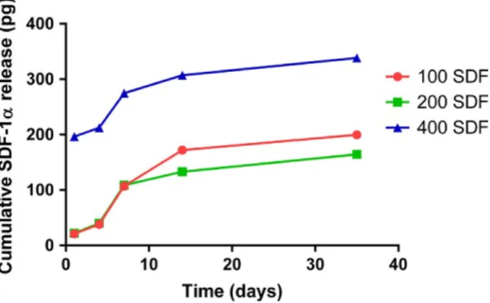
Figure 2 shows the release profiles for the constructs loaded with dif-ferent amounts of SDF- 1α. For all three loading amounts, a similar re-lease pattern was observed. The cumulative rere-leased amount of SDF- 1α
increased gradually up to 35 days, where the 400 SDF group showed the highest release (338.6 pg). Nevertheless, for all three groups, the re-lease of SDF- 1α over time was limited in pg compared with loading in ng.
3.2
|
General in vivo observations
All animals had uneventful recovery after surgery and gained weight during the experimental period. No signs of infection or clinical in-flammation were observed. At euthanasia, visual inspection of the surgical sites showed an undisturbed wound healing.
3.3
|
Micro- CT analysis
Data on new bone formation and 3D reconstructed images of the periodontal defects are presented in Figure 3. After 1 week, defect areas were easily detected, and minor bone formation was observed
F I G U R E 2 Cumulative release profiles of SDF- 1α from different groups in 1 ml PBS
F I G U R E 3 (a) 3D reconstructed micro- CT images of periodontal defects with different groups after 1 and 6 weeks: empty control (EMP); unloaded material (M); SDF- 1α loaded gelatin sponge (SDF). Black asterisk indicates the position of the molar. (b) The histogram below represents the percentage of bone volume to tissue volume of different groups at Weeks 1 and 6
|
855 CAI etAl.predominantly at the defect edges in all experimental groups. After 6 weeks, EMP showed limited new bone formation, whereas more newly formed bone was observed for M and SDF.
The volumes of the newly formed bone at Week 1 were similar (1%) for all groups (p > 0.05). After 6 weeks, all groups presented a significant increase in the amount of new bone formation (6.8%, 9.6% and 17.1%, respectively); SDF showed significantly higher new bone formation compared with EMP (p < 0.01) and M (p < 0.05). Only for SDF, bone ingrowth from the bottom of the defect was observed.
3.4
|
Descriptive histology
3.4.1
|
One- week results
After 1 week, the defects were easily detected by the removed ce-mentum from the root surface and absence of supportive alveolar bone. This occasionally resulted in penetration into the glands api-cally (Figure 4a–c). For M and SDF, the implanted material was found in place with hardly any signs of degradation and surrounded by thin fibrous tissue capsule. Above them subepithelial inflammatory infil-trate was found (Figure 4d–f). For EMP, the defects were filled with connective tissue and immense inflammatory infiltrate. Besides the presence of extensive inflammation, other common findings observed in all groups were epithelial down- growth (to the level to which the ce-mentum was removed), minor start of bone formation from the defect edges and no signs of PDL formation or cementum formation.
3.4.2
|
Six- week results
After 6 weeks, the edges of the periodontal defects were still visible. All materials were found completely degraded, and the
inflammatory infiltrate in the defect region was considerably reduced (Figure 5a–c). For SDF, bone formation was more pro-nounced than for EMP and M, and in 3/8 of samples, the alveolar bone in the defect area was completely regenerated (Figure 5d–f). Epithelial down- growth remained at the same level as at Week 1, with no obvious differences among the experimental groups, which proves that a barrier membrane is not required to prevent soft tissue ingrowth. As such, we opted to leave out a barrier membrane in the current study. Limited formation of periodon-tal ligament with new cementum and new bone was observed, which was significant in SDF compared with the other two groups (Figure 5g–i). For the pan- macrophage anti- CD68 staining, an evi-dent decrease in inflammatory response was observed for all ex-perimental groups compared with Week 1. SDF demonstrated a significantly lower level of inflammatory response compared with EMP and M (Figure 5j–l). In addition, anti- GFP staining was per-formed to track the recruitment of GFP- positive rat BMSCs to the periodontal defect in vivo, but no positive result could be obtained for all groups (data not shown).
3.5
|
Histomorphometry
For alveolar bone height, a substantial increase with time was ob-served in all groups (Figure 6). The SDF demonstrated a signifi-cantly higher alveolar bone height compared with the EMP and M at Week 6 (p < 0.01), with no statistical difference among all groups at Week 1 (p > 0.05), which was in accordance with the micro- CT results. Similar results could also be found in the bone area measurement, where the SDF had significantly more new bone formation in the defect area than the M at Week 6 (p < 0.05). For the epithelial down- growth, no statistical difference could be
F I G U R E 4 Histological overview after 1 week of implantation. (a) EMP group (HE staining; original magnification 10 × ), no obvious periodontal regeneration could be observed; (b) M group, scaffold remnants could be found in the defect site; (c) SDF group, scaffold remnants could also be found; black asterisk indicates the defect site. (d) Higher magnification of the defect site in (a), positive staining could be found in the whole defect area (anti- CD68 staining; original magnification 10 × ); (e) higher magnification of the defect site in (b); (f) higher magnification of the defect site in (c), relatively less positive staining could be found in the defect area
found among all groups at both Week 1 and 6 (p > 0.05). As to the functional ligament length, the SDF had significantly higher score than the M at Week 6 (p < 0.05), with no statistical differ-ence among all groups at Week 1 (p > 0.05). Although the SDF had higher scores than the EMP in the new bone area and functional ligament measurements at Week 6, no statistical difference had been obtained between them (p > 0.05). Finally, for the inflamma-tory response, it was around 28% for all groups at Week 1, with no statistical difference among them (p > 0.05). At Week 6, the SDF (7.7%) showed significantly lower level of inflammatory response than the M (14.8%, p < 0.05), with no statistical difference to the EMP (11.8%, p > 0.05).
4
|
DISCUSSION
The objective of this study was to develop a construct for periodon-tal regeneration based on cell recruitment using SDF- 1α and to eval-uate the feasibility and biological performance of this construct. The results showed that SDF- 1α constructs significantly improved peri-odontal wound healing at Week 6 in terms of alveolar bone height, new bone area and functional ligament length. However, in vitro re-lease of SDF from the scaffold was suboptimal.
Our in vitro release results corroborate those of Takayama et al., whose used a collagen membrane as a carrier for SDF- 1α with only 10% SDF- 1α release after 3 weeks (Takayama et al., 2017). In our
F I G U R E 5 Histological overview after 6 weeks of implantation. (a) EMP group (HE staining; original magnification 10 × ); (b) M group; (c) SDF group; (d) higher magnification of the defect area in (a), new bone formation in the defect was marked with arrows, and the dashed line presents the boundary between new bone and old bone (EVG staining; original magnification 10 × ); (e) higher magnification of the defect area in (b), new bone formation deriving from the bone margin of the defect (arrow); (f) higher magnification of the defect area in (c), the defect site was fully recovered by new bone formation (arrow); (g) higher magnification of the root surface in (a), collagen fibres were attached obliquely to the denuded root surface (Azan staining; original magnification 10 × ); (h) higher magnification of the root surface in (b), very limited amount of fibres obliquely oriented to the root surface on the coronal side; (i) higher magnification of the root surface in (c), collagen fibres obliquely oriented to the root surface; (j) higher magnification of the defect area in (a), positive staining could be found in the defect area (anti- CD68 staining; original magnification 10 × ); (k) higher magnification of the defect area in (b); (l) higher magnification of the defect area in (c), almost on positive staining could be found in the defect area. Black asterisk indicates the defect site; D: dentin of the root
|
857 CAI etAl.study, also limited amount of SDF- 1α was released in vitro up to 35 days. The most part of SDF- 1α remained absorbed on the gela-tin sponge, or deactivated during the test. It seems that the bonding force between SDF- 1α and gelatin/collagen might be too strong, and/
or physical adsorption of SDF- 1α on the gelatin sponge (Spongostan®)
is not an efficient approach for SDF- 1α delivery. It has to be noticed that, Spongostan® is a commercial product, and besides gelatin,
the rest of its components are unknown. Other carriers, such as a
F I G U R E 6 Histomorphometrical measurement at Weeks 1 and 6: (a, b) relative alveolar height; (c, d) relative new bone area; (e, f) relative epithelial down- growth; (g, h) relative functional ligament length; (i, j) relative amount of inflammation in percentage for all evaluation periods. *p < 0.05, **p < 0.01; error bars represent standard error of the mean (n = 8)
hydrogel (Kimura & Tabata, 2010), PLGA (Thevenot et al., 2010), al-ginate (Rabbany et al., 2010) and radially oriented collagen scaffold (Chen et al., 2015) have been utilized with various success to contin-uously deliver SDF- 1α to accelerate blood vessel, cartilage and bone regeneration. More extensive research with other carriers (such as polymer scaffolds) for SDF- 1α delivery, or using gene transduction approaches to up- regulate SDF- 1α expression should be explored in the future.
Despite the suboptimal in vitro release of SDF- 1α from the scaf-fold, the SDF- 1α construct significantly increased new bone forma-tion in rat periodontal defect after 6 weeks of implantaforma-tion. This effect was not found after 1 week of implantation, and this could be explained with the retention of SDF- 1α within the scaffold. After 6 weeks the gelatin was found completely degraded within our de-fect, and obviously during this degradation process, SDF- 1α was released and managed to exert its effect. Our histomorphomteri-cal and micro- CT results are in accordance with a previous study made by our group (Ji et al., 2013), in which a sixfold increase in bone formation was observed in a SDF- 1α mediated GBR procedure. Takayama et al. applied a SDF- 1α loaded collagen membrane in a rat mandibular bone defect model and got similar results to our find-ings on the positive effect of SDF- 1α on bone formation (Takayama et al., 2017). Their micro- CT analysis showed similar amounts of new formed bone (i.e., volume and area) as in our study. Although they used a model for bone augmentation, different scaffold (i.e., collagen membrane), higher doses of SDF- 1α (3 and 6 μg) and implantation period of 4 weeks, the results were still comparable.
Similar to the new bone formation, the SDF- 1α group also demonstrated significantly longer functional ligament length than the unloaded group after 6 weeks of implantation. This finding is in accordance with other studies that utilize SDF- 1α incorporated collagen membrane (Wang et al., 2016), endochondral approach (Cai et al., 2015) or PDL cells implantation with gelatin sponge (Yu et al., 2013) to regenerate periodontium. In these studies, the regeneration of periodontal ligament as well as alveolar bone could be detected. It can even be hypothesized that the processes of bone and ligament formation are related to each other, similar to the development of the natural periodontium during teeth eruption.
Regarding our experimental design, no barrier membrane was included, which was widely used in the traditional GTR procedure. Based on our previous work (Cai et al., 2015; Yu et al., 2013), gelatin sponge and electrospun scaffolds have been successfully applied in rat periodontal defect without the use of barrier membranes and showing evident periodontal regeneration. Moreover, no significant differences in epithelial down- growth were observed among exper-imental groups at different time points, which proves that a barrier membrane is not required to prevent soft tissue ingrowth. As such, we opted to leave out a barrier membrane in the current study.
For the in vivo rat BMSCs recruitment test, no GFP- positive rat BMSCs could be found in the periodontal defect area. This might be explained by the slow degradation of gelatin sponge in vivo, which resulted in the very delayed release of SDF- 1α, and/or by the low
dose of SDF- 1α used. This mismatched timeline might resulted in the immobilization of GFP- positive cell in the other organs rather than the periodontal defect. Similar result had also been reported by another research group (Huang, Gronthos, & Shi, 2009). However, other studies using constructs with burst release of SDF- 1α in the early stage demonstrated successfully the recruitment of BMSCs (Dashnyam et al., 2014; He, Ma, & Jabbari, 2010; Ji et al., 2013). Therefore, further investigation is needed to confirm the recruit-ment of GFP- positive rat BMSCs in the periodontal defect, and higher dose of SDF- 1α might be considered.
The enhanced periodontal regeneration in response to local SDF- 1α stimuli could be explained such as follows. Firstly, the bone marrow- derived osteoblast progenitor cells existed in the circulating blood, might be recruited to the defect site in response to local re-lease of SDF- 1α via SDF- 1α/CXCR4 axis (Ji et al., 2013; Otsuru, Tamai, Yamazaki, Yoshikawa, & Kaneda, 2008). Those recruited cells contrib-uted to the periodontal healing process by not only exerting their mul-tilineage differentiation capacity in the defect site, but also secreting a number of cytokines and growth factors to promote tissue regener-ation (Bryan, Walker, Ferguson, & Thorpe, 2005). Secondly, the local release of SDF- 1α might also generate proangiogenic environment in the defect site by mobilizing other progenitor cells resident in bone marrow, such as hematopoietic stem cell and endothelial progenitor cell, to the defect site through SDF- 1α/CXCR4 axis (Hattori, Heissig, & Rafii, 2003; Petit, Jin, & Rafii, 2007). Those cells could have enhanced angiogenesis in the defect area, hence indirectly enhancing periodon-tal regeneration. The effect of SDF- 1α on angiogenesis was not inves-tigated in our study as a haemostatic material (i.e., Spongostan) was used for its delivery into the defect. However, this mechanism should be explored when SDF- 1α is further investigated with a more suitable scaffold for periodontal regeneration.
Regarding the inflammatory response, the SDF showed sig-nificantly lower inflammatory response than the M at Week 6. Inhibitory effect on inflammation of SDF- 1α was also seen in other studies. Thevenot et al. reported that SDF- 1α could reduce but not reverse the inflammatory responses (Thevenot et al., 2010). There was a substantial decrease in density and activation of mast cells and inflammatory (CD11b+) cells in the space surrounding the SDF- 1- loaded PLGA scaffold in mice. Meanwhile, in a rat mandibular bone defect, SDF- 1α significantly reduced the CD11b+ inflammatory cell response (Liu, Li, Du, Yang, & Ge, 2015).
5
|
CONCLUSION
Based on our findings, and within the limitations of this study, it can be concluded that the chemoattractive constructs signifi-cantly improve periodontal wound healing at 6 weeks in terms of alveolar bone height, new bone area and functional ligament length. Chemoattractive constructs based on SDF- 1α were proven effective and should be considered for further development towards clinical use, in the treatment of periodontology.
|
859 CAI etAl.CONFLIC T OF INTEREST
The authors declare that there are no conflict of interests in this study.
ORCID
Xinjie Cai http://orcid.org/0000-0003-0179-7181
Yining Wang http://orcid.org/0000-0002-6749-4362
REFERENCES
Aiuti, A., Webb, I. J., Bleul, C., Springer, T., & Gutierrez-Ramos, J. C. (1997). The chemokine SDF- 1 is a chemoattractant for human CD34 + hematopoietic progenitor cells and provides a new mecha-nism to explain the mobilization of CD34 + progenitors to peripheral blood. Journal of Experimental Medicine, 185, 111–120. https://doi. org/10.1084/jem.185.1.111
Arias-Gallo, J., Chamorro-Pons, M., Avendano, C., & Gimenez-Gallego, G. (2013). Influence of acidic fibroblast growth factor on bone regen-eration in experimental cranial defects using spongostan and Bio- Oss as protein carriers. Journal of Craniofacial Surgery, 24, 1507–1514. https://doi.org/10.1097/SCS.0b013e31828f2469
Bryan, D., Walker, K. B., Ferguson, M., & Thorpe, R. (2005). Cytokine gene expression in a murine wound healing model. Cytokine, 31, 429– 438. https://doi.org/10.1016/j.cyto.2005.06.015
Cai, X., Yang, F., Yan, X., Yang, W., Yu, N., Oortgiesen, D. A., … Walboomers, X. F. (2015). Influence of bone marrow- derived mes-enchymal stem cells pre- implantation differentiation approach on periodontal regeneration in vivo. Journal of Clinical Periodontology,
42, 380–389. https://doi.org/10.1111/jcpe.12379
Cao, J., Wang, L., Du, Z. J., Liu, P., Zhang, Y. B., Sui, J. F., … Lei, D. L. (2013). Recruitment of exogenous mesenchymal stem cells in man-dibular distraction osteogenesis by the stromal cell- derived fac-tor- 1/chemokine recepfac-tor- 4 pathway in rats. British Journal of Oral
and Maxillofacial Surgery, 51, 937–941. https://doi.org/10.1016/j.
bjoms.2013.05.003
Chen, P., Tao, J., Zhu, S., Cai, Y., Mao, Q., Yu, D., … Ouyang, H. (2015). Radially oriented collagen scaffold with SDF- 1 promotes osteo-chondral repair by facilitating cell homing. Biomaterials, 39, 114–123. https://doi.org/10.1016/j.biomaterials.2014.10.049
Chen, F. M., Wu, L. A., Zhang, M., Zhang, R., & Sun, H. H. (2011). Homing of endogenous stem/progenitor cells for in situ tissue regeneration: Promises, strategies, and translational perspec-tives. Biomaterials, 32, 3189–3209. https://doi.org/10.1016/j. biomaterials.2010.12.032
Dashnyam, K., Perez, R., Lee, E. J., Yun, Y. R., Jang, J. H., Wall, I. B., & Kim, H. W. (2014). Hybrid scaffolds of gelatin- siloxane releasing stromal derived factor- 1 effective for cell recruitment. Journal of Biomedical
Materials Research Part A, 102, 1859–1867. https://doi.org/10.1002/
jbm.a.34838
Dickinson, D. P., Coleman, B. G., Batrice, N., Lee, J., Koli, K., Pennington, C., … Wikesjo, U. M. (2013). Events of wound healing/regenera-tion in the canine supraalveolar periodontal defect model. Journal
of Clinical Periodontology, 40, 527–541. https://doi.org/10.1111/
jcpe.12055
Du, L., Yang, P., & Ge, S. (2012). Stromal cell- derived factor- 1 signifi-cantly induces proliferation, migration, and collagen type I ex-pression in a human periodontal ligament stem cell subpopulation.
Journal of Periodontology, 83, 379–388. https://doi.org/10.1902/
jop.2011.110201
French, K. M., Somasuntharam, I., & Davis, M. E. (2016). Self- assembling peptide- based delivery of therapeutics for myocardial infarction.
Advanced Drug Delivery Reviews, 96, 40–53. https://doi.org/10.1016/j.
addr.2015.04.023
Hattori, K., Heissig, B., & Rafii, S. (2003). The regulation of hema-topoietic stem cell and progenitor mobilization by chemok-ine SDF- 1. Leukemia and Lymphoma, 44, 575–582. https://doi. org/10.1080/1042819021000037985
He, X., Ma, J., & Jabbari, E. (2010). Migration of marrow stromal cells in response to sustained release of stromal- derived factor- 1alpha from poly(lactide ethylene oxide fumarate) hydrogels. International
Journal of Pharmaceutics, 390, 107–116. https://doi.org/10.1016/j.
ijpharm.2009.12.063
Huang, G. T., Gronthos, S., & Shi, S. (2009). Mesenchymal stem cells de-rived from dental tissues vs. those from other sources: Their biol-ogy and role in regenerative medicine. Journal of Dental Research, 88, 792–806. https://doi.org/10.1177/0022034509340867
Hynes, K., Menicanin, D., Gronthos, S., & Bartold, P. M. (2012). Clinical utility of stem cells for periodontal regen-eration. Periodontology 2000 59, 203–227. https://doi. org/10.1111/j.1600-0757.2012.00443.x
Ji, W., Yang, F., Ma, J., Bouma, M. J., Boerman, O. C., Chen, Z., … Jansen, J. A. (2013). Incorporation of stromal cell- derived factor- 1alpha in PCL/gelatin electrospun membranes for guided bone re-generation. Biomaterials, 34, 735–745. https://doi.org/10.1016/j. biomaterials.2012.10.016
Kaku, M., Kitami, M., Rosales Rocabado, J. M., Ida, T., Akiba, Y., & Uoshima, K. (2017). Recruitment of bone marrow- derived cells to the periodontal ligament via the stromal cell- derived factor- 1/C- X- C chemokine receptor type 4 axis. Journal of Periodontal Research, 52, 686–694. https://doi.org/10.1111/jre.12433
Kimura, Y., & Tabata, Y. (2010). Controlled release of stromal- cell- derived
factor- 1 from gelatin hydrogels enhances angiogenesis. Journal
of Biomaterials Science, Polymer Edition, 21, 37–51. https://doi.
org/10.1163/156856209X410193
Liu, H., Li, M., Du, L., Yang, P., & Ge, S. (2015). Local administration of stromal cell- derived factor- 1 promotes stem cell recruitment and bone regeneration in a rat periodontal bone defect model. Materials
Science and Engineering C: Materials for Biological Applications, 53,
83–94. https://doi.org/10.1016/j.msec.2015.04.002
Oortgiesen, D. A., Plachokova, A. S., Geenen, C., Meijer, G. J., Walboomers, X. F., van den Beucken, J. J., & Jansen, J. A. (2012). Alkaline phospha-tase immobilization onto Bio- Gide(R) and Bio- Oss(R) for periodontal and bone regeneration. Journal of Clinical Periodontology, 39, 546– 555. https://doi.org/10.1111/j.1600-051X.2012.01877.x
Otsuru, S., Tamai, K., Yamazaki, T., Yoshikawa, H., & Kaneda, Y. (2008). Circulating bone marrow- derived osteoblast progenitor cells are re-cruited to the bone- forming site by the CXCR4/stromal cell- derived factor- 1 pathway. Stem Cells, 26, 223–234. https://doi.org/10.1634/ stemcells.2007-0515
Petit, I., Jin, D., & Rafii, S. (2007). The SDF- 1- CXCR4 signaling pathway: A molecular hub modulating neo- angiogenesis. Trends in Immunology,
28, 299–307. https://doi.org/10.1016/j.it.2007.05.007
Rabbany, S. Y., Pastore, J., Yamamoto, M., Miller, T., Rafii, S., Aras, R., & Penn, M. (2010). Continuous delivery of
stro-mal cell- derived factor- 1 from alginate scaffolds accelerates
wound healing. Cell Transplantation, 19, 399–408. https://doi. org/10.3727/096368909X481782
Susin, C., & Wikesjo, U. M. (2013). Regenerative periodontal therapy: 30 years of lessons learned and unlearned. Periodontology 2000, 62, 232–242. https://doi.org/10.1111/prd.12003
Takayama, T., Dai, J., Tachi, K., Shohara, R., Kasai, H., Imamura, K., & Yamano, S. (2017). The potential of stromal cell- derived fac-tor- 1 delivery using a collagen membrane for bone regeneration.
Journal of Biomaterials Applications, 31, 1049–1061. https://doi.
org/10.1177/0885328216686727
Thevenot, P. T., Nair, A. M., Shen, J., Lotfi, P., Ko, C. Y., & Tang, L. (2010). The effect of incorporation of SDF- 1alpha into PLGA scaffolds on stem cell recruitment and the inflammatory re-sponse. Biomaterials, 31, 3997–4008. https://doi.org/10.1016/j. biomaterials.2010.01.144
Villar, C. C., & Cochran, D. L. (2010). Regeneration of periodontal tis-sues: Guided tissue regeneration. Dental Clinics of North America, 54, 73–92. https://doi.org/10.1016/j.cden.2009.08.011
Wang, F., Du, L., & Ge, S. (2016). PTH/SDF- 1alpha cotherapy induces CD90 + CD34- stromal cells migration and promotes tissue regener-ation in a rat periodontal defect model. Scientific Reports, 6, 30403. https://doi.org/10.1038/srep30403
Yu, N., Oortgiesen, D. A., Bronckers, A. L., Yang, F., Walboomers, X. F., & Jansen, J. A. (2013). Enhanced periodontal tissue regeneration by periodontal cell implantation. Journal of Clinical Periodontology, 40, 698–706. https://doi.org/10.1111/jcpe.12113
Zhao, W., Jin, K., Li, J., Qiu, X., & Li, S. (2017). Delivery of
stro-mal cell- derived factor 1alpha for in situ tissue regeneration.
Journal of Biological Engineering, 11, 22. https://doi.org/10.1186/
s13036-017-0058-3
Zhou, J., Shi, S., Shi, Y., Xie, H., Chen, L., He, Y., … Jin, Y. (2011). Role of bone marrow- derived progenitor cells in the maintenance and regen-eration of dental mesenchymal tissues. Journal of Cellular Physiology,
226, 2081–2090. https://doi.org/10.1002/jcp.22538
How to cite this article: Cai X, Yang F, Walboomers XF, et al. Periodontal regeneration via chemoattractive constructs.
J Clin Periodontol. 2018;45:851–860. https://doi.org/10.1111/