1520
MS
Lateralization of Cortical Function in Swallowing:
A Functional MR Imaging Study
Kristine M. Mosier, Wen-Ching Liu, Joseph A. Maldjian, Rasesh Shah, and Bijal Modi
BACKGROUND AND PURPOSE: While functional MR imaging and other techniques have
contributed to our knowledge of functional brain localization, these methods have not been extensively applied to the complex and incompletely understood task of swallowing. We used functional MR imaging to investigate motor cortex activity during swallowing in healthy human adults.
METHODS: Eight subjects were imaged on a 1.5-T MR system using blood oxygen level–
dependent contrast mechanisms. Subjects performed three swallowing tasks and a finger-tap-ping task. Areas of activation in the cortex and subcortical areas were tabulated, and a later-ality index, defined as LI5 [Ss left2Ss right]/[Ss left 1 Ss right]3100, was computed for the three tasks.
RESULTS: Activation was observed in the primary motor and sensory cortices, motor
pro-cessing and association areas, and subcortical sites. This activity was dominant for one hemi-sphere with left hemispheric dominance more prevalent among the subjects. Right hemispheric dominance, however, showed stronger lateralization than the left hemisphere.
CONCLUSION: Our data indicate that specific sites in the motor cortex and other cortical
and subcortical areas are activated with swallowing tasks and that hemispheric dominance is a feature of swallowing under these conditions. In addition, we demonstrate the utility of func-tional MR imaging in the study of the cortical representation of swallowing and suggest a role for functional MR imaging in the diagnosis of dysphagia of cerebral origin.
Swallowing is a complex physiologic function, re-quiring control or regulation at the cortical, brain stem, and peripheral levels. It follows that an un-derstanding of the cortical control mechanisms in swallowing would facilitate the ability to predict dysfunction following cerebral injury or disease. Nevertheless, efforts to correlate vascular territory or size (large or small vessels) of cerebrovascular accidents with specific oral, pharyngeal, or
laryn-Received February 27, 1998; accepted after revision March 29, 1999.
Supported by Foundation of UMDNJ Grant #15–97. Presented in part at the fifth annual meeting of the Inter-national Society for Magnetic Resonance in Medicine, Van-couver, April 1997, and at the annual meeting of the American Society of Neuroradiology, Toronto, May 1997.
From the Department of Oral Pathology Biology and Di-agnostic Sciences, New Jersey Dental School (K.M.M.), the Department of Radiology, New Jersey Medical School (K.M.M., W-C.L.), and the New Jersey Medical School (R.S., B.M.), University of Medicine and Dentistry of New Jersey; and the Department of Radiology, Hospital of the University of Pennsylvania, Philadelphia (J.A.M.).
Address reprint requests to Kristine M. Mosier, DMD, PhD, Department of Oral Pathology Biology and Diagnostic Sci-ences, Division of Oral & Maxillofacial Radiology, Room C-829, New Jersey Dental School, University of Medicine and Dentistry of New Jersey, 110 Bergen St, Newark, NJ 07103.
geal deficits have been disappointing (1–4). This inability to correlate cerebral vascular disease with peripheral dysfunction may relate to a fundamental lack of understanding of cortical function in human swallowing.
The advent of functional MR imaging techniques has permitted measurements of cortical activity during the performance of specific motor tasks (14). Functional MR imaging has been used to study simple movements, such as single-plane fin-ger movements, and even more complex functions, including the motor processing of speech (15). The purpose of this study was to use functional MR imaging techniques to investigate the motor control of swallowing in a preliminary effort to gain in-sight into the control scheme of this complex phys-iologic function.
Methods
Eight healthy adult subjects (four men and four women; av-erage age, 34 years; seven right-handed, one left-handed) were imaged in the axial and coronal planes using standard func-tional MR imaging blood oxygen level–dependent (BOLD) techniques. The subjects were imaged on a 1.5-T MR system using gradient-echo echo-planar sequences with the following acquisition parameters: 2000/60 (TR/TE), 64364 matrix, 24-cm field of view, 5-mm-thick contiguous sections, and a 908 flip angle. A total of 140 images at each of 14 section locations were obtained using a standard quadrature bird cage head coil. Spin-echo (450/14) high-resolution anatomic images were ac-quired in the axial and coronal planes in the same section lo-cations during the same imaging session. Subjects’ heads were immobilized with foam and taped to the head holder to prevent motion. Institutionally approved written informed consent was obtained for all subjects.
Task Paradigms
To stimulate cortical activation associated with swallowing, the following paradigm was employed: subjects performed four repetitions of swallowing for 10 or 15 seconds alternating with rest for 30 seconds. Subjects first performed repeated dry swallows (swallowing their own saliva) for 10 seconds, then repeatedly swallowed a small bolus (approximately 3 mL) of self-administered room-temperature sterile water supplied through a plastic catheter for 10 seconds. Hand movement was restricted during self-administration of the water bolus to avoid contamination of the signal by motion artifacts. Last, subjects performed repeated dry swallows for 15 seconds. For each swallow task, subjects were instructed to begin swallowing on verbal command and to swallow as fast as possible until in-structed to stop. The 10- and 15-second activation cycles were chosen to permit adequate sampling of motor activity during swallowing, given the considerable intrasubject variability in peripheral motor output for swallowing (11).
As a positive control for motor cortex activation, subjects additionally performed a self-paced finger-tapping task as a standard measure of motor cortical activation on functional MR imaging examinations (16). Subjects tapped the fingers against the opposing thumb on both hands simultaneously, at maximum speed for 30 seconds, with a rest period of 30 sec-onds, repeated twice.
Data Analysis
Image reconstruction and analysis were performed using software routines written in Interactive Data Language (Re-search Systems, Inc, Boulder, CO). Motion correction was ex-ecuted with an in-plane least-squares motion correction algo-rithm. Spurious signals resulting from motion or cardiac or respiratory cycles were removed via a section-wise method that removes spatially coherent signal changes via the appli-cation of a partial correlation method to each section in time (17). A cross-correlational analysis was applied to create
sta-tistical maps of activation associated with each motor task, and the resultant images were overlaid on the spin-echo anatomic images (18). Statistical maps of swallowing and finger-tapping were generated using P, .03, r50.60. Localization of ac-tivated pixels for comparison of different tasks within subjects was performed relative to the central sulcus. The central sulcus was identified according to the method of Meyer et al (19).
To verify that the subjects swallowed during the activation periods and remained motionless during rest periods, MR-com-patible surface electrodes were placed over the thyrohyoid muscle and thyroid cartilage to measure movement of the hy-oid/laryngeal complex. Signals were recorded on a monitor (Model 3100, In Vivo Research Systems, Orlando, FL). Tim-ing of the electrode recordTim-ings was synchronized to the swal-lowing task paradigm.
The location of activated areas for each task in each subject was determined by identifying the location of activation in Brodmann’s areas using established neuroanatomic landmarks (20, 21).
To differentiate areas of activation associated with swallow-ing from those associated with fswallow-inger tappswallow-ing within subjects, ax2analysis was performed comparing site (location of
acti-vated pixels relative to the central sulcus and intrahemispheric fissure in the axial plane, and the sylvian fissure and intra-hemispheric fissure in the coronal plane), location (section number), volume, and percentage of activation.
Swallowing movements are thought to produce motion ar-tifacts resulting in spurious activation. To ensure elimination of data compromised by motion, echo-planar acquisitions were inspected visually in cine mode. No gross head motion oc-curred for the data sets used in this investigation. In addition, the data sets were analyzed for translational and rotational movement in each plane using SPM96 (Wellcome Department of Cognitive Neurology). Mean translation movement in the z,
y, and x planes for swallowing were 0.28 mm (60.6 mm SD),
20.28 mm (60.7 mm SD), and20.05 mm (60.2 mm SD), respectively. Mean translation in the z, y, and x planes for finger-tapping were 0.3 mm (60.3 mm SD), 0.26 mm (60.4 mm SD), and 0.18 mm (6 0.3 mm SD), respectively. Mean rotational movements for swallowing were as follows: pitch (20.128 61.08SD), yaw (0.238 60.48SD), and roll (20.138
6 0.78 SD). Mean rotational movements for finger-tapping were as follows: pitch (0.128 60.38SD), yaw (0.118 60.138 SD), and roll (0.068 60.048SD). An unpaired Student’s t-test was performed (CI595; Bonferroni-Dunn correction) to de-termine differences in the amount of movement between the swallowing and finger-tapping tasks. No significant differences between the tasks were found in the three planes for translation movements (z, t50.87, P5.45; y, t5 21.91, P5.15; x,
t5 21.62, P5 .20) or in the three directions for rotational movements (pitch, t5 21.09, P5.36; yaw, t 5 21.22, P
5.31; roll, y5 21.54, P5.22).
Results
FIG 1. A and B, Coronal functional MR
[image:3.612.46.528.266.521.2]images overlaid to T1-weighted (450/14/ 0.75) anatomic MR images show activa-tion of the primary motor cortex (arrows) and superior temporal gyrus (arrowhead) during a 10-second dry swallow task for subjects 4 (A) and 6 (B).
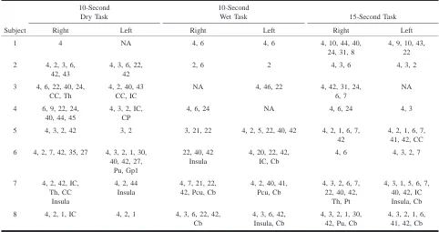
TABLE 1: Distribution of activation during swallowing tasks in Brodmann’s areas (area number designation)
Subject
10-Second Dry Task Right Left
10-Second Wet Task Right Left
15-Second Task Right Left 1 4 NA 4, 6 4, 6 4, 10, 44, 40,
24, 31, 8
4, 9, 10, 43, 22 2 4, 2, 3, 6,
42, 43
4, 3, 6, 22, 42
2, 6 2 4, 3, 6 4, 3, 2
3 4, 6, 22, 40, 24, CC, Th
4, 2, 40, 43 CC, IC
NA 4, 46, 22 4, 42, 31, 24, 6, 7
NA
4 6, 9, 22, 24, 40, 44, 45
4, 3, 2, IC, CP
4, 6, 24 NA 4, 6, 24 4, 3
5 4, 3, 2, 42 3, 2 3, 21, 22 4, 2, 5, 22, 40, 42 4, 2, 1, 6, 7, 42
4, 2, 1, 6, 7, 41, 42, CC 6 4, 2, 7, 42, 35, 27 4, 3, 2, 1, 30,
40, 42, 27, Pu, Gp1
22, 40, 42 Insula
4, 20, 22, 42, IC, Cb
4, 6 4, 3, 2, 7
7 4, 2, 42, IC, Th, CC
Insula
4, 2, 44 Insula
4, 7, 21, 22, 42, Pcu, Cb
4, 2, 40, 41, Pcu, Cb
4, 3, 2, 6, 7, 22, 40, 42,
Th, Pt
4, 3, 1, 5, 6, 7, 40, 42, IC Insula, Cb 8 4, 2, 1, IC 4, 2, 1 4, 3, 6, 22, 42,
Cb
4, 3, 6, 42, Insula, Cb
4, 3, 2, 1, 30, 42, Pu, Cb
4, 3, 2, 1, 6, 41, 42, Cb Note.—Cb indicates cerebellum; CC, corpus callosum; CP, cerebral peduncle; IC, internal capsule; Gp1, globus pallidus (medial); NA, no activation identified; Pc, precuneus; Pt, putamen; Pu, pulvinar; Th, thalamus.
Table 1 shows the distribution of activation in cortical and subcortical sites (designated in Brod-mann’s areas) during the swallowing paradigms. The activation recorded for these three tasks, for most of the subjects, was generally diffuse, occur-ring in several locations in the frontal, parietal, and temporal lobes. Specifically, Table 1 shows acti-vation occurring predominately in Brodmann’s area 4 (the primary motor cortex), areas 3, 2, and 1 (the primary somatosensory cortex), area 6 (the supple-mentary motor cortex), areas 9 and 10 (the pre-frontal cortex), area 42 (the transverse temporal gy-rus), areas 24 and 31 (the cingulate gyrus, insular cortex, and internal capsule), areas 44 and 45 (the speech areas), as well as in areas 39 and 40 (other association areas), area 22 (the superior temporal
gyrus), and areas 5 and 7 (the sensory-motor inte-gration areas).
ac-TABLE 2: Laterality index (LI) for different swallowing tasks
Subject Sex
Right or Left Hand Dominance
Task
10-Second Dry 10-Second Wet 15-Second 1
2 3 4 5 6 7 8
F M M M F F F M
Right Right Left Right Right Left Right Right
252
20.9 20
284 10 39 38 24
213 2 36 12
262
20.6 7 14
246 77
270 32 8 4 39.8
225 LI index (mean6SD)
Right* Left* Average†
246642 26612
20.74644.4
225633 14613
20.58626.5
247622 32629 4648.1 * Mean LI value for the right side (negative values) or left side (positive values) for that task.
† Mean LI value for both the left and right side, pooled across eight subjects for that task.
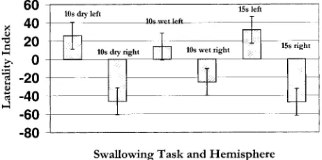
FIG2. Graph of the mean LI for three swallowing tasks in the
right and left hemispheres. Each bar represents pooled data from all eight subjects. Positive values5 left hemisphere; negative values5right hemisphere. Length of the bar represents the de-gree of laterality for that hemisphere.
tivation (cases 1 and 8: 10-second wet task; cases 2 and 8: 15-second tasks).
This apparent predominance of activity on one side of the motor cortex within subjects during swallowing tasks suggests that the motor control of swallowing may be characterized by hemispheric dominance, as has been documented for speech (10, 22). Hemispheric dominance has been quan-tified in other studies using a laterality index (LI), defined by the ratio
LI5 [Ss left 2 Ss right] / [Ss left 1 Ss right] 3100
where s5(percentage of activation)3(number of activated pixels). A positive LI indicates left hemi-spheric dominance while a negative LI indicates right hemispheric dominance. Ratios at or close to 0 are thought to represent an indeterminate domi-nance (15, 23).
Table 2 shows the LI for each of our eight sub-jects during the 10-seconds dry, 10-seconds wet, and 15-seconds dry swallowing tasks. Five of the eight subjects (cases 3, 5, 6, 7, and 8) showed left hemispheric dominance during the 10-second dry swallow task; two (cases 1 and 4) were right dom-inant, and one (case 2) was indeterminate or slight-ly right dominant. For the 15-second task, five sub-jects (cases 2, 4, 5, 6, and 7) were left dominant and three (cases 1, 3, and 8) were right dominant. Five of the subjects (cases 2, 3, 4, 7, and 8) were left dominant during the 10-second wet task, whereas subjects 1 and 5 were right dominant; sub-ject 6 was indeterminate. Examination of the data for the three swallowing tasks individually, as well as for the three tasks pooled, consistently showed that five (63%) of the eight subjects were left dom-inant. Within subjects, however, left or right hemi-spheric dominance was not consistent through the three tasks for six of the eight subjects. Thus, the LI changed for one out of the three tasks in 75% of the subjects.
[image:4.612.60.545.74.239.2] [image:4.612.311.542.280.395.2]Last, the relationship between handedness and hemispheric dominance for swallowing tasks was examined with a Pearson correlation analysis. There was no significant relationship between handedness and hemispheric dominance for swal-lowing in this study (r 5 20.12, 95% confidence interval), consistent with findings from other stud-ies (24).
Discussion
The purpose of this investigation was to identify sites of cortical activation during swallowing by us-ing functional MR imagus-ing techniques. Swallowus-ing in healthy adult subjects resulted in activation of the primary motor cortex, with activation also iden-tified in the primary somatosensory cortex, the sup-plementary motor cortex, the prefrontal cortex, the transverse temporal gyrus, the insular cortex, the internal capsule, the cingulate gyrus, the speech ar-eas, as well as other association arar-eas, the auditory association area, and the sensory-motor integration areas.
The activation identified in this study corre-sponds to sites of cortical stimulation or evoked responses reported in other studies (7, 13, 24) as well as to sites not identified with traditional cor-tical stimulation techniques. Multiple sites of acti-vation, including those associated with motor pro-cessing, suggest that the motor control of swallowing may involve several cortical sites to initiate, process, and execute the necessary output for swallowing. Activation of other cortical sites, such as the supplementary motor area, represented in the superior and middle frontal gyri, is believed to be associated with motor planning and, in par-ticular, with planning of sequential movements, as occurs with swallowing (25). Moreover, activation in the various association areas, including the speech areas and the sensory-motor integration ar-eas, is consistent with earlier studies reporting swallowing responses evoked via stimulation of these areas and activity of prefrontal and parietal association areas (7, 10).
In subcortical areas, activation of the posterior limb of the internal capsule, the insular cortex, and the thalamus was observed in the subjects during swallowing tasks. Activation of the internal capsule is an expected and important functional feature in swallowing, as the internal capsule serves to func-tionally connect cortical and brain stem nuclei via the cortical bulbar tracts. Vascular injury to these white matter tracts has been associated with oro-pharyngeal deficits in swallowing (26). Activation in the white matter, however, raises the issue of the BOLD effect in the absence of synaptic activity, since the white matter consists of axons. It is well established that neuronal activity is accompanied by increasing metabolic activity. BOLD techniques do not directly measure metabolic activity; rather BOLD contrast is a reflection of the hemodynamic response associated with increases in metabolic
ac-tivity (27, 28). Both synaptic acac-tivity and axonal conduction require the active maintenance of ionic (Na1 2 K1) gradients. The metabolic processes that drive these gradients involve the same sub-strates (eg, glutamate transport) in both the synaptic cleft and in the axon (29–31). Thus, synaptic ac-tivity and axonal acac-tivity rely on fundamentally similar metabolic processes. Aside from metabolic requirements, other explanations for the white mat-ter activation observed in these experiments may include spatial averaging from the basal ganglia in some cases. Regardless of the specific source(s) of the white matter signals, further investigation into the relationship between white matter function and hemodynamic response is warranted.
In other areas, the insular cortex is thought to serve a variety of functions, including sensory and motor integration between primary cortex and other subcortical (thalamic) nuclei or limbic areas. Other functions include a role in gustatory sensation, vis-ceral motor activity, and motor association, as well as the processing of ‘‘routine’’ or ‘‘overlearned’’ motor associated tasks. Thus, activation of the in-sular cortex during swallowing tasks may be linked to its visceral motor and integrative functions (32). The nuclei of the thalamus are heterogeneous in their functional roles, with some nuclei serving as relays for cortical areas and others generally serv-ing as association areas (33, 34). The globus pal-lidus projects to the ventral anterior group of the thalamus, which then projects to the primary motor and supplementary motor cortices. The ventral pos-terior group is primarily involved with somatosen-sory integration, receiving input from the spinotha-lamic and trigeminal tracts and projecting to the primary somatosensory cortex. The pulvinar, as an association nucleus, maintains reciprocal connec-tions to the temporal, parietal, and occipital lobes for integration of sensory information and cogni-tive and visual association functions. Activation of thalamic nuclei during swallowing tasks indicates the necessary role of sensory and motor input pro-cessing via thalamocortical or thalamostriatal path-ways in swallowing.
cortical control processes for speech and swallow-ing in the same cohort of subjects.
Hemispheric dominance, however, appeared to be variable in some subjects depending on the swallowing task. As shown in Table 2, six of the eight subjects alternated hemispheric dominance during the three swallowing tasks. For example, subject 3 was left-hemisphere dominant during the 10-second dry and 10-second wet swallow tasks, while during the 15-second task, the subject showed right hemispheric dominance. This tenden-cy toward alternate hemispheric dominance during different swallowing tasks could be attributed to a variable attention state. Other investigations have found differential activation in the motor cortex during motor activity with different attention states (37). Alternatively, previous studies have shown that subjects use highly variable neuromuscular strategies in different swallowing sensory and mo-tor tasks (11, 38). Hamdy et al (24) used transcran-ial magnetic stimulation in a group of 20 healthy subjects to show that evoked responses from the pharynx and esophagus were lateralized to either the right or left hemisphere, but there was no con-sistent pattern of lateralization from these sites. Thus, alternate hemispheric dominance may reflect a cortical organization scheme for swallowing that facilitates the diverse neuromuscular demands of different swallowing tasks.
In addition, as Figure 2 shows, lateralization in the right hemisphere tended to be greater than in the left hemisphere, although, as discussed previ-ously, the majority of subjects were left-hemisphere dominant for swallowing tasks. The average LI (Table 2) for any of the swallowing tasks, however, was less than 5 or close to 0. These results suggest that the hemispheric control of swallowing, when averaged across subjects and tasks, is indeterminate or bilateral. Why the right hemisphere shows stron-ger lateralization is unclear and remains to be de-termined; however, recent studies have provided in-sight into the developmental dichotomy of the two hemispheres. Swallowing is a phylogenetically old physiologic function and in an investigation of functional brain asymmetry during childhood, Chi-ron and colleagues (39) showed that human infants are right-hemisphere dominant up to the age of 3, at which point asymmetric shifts to the left occur. One possibility is that stronger lateralization in the right hemisphere during swallowing tasks may rep-resent a primal cortical organizational structure.
Nonetheless, there are experimental considera-tions and limitaconsidera-tions to this investigation. First, the activation observed during these tasks could rep-resent the cortical activity associated with swallow initiation (the oral phase of swallowing, thought to be under voluntary control) and not activation as-sociated with motor control of the reflexive or au-tomatic phases of swallowing: the pharyngeal and esophageal phases (40). This was addressed during the 10-second wet task by requiring the subjects to swallow a small bolus. Delivery of a bolus to the
pharyngeal inlet (tonsillar pillars) is believed to stimulate the pharyngeal phase of swallowing (40). Apart from differences in laterality measures, sites of activation during the 10-second wet task were very similar to the 10- or 15-second dry tasks for each subject. Thus, the cortical activation observed in this investigation likely represents the cortical activity associated with all phases of swallowing.
Furthermore, subjects were required to swallow repeatedly while lying supine over the course of several seconds. Although this does not represent ‘‘normal’’ swallowing conditions, subjects com-pleted swallows of normal duration for all swallow-ing tasks, as measured by the number of swallows in each activation period. Cortical activation ob-served under these conditions appears to be asso-ciated with swallows of normal duration (mean, 1.8 seconds).
Last, the subjects’ attention state was not cor-rected for in any of the swallowing tasks. Statistical analysis, however, showed no effect of task se-quence, which would indicate that differing cog-nitive attention states due to task sequence did not significantly contribute to the observed variability in hemispheric dominance. Rushworth and col-leagues (41) examined cued sequential hand move-ments and proposed that the left parietal cortex may play a role in motor attention, specifically, the shift from one movement to the next in sequential motor movements. Future studies are indicated to deter-mine the hemispheric role of motor attention states for complex movements such as swallowing.
Conclusion
The results of this investigation confirm earlier studies of cortical activity in swallowing and con-tribute additional information on the cortical or-ganization for swallowing. Although we found hemispheric dominance for different swallowing tasks under the conditions of this investigation, examination of the role of hemispheric dominance in the cortical control of swallowing and the im-plications for normal and abnormal swallowing is necessary.
Acknowledgment
We thank Andrew Kalnin for his helpful comments on the manuscript.
References
1. Alberts MJ, Horner J, Gray L, Brazer SR. Aspiration after
stroke: lesion analysis by brain MRI. Dysphagia 1992;7:
170–173
2. Crary M. A direct intervention program for chronic
neurogen-ic dysphagia secondary to brainstem stroke. Dysphagia 1995;
10:6–18
3. Horner J, Massey EW, Riski JE, Lathrop DL, Chase KN.
Aspi-ration following stroke: clinical correlates and outcome. Neu-rology 1988;38:1359–1362
4. Horner J, Buoyer FG, Alberts MJ, Helms MJ. Dysphagia
follow-ing brain-stem stroke clinical correlates and outcome. Arch Neurol 1991;48:1170–1173
5. Kirshner H. Causes of neurogenic dysphagia. Dysphagia 1989; 3:184–188
6. Miller AJ. Neurophysiological basis of swallowing. Dysphagia 1986;1:91–100
7. Martin R, Sessle B. The role of the cerebral cortex in
swallow-ing. Dysphagia 1993;8:195–202
8. Veis SL, Logemann JA. Swallowing disorders in persons with
cerebrovascular accident. Arch Phys Med Rehabil 1985;66:
372–375
9. Robbins J, Levine R, Maser A, Rosenbek JC, Kempster G.
Swal-lowing after unilateral stroke of the cerebral cortex. Arch Phys Med Rehabil 1993;74:1295–1300
10. Penfield W, Boldery E. Somatic motor and sensory
represen-tation in the cerebral cortex of man as studied by electrical stimulation. Brain 1937;60:389–443
11. Mosier K. The Motor Control of Swallowing. Ann Arbor, MI: UMI Publications; 1997;71–109
12. Martin RE, Murray GM, Sessle BJ. Cerebral cortical control of
primate orofacial movements: role of face motor cortex in trained and semi-automatic motor behaviors. In: Alpha and
Gamma Motor Systems. New York: Plenum Press; 1995;350–355 13. Martin RE, Murray GM, Kemppainen P, Masuda Y, Sessle BJ.
Functional properties of neurons in the primate tongue pri-mary motor cortex during swallowing. J Neurophysiol 1997;78:
1516–1530
14. Orrison WW, Lewine JD, Sanders J, Hartshorne MF. Functional
Brain Imaging. St Louis: Mosby; 1995;239–326
15. Yetkin FZ, Hammeke TA, Swanson SJ, et al. A comparison of
functional MR activation patterns during silent and audible language tasks. AJNR Am J Neuroradiol 1995;16:1087–1092
16. Yousry T, Schmid U, Jassoy AG, et al. Topography of the
cor-tical motor hand area: prospective study with functional MR imaging and direct motor mapping at surgery. Radiology 1995;
195:23–29
17. Zarahn E, Aguire GK, D’Esposito M. Empirical analyses of
BOLD fMRI statistics. Neuroimage 1997;5:179–197
18. Maldjian J, Howard R, van Buchem M, et al. Functional
mag-netic resonance imaging of regional brain activity in patients with intracerebral arteriovenous malformation before surgical or endovascular therapy. J Neurosurg 1996;84:477–483
19. Meyer JR, Roychowdhury S, Russel EJ, Callahan C, Gitelman D, Mesulam MM. Location of the central sulcus via cortical
thick-ness of the precentral and postcentral gyri on MR. AJNR Am J Neuroradiol 1996;17:1699–1706
20. Talairach J, Tournoux P. Co-Planar Stereotaxic Atlas of the
Hu-man Brain. New York: Thieme; 1988;37–110
21. Truwit CL, Lempert TE. High Resolution Atlas of Cranial
Neu-roanatomy. Baltimore: Williams & Wilkins; 1994;2–302
22. Ojemann GA. Speech representation in ventrolateral thalamus.
Brain 1971;94:669–680
23. Binder JR, Swanson SJ, Hammeke TA, et al. Determination of
language dominance using functional MRI: a comparison with the Wada test. Neurology 1996;46:978–984
24. Hamdy S, Aziz Q, Rothwell JC, et al. The cortical topography
of human swallowing musculature in health and disease. Nat Med 1996;2:1217–1224
25. Tanji J, Shima K, Mushiake H. Multiple cortical motor areas
and temporal sequencing of movements. Cog Brain Res 1996;
5:117–122
26. Logemann JA. Oropharyngeal swallowing after stroke in the
left basal ganglion/internal capsule. Dysphagia 1993;8:230–234
27. Ogawa S, Lee TM, Kay AR, Tank DW. Brain magnetic
reso-nance imaging with contrast dependent on blood oxygenation.
Proc natl. Acad. Sci. USA 1990;87:9868–9872
28. Ogawa S, Menon RS, Tank DW et al. Functional brain mapping
by blood oxygenation level-dependent contrast magnetic res-onance imaging. Biophys J. 1993;64:803–812
29. Magistretti PJ, Pellerin L, Rothman D, Schulman RG. Energy on
Demand. Science 1999;283:496–497
30. Kriegler S, Chiu SY. Calcium Signaling of Glial Cells along
Mammalian Axons. J Neuroscience 1993;13:10:4229–4245
31. Chiu SY, Kriegler S. Neurotransmitter-mediated signaling
be-tween axons and glial cells. Glia 1994;11:2:191–200
32. Augustine JR. Circuitry and functional aspects of the insular
lobe in primates including humans. Brain Res Rev 1996;22:
229–244
33. Heimer L. The Human Brain and Spinal Cord: Functional
Neuroanatomy and Dissection Guide. New York: Springer;
1983;331–352
34. Netter FH. The Ciba Collection of Medical Illustrations, Vol. 1: Nervous System, Part 1: Anatomy and Physiology, West Cald-well, NJ: Ciba Pharmaceuticals; 1995;192–193
35. Geschwind N, Levitsky W. Human brain: left-right
asymme-tries in temporal speech region. Science 1968;161:186–187
36. Geschwind N, Galabruder A. Cerebral lateralization: biological
mechanisms, associations, and pathology, I: a hypothesis and a program for research. Arch Neurol 1985;42:428–459
37. Binkofski F, Buccino G, Taylor JG, et al. Attention modulates
motor cortex activation: an fMRI study. Neuroimage 1998;7:S84
38. Gay T, Rendell J, Spiro J, Mosier K, Lurie A. Coordination of
oral cavity and laryngeal movements during swallowing. J Appl Physiol 1994;77:357–365
39. Chiron C, Jambaque I, Nabbout R, Lounes R, Syrota A, Dulac O.
The right brain hemisphere is dominant in human infants. Brain 1997;120:(Pt 6):1057–1065
40. Jones B, Donner M. Normal and Abnormal Swallowing:
Im-aging in Diagnosis and Therapy. New York: Springer; 1991;
7–32
41. Rushworth MF, Nixon PD, Renowden S, Wade DT, Passingham RE. The left parietal cortex and motor attention.