(
E
)-2-Phenyl-
N
-tosylnon-2-en-4-ynamide
Xiang-Zhen Meng and Xin-Jun Wan*
Department of Chemistry and Life Science, Chaohu College, Anhui Province, People’s Republic of China
Correspondence e-mail: [email protected]
Received 22 November 2012; accepted 26 November 2012
Key indicators: single-crystal X-ray study;T= 293 K; mean(C–C) = 0.004 A˚; disorder in main residue;Rfactor = 0.048;wRfactor = 0.096; data-to-parameter ratio = 15.5.
The molecule of the title compound, C22H23NO3S, adopts anE
conformation about the C C bond. The dihedral angle between the benzene rings is 23.79 (5). In the crystal, pairs of
N—H O hydrogen bonds link the molecules, forming inversion dimers. The terminal butyl group is disordered over two sets of sites in a 0.559 (6):0.441 (6) ratio.
Related literature
For the synthesis of the titlw compound, see: Cheng et al.
(2012). For applications of conjugated enynes, see: Ochiaiet al.
(1999); Saitoet al.(2001).
Experimental
Crystal data
C22H23NO3S
Mr= 381.47
Triclinic,P1
a= 9.8186 (10) A˚
b= 9.8201 (9) A˚
c= 11.3352 (13) A˚
= 81.470 (8) = 76.308 (9)
= 75.042 (9)
V= 1021.46 (18) A˚3
Z= 2
MoKradiation
= 0.18 mm1
T= 293 K
0.420.380.32 mm
Data collection
Agilent Xcalibur (Atlas, Gemini ultra) diffractometer
Absorption correction: multi-scan (CrysAlis PRO; Agilent, 2011)
Tmin= 0.928,Tmax= 0.945
9118 measured reflections 4425 independent reflections 3005 reflections withI> 2(I)
Rint= 0.028
Refinement
R[F2> 2(F2)] = 0.048
wR(F2) = 0.096
S= 1.00 4425 reflections 285 parameters
170 restraints
H-atom parameters constrained max= 0.19 e A˚
3 min=0.28 e A˚
3
Table 1
Hydrogen-bond geometry (A˚ ,).
D—H A D—H H A D A D—H A
N1—H1 O2i
0.86 2.32 2.947 (2) 130
Symmetry code: (i)xþ1;yþ1;z.
Data collection: CrysAlis PRO(Agilent, 2011); cell refinement: CrysAlis PRO; data reduction: CrysAlis PRO; program(s) used to solve structure: SHELXTL (Sheldrick, 2008); program(s) used to refine structure: SHELXTL; molecular graphics: SHELXTL; soft-ware used to prepare material for publication:SHELXTL.
The work was supported financially by the Chaohu College Project (XLY–201105).
Supplementary data and figures for this paper are available from the IUCr electronic archives (Reference: XU5655).
References
Agilent (2011).CrysAlis PRO. Agilent Technologies, Yarnton, England. Cheng, D., Ling, F., Li, Z.-X., Yao, W.-J. & Ma, C. (2012).Org. Lett.14, 3146–
3149.
Ochiai, B., Tomita, I. & Endo, T. (1999).Macromolecules,32, 238–240. Saito, S., Kawasaki, T., Tsuboya, N. & Yamamoto, Y. (2001).J. Org. Chem.66,
796–802.
Sheldrick, G. M. (2008).Acta Cryst.A64, 112–122. Acta Crystallographica Section E
Structure Reports
Online
supporting information
Acta Cryst. (2013). E69, o78 [https://doi.org/10.1107/S1600536812048489]
(
E
)-2-Phenyl-
N
-tosylnon-2-en-4-ynamide
Xiang-Zhen Meng and Xin-Jun Wan
S1. Comment
Conjugated enynes can be used in the synthesis of polymers (Ochiai et al., 1999) and in the selective construction of aromatic frameworks (Saito et al., 2001). Here, we report the crystal structure of the title enyne compound.
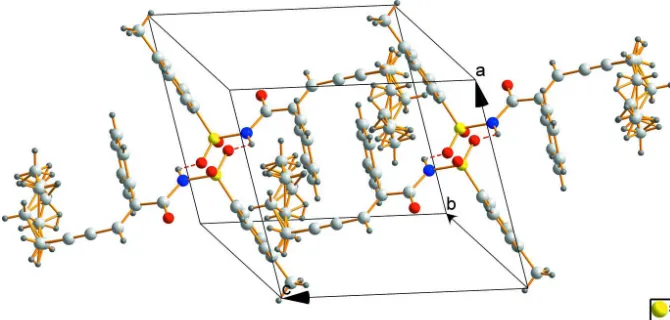
The molecular structure of the title compound is shown in Figure 1, the ORTEP diagram shows that the structure adopts the E isomer, the double bond and triple bond are within normal ranges. The benzene C2–C7 and C10–C15 rings are tilted relative to each other by 23.79 (5)°. The chain C19—C22 is disorder. A view of the crystal packing for the title compound is illustrated in Fig. 2, the crystal structure is stabilized by N—H···O hydrogen bonds.
S2. Experimental
The compound was prepared according to the reference (Cheng et al., 2012). 4-Methylbenzenesulfonyl azide (0.45 mmol), CuI (5.7 mg, 0.03 mmol), ethynylbenzene (0.45 mmol), and hept-2-ynal (0.3 mmol) were suspended in THF in a 10 ml Schlenk tube at room temperature at N2 atmosphere. Cs2CO3 (8.64 mg, 0.36 mmol) was then added, and the
resulting solution was stirred at this temperature for 24 h. The reaction was quenched by saturated aqueous NH4Cl (5 ml)
and extracted with CH2Cl2 (15 ml × 3). The combined organic layers were dried over anhydrous Na2SO4 and concentrated in vacuo. The crude residue was purified by column chromatography on silica gel (n-hexane/EtOAc) to afford the title compound. The title compound was recrystallized from CH2Cl2 at room temperature to give the desired crystals suitable
for single-crystal X-ray diffraction.
S3. Refinement
H atoms were placed in geometrically idealized positions and constrained to ride on their parent atoms, with aromatic C —H = 0.93–0.97 Å and N—H = 0.86 Å, Uiso(H) = 1.5Ueq(C) for methyl H atoms and 1.2Ueq(C,N) for the others. The butyl
Figure 1
The molecular structure of the compound with displacement ellipsoids drawn at 30% probability level.
Figure 2
The crystal packing diagram.
(E)-2-Phenyl-N-tosylnon-2-en-4-ynamide
Crystal data
C22H23NO3S Mr = 381.47
Triclinic, P1 Hall symbol: -P 1
a = 9.8186 (10) Å
b = 9.8201 (9) Å
c = 11.3352 (13) Å
α = 81.470 (8)°
β = 76.308 (9)°
γ = 75.042 (9)°
V = 1021.46 (18) Å3
Z = 2
F(000) = 404
Dx = 1.240 Mg m−3
Mo Kα radiation, λ = 0.71073 Å Cell parameters from 2435 reflections
θ = 2.9–29.6°
[image:3.610.135.470.286.446.2]Data collection
Agilent Xcalibur (Atlas, Gemini ultra) diffractometer
Radiation source: fine-focus sealed tube Graphite monochromator
Detector resolution: 10.3592 pixels mm-1 ω scans
Absorption correction: multi-scan (CrysAlis PRO; Agilent, 2011)
Tmin = 0.928, Tmax = 0.945
9118 measured reflections 4425 independent reflections 3005 reflections with I > 2σ(I)
Rint = 0.028
θmax = 27.1°, θmin = 2.9° h = −12→11
k = −12→12
l = −14→13
Refinement
Refinement on F2
Least-squares matrix: full
R[F2 > 2σ(F2)] = 0.048 wR(F2) = 0.096 S = 1.00 4425 reflections 285 parameters 170 restraints
Primary atom site location: structure-invariant direct methods
Secondary atom site location: difference Fourier map
Hydrogen site location: inferred from neighbouring sites
H-atom parameters constrained
w = 1/[σ2(F
o2) + (0.0145P)2 + 0.450P]
where P = (Fo2 + 2Fc2)/3
(Δ/σ)max < 0.001
Δρmax = 0.19 e Å−3
Δρmin = −0.28 e Å−3
Extinction correction: SHELXTL (Sheldrick, 2008), Fc*=kFc[1+0.001xFc2λ3/sin(2θ)]-1/4
Extinction coefficient: 0.0511 (16)
Special details
Geometry. All e.s.d.'s (except the e.s.d. in the dihedral angle between two l.s. planes) are estimated using the full covariance matrix. The cell e.s.d.'s are taken into account individually in the estimation of e.s.d.'s in distances, angles and torsion angles; correlations between e.s.d.'s in cell parameters are only used when they are defined by crystal symmetry. An approximate (isotropic) treatment of cell e.s.d.'s is used for estimating e.s.d.'s involving l.s. planes.
Refinement. Refinement of F2 against ALL reflections. The weighted R-factor wR and goodness of fit S are based on F2,
conventional R-factors R are based on F, with F set to zero for negative F2. The threshold expression of F2 > σ(F2) is used
only for calculating R-factors(gt) etc. and is not relevant to the choice of reflections for refinement. R-factors based on F2
are statistically about twice as large as those based on F, and R- factors based on ALL data will be even larger.
Fractional atomic coordinates and isotropic or equivalent isotropic displacement parameters (Å2)
x y z Uiso*/Ueq Occ. (<1)
C4 0.1106 (3) 0.2494 (3) −0.0651 (2) 0.0753 (8) C5 0.1929 (3) 0.1314 (3) −0.0146 (3) 0.0774 (8) H5 0.1695 0.0446 −0.0105 0.093* C6 0.3097 (3) 0.1372 (2) 0.0305 (2) 0.0608 (6) H6 0.3638 0.0557 0.0652 0.073* C7 0.3449 (2) 0.26491 (19) 0.02339 (17) 0.0464 (5) C8 0.3496 (2) 0.3491 (2) 0.29221 (19) 0.0529 (5) C9 0.3001 (2) 0.4641 (2) 0.37598 (18) 0.0483 (5) C10 0.3535 (2) 0.5957 (2) 0.34604 (17) 0.0461 (5) C11 0.2602 (3) 0.7245 (2) 0.3255 (2) 0.0645 (6) H11 0.1639 0.7281 0.3285 0.077* C12 0.3082 (4) 0.8474 (3) 0.3008 (2) 0.0830 (9) H12 0.2442 0.9333 0.2871 0.100* C13 0.4484 (4) 0.8444 (3) 0.2964 (2) 0.0825 (9) H13 0.4803 0.9278 0.2790 0.099* C14 0.5422 (3) 0.7185 (3) 0.3175 (2) 0.0743 (7) H14 0.6379 0.7163 0.3158 0.089* C15 0.4949 (3) 0.5942 (2) 0.3415 (2) 0.0576 (6) H15 0.5597 0.5086 0.3547 0.069* C16 0.2024 (3) 0.4427 (2) 0.4767 (2) 0.0615 (6) H16 0.1715 0.3588 0.4888 0.074* C17 0.1426 (3) 0.5407 (3) 0.5667 (2) 0.0658 (7) C18 0.0934 (3) 0.6244 (3) 0.6390 (2) 0.0734 (7)
H22F 0.1266 1.0864 0.5580 0.239* 0.441 (6)
Atomic displacement parameters (Å2)
U11 U22 U33 U12 U13 U23
S1 0.0597 (3) 0.0411 (3) 0.0472 (3) −0.0092 (2) −0.0053 (3) −0.0148 (2) O1 0.0807 (11) 0.0439 (8) 0.0695 (11) 0.0011 (7) −0.0232 (9) −0.0118 (7) O2 0.0599 (9) 0.0561 (8) 0.0546 (9) −0.0175 (7) 0.0046 (7) −0.0182 (7) O3 0.1151 (15) 0.0594 (9) 0.0603 (10) −0.0392 (10) 0.0058 (10) −0.0144 (8) N1 0.0683 (12) 0.0441 (9) 0.0449 (10) −0.0186 (8) −0.0025 (9) −0.0154 (8) C1 0.081 (2) 0.166 (3) 0.153 (3) −0.021 (2) −0.040 (2) −0.074 (3) C2 0.0676 (16) 0.0556 (13) 0.0668 (16) −0.0143 (12) −0.0147 (13) −0.0064 (11) C3 0.0676 (17) 0.0838 (18) 0.0725 (18) −0.0055 (14) −0.0183 (14) −0.0122 (14) C4 0.0601 (16) 0.099 (2) 0.0725 (17) −0.0169 (15) −0.0041 (14) −0.0429 (16) C5 0.0717 (18) 0.0737 (17) 0.094 (2) −0.0262 (15) −0.0009 (16) −0.0413 (15) C6 0.0693 (16) 0.0480 (12) 0.0651 (15) −0.0148 (11) −0.0040 (12) −0.0193 (11) C7 0.0553 (13) 0.0445 (11) 0.0385 (11) −0.0129 (9) 0.0000 (9) −0.0141 (9) C8 0.0640 (14) 0.0505 (12) 0.0462 (12) −0.0160 (11) −0.0087 (11) −0.0104 (10) C9 0.0536 (13) 0.0529 (11) 0.0409 (12) −0.0134 (10) −0.0089 (10) −0.0116 (9) C10 0.0568 (13) 0.0477 (11) 0.0338 (10) −0.0093 (10) −0.0079 (9) −0.0112 (8) C11 0.0732 (16) 0.0630 (14) 0.0511 (14) −0.0033 (12) −0.0158 (12) −0.0035 (11) C12 0.117 (3) 0.0498 (14) 0.0658 (17) −0.0027 (15) −0.0091 (17) 0.0021 (12) C13 0.131 (3) 0.0585 (16) 0.0590 (16) −0.0407 (18) 0.0002 (17) −0.0082 (12) C14 0.0855 (19) 0.0792 (18) 0.0674 (17) −0.0391 (15) −0.0066 (14) −0.0155 (14) C15 0.0634 (15) 0.0532 (12) 0.0585 (14) −0.0144 (11) −0.0116 (12) −0.0122 (10) C16 0.0682 (15) 0.0689 (14) 0.0531 (14) −0.0278 (12) −0.0043 (12) −0.0164 (11) C17 0.0645 (15) 0.0796 (16) 0.0535 (14) −0.0271 (13) 0.0059 (12) −0.0179 (13) C18 0.0740 (17) 0.0854 (17) 0.0586 (15) −0.0279 (14) 0.0098 (13) −0.0216 (14) C19 0.118 (6) 0.085 (5) 0.071 (5) −0.028 (4) 0.022 (4) −0.029 (4) C20 0.101 (4) 0.097 (4) 0.096 (5) −0.024 (4) 0.005 (4) −0.041 (4) C21 0.089 (4) 0.089 (4) 0.066 (3) −0.018 (3) −0.024 (3) −0.016 (3) C22 0.143 (7) 0.104 (6) 0.130 (6) −0.056 (5) −0.011 (6) −0.037 (5) C19A 0.095 (6) 0.092 (6) 0.058 (5) −0.026 (5) 0.018 (4) −0.030 (5) C20A 0.117 (6) 0.110 (5) 0.063 (4) −0.030 (4) −0.006 (4) −0.031 (4) C21A 0.139 (7) 0.145 (7) 0.099 (6) −0.068 (6) 0.015 (6) −0.042 (5) C22A 0.197 (11) 0.108 (7) 0.121 (8) 0.018 (8) −0.002 (9) 0.009 (6)
Geometric parameters (Å, º)
S1—O1 1.4179 (15) C14—C15 1.384 (3) S1—O2 1.4329 (14) C14—H14 0.9300 S1—N1 1.6488 (15) C15—H15 0.9300 S1—C7 1.743 (2) C16—C17 1.424 (3) O3—C8 1.205 (2) C16—H16 0.9300 N1—C8 1.387 (3) C17—C18 1.178 (3)
N1—H1 0.8600 C18—C19 1.472 (6)
C1—H1B 0.9600 C19—H19A 0.9700
C1—H1C 0.9600 C19—H19B 0.9700
C2—C3 1.368 (3) C20—C21 1.595 (6) C2—C7 1.380 (3) C20—H20A 0.9700
C2—H2 0.9300 C20—H20B 0.9700
C3—C4 1.377 (3) C21—C22 1.374 (6)
C3—H3 0.9300 C21—H21A 0.9700
C4—C5 1.365 (4) C21—H21B 0.9700 C5—C6 1.378 (3) C22—H22A 0.9600
C5—H5 0.9300 C22—H22B 0.9600
C6—C7 1.371 (3) C22—H22C 0.9600 C6—H6 0.9300 C19A—C20A 1.567 (9) C8—C9 1.493 (3) C19A—H19C 0.9700 C9—C16 1.335 (3) C19A—H19D 0.9700 C9—C10 1.482 (3) C20A—C21A 1.570 (8) C10—C15 1.373 (3) C20A—H20C 0.9700 C10—C11 1.382 (3) C20A—H20D 0.9700 C11—C12 1.373 (3) C21A—C22A 1.321 (8) C11—H11 0.9300 C21A—H21C 0.9700 C12—C13 1.358 (4) C21A—H21D 0.9700 C12—H12 0.9300 C22A—H22D 0.9600 C13—C14 1.365 (4) C22A—H22E 0.9600 C13—H13 0.9300 C22A—H22F 0.9600
C4—C5—C6 121.8 (2) C22—C21—H21A 108.8 C4—C5—H5 119.1 C20—C21—H21A 108.8 C6—C5—H5 119.1 C22—C21—H21B 108.8 C7—C6—C5 119.1 (2) C20—C21—H21B 108.8 C7—C6—H6 120.5 H21A—C21—H21B 107.7 C5—C6—H6 120.5 C18—C19A—C20A 110.9 (8) C6—C7—C2 120.2 (2) C18—C19A—H19C 109.5 C6—C7—S1 120.43 (17) C20A—C19A—H19C 109.5 C2—C7—S1 119.29 (16) C18—C19A—H19D 109.5 O3—C8—N1 121.30 (18) C20A—C19A—H19D 109.5 O3—C8—C9 124.1 (2) H19C—C19A—H19D 108.0 N1—C8—C9 114.59 (17) C19A—C20A—C21A 97.2 (11) C16—C9—C10 122.48 (18) C19A—C20A—H20C 112.3 C16—C9—C8 116.03 (19) C21A—C20A—H20C 112.3 C10—C9—C8 121.45 (17) C19A—C20A—H20D 112.3 C15—C10—C11 118.2 (2) C21A—C20A—H20D 112.3 C15—C10—C9 121.44 (19) H20C—C20A—H20D 109.9 C11—C10—C9 120.4 (2) C22A—C21A—C20A 135.1 (11) C12—C11—C10 120.7 (3) C22A—C21A—H21C 103.4 C12—C11—H11 119.6 C20A—C21A—H21C 103.4 C10—C11—H11 119.6 C22A—C21A—H21D 103.4 C13—C12—C11 120.5 (3) C20A—C21A—H21D 103.4 C13—C12—H12 119.8 H21C—C21A—H21D 105.2 C11—C12—H12 119.8 C21A—C22A—H22D 109.5 C12—C13—C14 119.8 (2) C21A—C22A—H22E 109.5 C12—C13—H13 120.1 H22D—C22A—H22E 109.5 C14—C13—H13 120.1 C21A—C22A—H22F 109.5 C13—C14—C15 120.0 (3) H22D—C22A—H22F 109.5 C13—C14—H14 120.0 H22E—C22A—H22F 109.5
Hydrogen-bond geometry (Å, º)
D—H···A D—H H···A D···A D—H···A
N1—H1···O2i 0.86 2.32 2.947 (2) 130