C
OMPARING AND
C
ONTRASTING THE
E
FFECTS OF
A
EROBIC
E
XERCISE AND THE
E
XERCISE
M
IMETIC
,
R
ESVERATROL
,
ON
N
EUROCOGNITIVE
F
UNCTION
R
OSALINDC
LAREH
USSEYSEPTEMBER 2013
Trinity College Institute of Neuroscience, School of Psychology,
Trinity College, University of Dublin,
Dublin 2, Ireland.
D
ECLARATION
I declare that this thesis has not been submitted as an exercise for a degree at this or any other university and it is entirely my own work.
I agree to deposit this thesis in the University’s open access institutional repository or allow the library to do so on my behalf, subject to Irish Copyright Legislation and Trinity College Library conditions of use and acknowledgement.
Signed
A
CKNOWLEDGEMENTS
Thanks go out first and foremost to my parents, Dr. Séamus Hussey and Dr. Kathleen Cassidy, for all the help and love you have provided to me throughout my life. It has been my life goal to do you proud and hopefully the completion of this thesis will add another notch to this post. Mum, I think of you often. I am also happy for all the light relief provided by my family and my fiancé’s family over the years: welcoming spouses into the family, and my four wonderful nieces and nephews. I love you all.
Thanks to Prof. Shane O’Mara for providing me with the opportunity to work under your guidance. It was your good humour and ideas that have made this journey, on the whole, a pleasure. Although we may have started down a very different path together, I’m very happy with the outcome of four years of ups and downs. Thanks to GlaxoSmithKline for providing the studentship for me to carry out this work and Richard Porter for the opportunity to work in his laboratory.
I would like to thank all the members that have passed through the Shane O’Mara lab throughout the years, providing help and happy times: Charlotte Callaghan, Jen Rouine, Maciej Jankowski, Marian Tsanov, Wen Whelan, Paul Wynne, Joanne Feeney, Ehsan Chah, Andrea Della Chiesa, Colleen Warren, as well as those members here for shorter projects and those outwith our own lab. Thanks also, for the support provided by my college appraisers, Prof. Ian Robertson and Barbara Hannigan. My appreciation is heartily extended to all the TCIN administration staff that helped me sort college necessities, experimental deliveries, conference travels and claim expenses!
Thanks to all of my friends that have made these college days a joy especially through orienteering, running, and those I have lived with over the years. I also appreciate the fun times spent with those in the TCD Geology Department. My gratitude goes out to all those within Irish Orienteering, Irish Mountain Running and DUCAC that have encouraged my sporting life throughout these years in Dublin.
T
ABLE OF
C
ONTENTS
I Title Page II Declaration III Summary
IV Acknowledgements V Table of Contents VI Abbreviations
CHAPTER ONE: Literature Review . . . 1
1.1 Introduction . . . 1
1.2 The Ageing Body and Brain. . . 3
1.2.1 Degeneration of Ageing . . . 4
1.2.2 Memory Decline Associated with Ageing . . . 17
1.2.3 Memory Decline Associated with Noncommunicable Diseases. . . 27
1.2.4 A Healthy Mind in a Healthy Body . . . 30
1.3 Aerobic Exercise Benefits Body and Brain . . . 31
1.3.1 Therapeutic Action of Aerobic Exercise on Ageing . . . 31
1.3.2 Therapeutic Action of Aerobic Exercise on Noncommunicable Diseases . 36 1.3.3 Therapeutic Potential of Aerobic Exercise on Memory Decline. . . 38
1.4 Resveratrol Action on Body and Brain . . . 40
1.4.1 Resveratrol – understanding the compound . . . 40
1.4.2 Therapeutic Action of Resveratrol on Ageing . . . 41
1.4.3 Therapeutic Action of Resveratrol on Noncommunicable Diseases . . . 45
1.4.4 Therapeutic Potential of Resveratrol on Memory Decline . . . 48
CHAPTER TWO: Aerobic Exercise and Resveratrol Improve Long-Term Memory through an
AMPK/SIRT1-independent Pathway . . . 52
2.1 Abstract . . . 52
2.2 Introduction . . . 54
2.3 Materials and Methods . . . 58
2.3.1 Animals . . . 58
2.3.2 Drug and Dosing Regime . . . 58
2.3.3 Exercise Programme . . . 58
2.3.4 Psychomotor Behaviour . . . 59
2.3.5 Open Field Exploration . . . 60
2.3.6 Novel Object Recognition Task . . . 60
2.3.7 Tissues and Serum Samples . . . 61
2.3.8 Analysis of Protein Levels by Enzyme-Linked Immunosorbent Assay (ELISA) . . . 62
2.3.9 Analysis of Protein Expression by Real-Time Polymerase Chain Reaction (RT-PCR) . . . 62
2.3.10 Statistical Analysis . . . 63
2.4 Results . . . 64
2.4.1 Psychomotor Behaviour Comparisons for Group Matching . . . 64
2.4.2 Effects of Exercise Compared to Resveratrol on Open Field Behaviour . . 65
2.4.3 Treadmill Running and Resveratrol Ingestion have Similar Effects on NOR Performance . . . 67
2.4.4 Effect on Protein Expression in Hippocampus and Perirhinal Cortex . . . . 73
2.4.5 Effect on BDNF and NGF Levels in Hippocampus and Perirhinal Cortex . . . . . . 78
2.5 Discussion . . . 82
CHAPTER THREE: Orally Administered Resveratrol Alleviates Scopolamine-Induced Amnesia . . . 88
3.1 Abstract . . . 88
3.2 Introduction . . . 90
3.3.1 Animals . . . 94
3.3.2 Drug and Dosing Regime . . . 94
3.3.3 Exercise Programme . . . 95
3.3.4 Open Field Exploration . . . 95
3.3.5 Novel Object Recognition Task . . . 96
3.3.6 Tissues and Serum Samples . . . 98
3.3.7 Analysis of Protein Levels by Enzyme-Linked Immunosorbent Assay (ELISA) . . . 99
3.3.8 Analysis of Protein Expression by Real-Time Polymerase Chain Reaction (RT-PCR) . . . 99
3.3.9 Statistical Analysis . . . 100
3.4 Results . . . 101
3.4.1 Experiment One: Effect of Exercise Compared to Resveratrol on Open Field Behaviour . . . 101
3.4.2 Treadmill Running and Resveratrol Ingestion have Similar Effects on NOR Performance . . . 102
3.4.3 Effect on Protein Expression in Hippocampus and Perirhinal Cortex . . . 105
3.4.4 Effect on BDNF and NGF Levels in Hippocampus and Perirhinal Cortex . . . . . . 108
3.4.5 Experiment Two: Effect of Exercise Compared to Resveratrol on Open Field Behaviour . . . 110
3.4.6 Effect of Scopolamine on Learning and Memory . . . 111
3.4.7 Improvements to Scopolamine-Induced Amnesia with Resveratrol Ingestion but not Treadmill Running . . . 112
3.5 Discussion . . . 115
CHAPTER FOUR: Oral Resveratrol and Aerobic Exercise Regimes Can Improve Memory Without Increasing General Mitochondrial Function . . . 123
4.1 Abstract . . . 123
4.2 Introduction . . . 125
4.3 Materials and Methods . . . 130
4.3.1 Animals . . . 130
4.3.3 Exercise Programme . . . 130
4.3.4 Tissues and Serum Samples . . . 131
4.3.5 Isolation of Mature Brown Adipocytes . . . 132
4.3.6 Measurement of Oxygen Consumption in Brown Adipocytes . . . 132
4.3.7 Preparation of BAT and SKM Mitochondria for Citrate Synthase . . . 133
4.3.8 Bicinchonic Acid (BCA) Assay to Determine Total Protein Concentration . . . . . 134
4.3.9 Citrate Synthase (CS) Assay to Determine Mitochondrial Abundance . . 134
4.3.10 Analysis of UCP-1 in BAT and UCP-3 in SKM Mitochondria using SDS-PAGE and Western Blot Analysis . . . 135
4.3.11 Statistical Analysis . . . 136
4.4 Results . . . 137
4.4.1 No Effect on Oxygen Consumption in Brown Adipocytes . . . 137
4.4.2 No Effect on Mitochondrial Abundance in BAT or SKM . . . 138
4.4.3 No Effect on UCP-1 expression in BAT and UCP-3 in SKM Mitochondria . . . . 139
4.5 Discussion . . . 142
CHAPTER FIVE: Aerobic Exercise and Resveratrol as Preventative Interventions in Working Memory Decline . . . 147
5.1 Abstract . . . 147
5.2 Introduction . . . 149
5.3 Materials and Methods . . . 152
5.3.1 Animals . . . 152
5.3.2 Drug and Dosing Regime . . . 152
5.3.3 Exercise Programme . . . 152
5.3.4 Delayed Non-Matching-to-Sample Apparatus . . . 153
5.3.5 Delayed Non-Matching-to-Sample Task Design . . . .153
5.3.6 Delayed Non-Matching-to-Sample Training Protocol . . . 154
5.3.7 Statistical Analysis . . . 157
5.4 Results . . . 158
5.4.2 Wheel Running and Resveratrol Ingestion Enhance Working Memory . .160 5.4.3 Wheel Running and Resveratrol Ingestion Improve Recovery in DNMS
Performance . . . 162
5.4.4 Effects of Resveratrol and Wheel Running Less Potent Following 8 Week Break . . . 167
5.5 Discussion . . . 171
CHAPTER SIX: Conclusions and Recommendations . . . 178
6.1 Synopsis of Results . . . 178
6.2 Future Work . . . 182
6.3 Concluding Remarks . . . 184
A
BBREVIATIONS
5-HPETE Arachidonic acid 5-hydroperoxide
5-LO Arachidonate 5-lipoxygenase
AA Arachidonic acid
AC Adenylate cyclise
AD Alzheimer’s disease
ADP Adenosine-5’-diphosphate
AMP Adenosine-5’-monophosphate
AMPK 5’ AMP-activated protein kinase
ANOVA Analysis of Variance
AP-1 Activator protein-1
ATP Adenosine-5’-triphosphate
BAT Brown adipose tissue BBB Blood-brain barrier
BCA Bicinchoninic Acid
BDNF Brain-derived neurotrophic factor
bFGF Basis fibroblast growth factor
BrdU Bromodeoxyuridine
BSA Bovine serum albumin
Ca2+ Calcium
CA1 Region I cornu ammonis
CA2 Region II cornu ammonis
CA3 Region III cornu ammonis
CAM Cell adhesion molecule
ICAM Intercellular cell adhesion molecule
VCAM Vascular cell adhesion molecule
CaMKII Ca2+/calmodulin-dependent protein kinases II
cAMP Cyclic adenosine monophosphate
cDNA Complementary deoxyribonucleic acid
C/EBPα CCAAT/enhancer binding α
cGMP Cyclic guanosine monophosphate
CNS Central nervous system
CoA-SH Coenzyme A
COX Cyclooxygenase
COX1 a cyclooxygenase isoenzyme
COX2 a cyclooxygenase isoenzyme
CR Calorie restriction
CREB Cyclic AMP response element-binding protein
CS Citrate synthase
DALY Disability-adjusted life year
DG Dentate gyrus
DHEA Dehydroepiandrosterone
DNA Deoxyribonucleic acid
DNMS Delayed-non-matching-to-sample DTNB Ellman’s reagent
ECL Enhanced chemiluminescence
ELISA Enxyme-linked immunosorbent assay
FaO a differentiated rat liver cancer line
FDG-PET Fludeoxyglucose-positron emission tomography
FOXO Forkhead box O
GFP Green fluorescent protein
GPCR G protein-coupled receptor
GPx Glutathione peroxidase
HDL High-density lipoprotein
HeLa immortal human cell line derived from cancer cells in patient, Henrietta Lacks
HIPP Hippocampus
H.M. a famous patient of Scoville and Milner
HO Haem oxygenase
HPA Hypothalamic-pituitary-adrenal
i.c.v. Intracerebroventricularly
IGF Insulin-like growth factor
IL Interleukin
i.p. Intraperitoneally
IQ Intelligence quotient
IRF-1 Interferon regulatory factor 1
ITI Intertrial interval
IKK IκB kinase
i.v. Intravenously
LDL Low-density lipoprotein
LKB1 Serine-threonine kinase B1
LPS Lipopolysaccahride
LTA4 Leukotriene A4
LTB4 Leukotriene B4
LTP Long-term potentiation
MAO Monoamine oxygenase
MAPK Mitogen-activated protein kinase
MCI Mild cognitive impairment
mRNA Messenger ribonucleic acid
mtDNA Mitochondrial DNA
mtTFA Mitochondrial transcription factor A
NA Noradrenaline
NAD+ Nicotinamide adenine dinucleotide
NBM Nucleus basalis magnocellularis
NCD Noncommunicable disease
NF-κB Nuclear factor kappa-light-chain-enhancer of activated B cells
NGF Nerve growth factor
NO Nitric oxide
NOR Novel object recognition
NOS Nitric oxide synthase
eNOS Endothelial nitric oxide synthase
iNOS Inducible nitric oxide synthase
NRF Nuclear respiratory factor
NSAIDs Non-steroidal anti-inflammatory drugs
NST Non-shivering thermogenesis
O2 Oxygen
O2- Superoxide
ONOO- Peroxynitrite
p75NTR p75 neurotrophin receptor
PC Perirhinal cortex
PDE4 Phosphodiesterase 4
PGC-1α Peroxisome proliferator-activated receptor γ coactivator-1α
PG Prostaglandin
PKA Protein kinase A
PKC Protein kinase C
PLA2 Phospholipase A2
p.o. Peroral
PPAR Peroxisome proliferator-activated receptor
PTK Protein tyrosine kinase
PTP Protein tyrosine phosphatise
PTZ Pentylenetetrazole
RM Repeated measures
RNS Reactive nitrogen species
ROS Reactive oxygen species
RT-PCR Real-time polymerase chain reaction
RunCTL Running control group
RunRES Running resveratrol-treated group
SDS-PAGE Sodium dodecyl sulphate-polyacrylamide gel electrophoresis
SedCTL Sedentary control group
SedRES Sedentary resveratrol-treated group
sir2 Silent information regulator 2
SIRT1 Silent information regulator two protein 1
SKM Skeletal muscle
SOD Superoxidase dismutase
CuSOD Copper superoxidase dismutase
MnSOD Manganese superoxidase dismutase
ZnSOD Zinc superoxidase dismutase
Thr Threonine residue
TNF-α Tumour necrosis factor-α
Trk Tropomyosin receptor kinase
TTR Transthyretin
TXA2 Thromboxane A2
UCP Uncoupling protein
UCP-1 Uncoupling protein 1 UCP-3 Uncoupling protein 3 YLD Years lived with disability YLL Years of life lost
VEGF Vascular endothelial growth factor
VEGFR-2 VEGF receptor-2
WAT White adipose tissue
WSCT Wisconsin Card Sorting Task
Chapter One
L
ITERATURE
R
EVIEW
“Mens sana in corpora sano”
1.1 I
NTRODUCTION
Although taken out of context from their use in the opening line of Juvenal’s
poem, Satire X, from the late 1st century, these words have often been repeated to
promote the belief that only a healthy body can produce or sustain a healthy mind – a
healthy mind in a healthy body. It is uncertain who introduced the use of this phrase
with its modern day interpretation; however, it extends long before scientific proof
was advanced enough to support such a theory. Luckily, we are presently in an epoch
that has made, and continues to make, leaps and bounds in research investigating the
connection between physical and mental health. There is compelling evidence that a
healthy mind is often linked to a healthy body. In humans it is difficult to interpret
whether a healthy body encourages mental wellbeing, or whether a healthy mind
promotes lifestyle choices that encourage fitness. However, animal studies indicate
that forced or voluntary exercise directly enhances cognitive function.
The concept of this thesis was to explore how elements of physical and
mental health are connected; particularly focusing on the effects that aerobic exercise
and resveratrol ingestion have on learning and memory. Research to date indicates
that factors that enhance the metabolic system also relieve certain elements of
vast number of scientists investigating different aspects of cognitive decline and
methods of altering metabolism. The aim of this thesis is to determine the extent of
cognitive enhancement associated with regular resveratrol ingestion and aerobic
exercise, and to explore and compare the pathway adjustments that are involved in
these actions. A direct comparison study of these two factors will be used in order to
highlight more clearly any differences between their actions on the metabolic system
and the varying outcome on cognition.
The following literature review will discuss the effects of ageing both
physiologically and neurologically, the growing problem of noncommunicable
diseases, and the therapeutic potential of aerobic exercise and resveratrol as tools
1.2 T
HE
A
GEING
B
ODY AND
B
RAIN
The leading risk factor for physical and neurological degeneration is ageing.
All humans experience some degree of physical and cognitive decline as we grow
older; for many these will become debilitating. Developments in scientific
understanding and medical care have improved both general health and average life
expectancy, particularly in developed countries. Average worldwide life expectancy
at birth has increased from 48 years in 1955, to 66 years in 2009, with predictions
that this figure will reach 73 years by 2025 (Wise, 1998). These values are greater in
developed countries, with life expectancy in Ireland at 80.5 years in 2011 (World
Bank, 2012). Life expectancy is commonly used as a measure of overall population
health, with predicted length of life being indicative of mortality rates at a given
time, and this dramatic improvement in mortality is seen as one of the most notable
achievements of the past century (World Health Organisation, 1998). However, with
these increases in the ageing population, mortality rates no longer present an
accurate picture of the population’s health status; indicators of morbidity, such as the
prevalence of chronic diseases and disabilities, present a more appropriate
impression of global health. One attempt to give a more accurate measure of the
global burden of disease is the disability-adjusted life year (DALY) that was created
as a measure of overall disease burden, expressed as number of years lost due to
ill-health, disability or early death (Murray and Lopez, 1997).
DALY = YLL + YLD
The swing from high to low mortality was largely facilitated by the
development of immunisations against infectious and parasitic diseases, such as
smallpox, polio and measles; with these previously the leading causes of disease and Disability-adjusted
life year
Years of life lost
death. Nowadays, the largest global health burden is the prevalence of chronic
noncommunicable diseases (NCD) (World Health Organisation, 2011). Although
often associated with ageing, these can affect both young and old, with 25% of
deaths in 2008 attributable to these disorders occurring in people under the age of 60.
However, age is certainly a key risk factor for these debilitating and life-threatening
conditions, such as cardiovascular disease, cancer and neurodegeneration; thus, these
are increasing in prevalence along with lengthened life expectancies. It is becoming
increasingly important for future medicine to improve mental and physical health in
old age.
1.2.1 Degeneration of Ageing
Ageing is the most prominent aetiological factor of physiological and
neurological decline. It is an accumulation of damaging alterations at molecular and
cellular levels that result in increased risk of morbidity and mortality. Ageing is
defined by (a) the increased probability of mortality with increasing age and (b) the
characteristic changes in phenotype that occur in all individuals due to the limiting of
certain processes over time (Johnson et al., 1999). These phenotypic changes
associated with ageing occur to a certain degree in all individuals of a population;
they are distinct from changes related to diseases of ageing which affect only a
Symptoms of Ageing
Associated NCD
Atherosclerosis, arteriosclerosis, hypertension Cardiovascular diseases
Loss of brain tissue, memory decline Neurodegenerative diseases
Demyelination Neurodegenerative diseases
Reduced gland size, hormone dysregulation Diabetes
Increased hyperplasia and macromolecular aggregates Cancer
Weakening of connective tissue Rheumatoid arthritis
Reduced bone mineral density Osteoporosis
Lung capacity and elasticity lessens Respiratory diseases
Higher autoimmunity
Decreased metabolism
Muscular atrophy
Anosmia
Presbycusis
Ageusia
Alopecia, canities
Wrinkling of skin
Reduced thermoregulation
Table. 1-1. The phenotypic changes associated with ageing and the key
noncommunicable disease that they increase the risk of attaining.
Although distinct from disease-related changes (Hayflick, 2007), these
modifications that occur with ageing leave the body more susceptible to
correlations between certain symptoms of ageing and specific NCDs, as outlined in
table. 1-1.Other symptoms of ageing, such as alopecia and wrinkling of skin, are
generally thought to be unrelated to an increased risk of mortality, but some argue
that the underlying mechanisms controlling these processes may also affect the
mortality of other organs (Schnohr et al., 1995). Ageing is a phenomenon that is not
fully understood; here several molecular models of ageing that have been developed
in attempts to explain this abating aspect of life will be discussed.
Age-related autoimmunity
Inflammation is involved in the development of most diseases because it puts
the entire body under metabolic stress, inducing symptoms and causing morbidity.
Targeting altered metabolic pathways in inflammation may enhance our
understanding of disease pathogenesis and point the way to new therapies. The
endogenous inflammatory response is initiated as a result of trauma or infections
which activate cellular mediators, such as macrophages. These cells release
pro-inflammatory cytokines, such as interleukins (IL) and tumour necrosis factor-α
(TNF-α), which are responsible for the progression of the response to a systemic
level. This inflammatory response is designed to destroy microbial pathogens,
initiate tissue repair processes, and promote a return to physiological homeostasis
(Gabay and Kushner, 1999). In response to the presence of pro-inflammatory
cytokines, a number of transcription factors are regulated to promote the synthesis of
the inducible isoform of nitric oxide synthase (iNOS) in many cells and produces
large levels of nitric oxide (NO) as a defence mechanism. Induction of iNOS usually
occurs in an oxidative environment, allowing the NO produced to react with
superoxide (O2-) to produce the anion, peroxynitrite (ONOO-), causing cell toxicity
(Fig. 1-1). The activated transcription factors also promote the synthesis of more
Fig. 1-1. The inflammatory mechanism by which the presence of cytokines encourages the production of nitric oxide as a defence mechanism in many cells.
In a parallel process, cell membrane phospholipids are hydrolysed by
phospholipase A2 (PLA
(Svensson and Yaksh, 2002). AA is then converted by cyclooxygenase (COX) to
prostaglandin H2 (PGH
called prostanoids. Prostanoids, such as prostaglandins and thromboxane
inflammatory molecules (Fig. 1
(NSAIDs), such as aspirin and ibuprofen, exert their anti
blocking this pathway through the inhibition of COX. The compounds produced
through these pathways lead to a number of pro
protect the body from invading pathogens and encourage the body to return to
homeostatic normality. As a result of production during an inflammatory response,
peroxynitrite enhances apoptosis
ammatory mechanism by which the presence of cytokines encourages the production of nitric oxide as a defence mechanism in many cells.
In a parallel process, cell membrane phospholipids are hydrolysed by
(PLA2) to produce arachidonic acid (AA) and lysophospholipids
(Svensson and Yaksh, 2002). AA is then converted by cyclooxygenase (COX) to
(PGH2), which is a precursor to important biological mediators
called prostanoids. Prostanoids, such as prostaglandins and thromboxane
inflammatory molecules (Fig. 1-2). Non-steroidal anti-inflammatory drugs
(NSAIDs), such as aspirin and ibuprofen, exert their anti-inflammatory effects by
blocking this pathway through the inhibition of COX. The compounds produced
e pathways lead to a number of pro-inflammatory mechanisms that
protect the body from invading pathogens and encourage the body to return to
homeostatic normality. As a result of production during an inflammatory response,
peroxynitrite enhances apoptosis and necrosis in order to fight off the invading ammatory mechanism by which the presence of cytokines encourages the production of nitric oxide as a defence mechanism in many cells.
In a parallel process, cell membrane phospholipids are hydrolysed by
d (AA) and lysophospholipids
(Svensson and Yaksh, 2002). AA is then converted by cyclooxygenase (COX) to
), which is a precursor to important biological mediators
called prostanoids. Prostanoids, such as prostaglandins and thromboxane, act as
pro-inflammatory drugs
inflammatory effects by
blocking this pathway through the inhibition of COX. The compounds produced
inflammatory mechanisms that
protect the body from invading pathogens and encourage the body to return to
homeostatic normality. As a result of production during an inflammatory response,
[image:21.595.115.501.71.343.2]pathogen, whilst prostanoids enhance angiogenesis and induce many of the
symptoms associated with inflammation, such as pain, fever, and hypertension.
Fig. 1-2. The inflammatory pathway targeted by non
drugs.
As we age, levels of pro
the absence of acute infection or other stressors. Studies investigating levels of
antibodies present in healthy elderly people, have found tha
specific (Manoussakis et al., 1987; Bruunsgaard et al., 2002) and organ specific
(Candore et al., 1997) antibodies are much higher than those found in younger
populations, with some antibody levels increased up to 25
response to ageing is a chronic, low level, subclinical process mediated by the same
molecules, but differing in degree (Tracy, 2003). Although this response is important
for preventing and neutralising dangerous infectious agents, in aged individuals pathogen, whilst prostanoids enhance angiogenesis and induce many of the
symptoms associated with inflammation, such as pain, fever, and hypertension.
The inflammatory pathway targeted by non-steroidal anti
As we age, levels of pro-inflammatory mediators typically increase, even in
the absence of acute infection or other stressors. Studies investigating levels of
antibodies present in healthy elderly people, have found that levels of non
specific (Manoussakis et al., 1987; Bruunsgaard et al., 2002) and organ specific
(Candore et al., 1997) antibodies are much higher than those found in younger
populations, with some antibody levels increased up to 25-fold.
response to ageing is a chronic, low level, subclinical process mediated by the same
molecules, but differing in degree (Tracy, 2003). Although this response is important
for preventing and neutralising dangerous infectious agents, in aged individuals pathogen, whilst prostanoids enhance angiogenesis and induce many of the
symptoms associated with inflammation, such as pain, fever, and hypertension.
teroidal anti-inflammatory
inflammatory mediators typically increase, even in
the absence of acute infection or other stressors. Studies investigating levels of
t levels of non-organ
specific (Manoussakis et al., 1987; Bruunsgaard et al., 2002) and organ specific
(Candore et al., 1997) antibodies are much higher than those found in younger
fold. The autoimmune
response to ageing is a chronic, low level, subclinical process mediated by the same
molecules, but differing in degree (Tracy, 2003). Although this response is important
[image:22.595.119.506.212.442.2]becomes a meaningful stress leading to altered immunoregulation and unbalanced
responses. The mechanisms and meaning of autoimmunity during ageing is not clear,
but it seems that this is a mere reflection of the advanced organ damage taking place
with ageing, resulting in a chronic immune response. One example is the adipose
tissue dysfunction associated with ageing. This results in impairment of adipogenesis
and an accumulation of senescent pre-adipocytes, which attract immune cells that
secrete pro-inflammatory cytokines, such as interleukins and TNF-α. This starts a
vicious cycle, as TNF-α and IL-6 repress the adipogenic transcription factors,
peroxisome proliferator activated receptor gamma (PPARγ) and CCAAT/enhancer
binding α (C/EBPα), whose key roles are involved in adipogenesis (Kirkland et al.,
2002). Ageing is associated with increased circulating levels of TNF-α, IL-6,
cytokine antagonists, and acute phase proteins (Bruunsgaard et al., 2001). Increased
levels of circulating pro-inflammatory mediators may be responsible for many
aspects of degeneration associated with ageing, with feedback loops explaining the
gradual increase over time.
The autoimmune process in the brain is unique due to the blood-brain barrier
(BBB). This layer of tightly packed endothelial cells prevents the permeation of
pro-inflammatory agents and only allows select nutrients and small molecules into the
central nervous system (CNS). However, the integrity of the BBB becomes
compromised with chronic systemic inflammation induced by stimuli such as ageing,
cigarette smoking, and poor diet (Mattson et al., 2002). This allows irritants to enter
the brain, leading to increased production of pro-inflammatory cytokines, such as the
interleukins, IL-6 and IL-18 which impair neurogenesis (Vallières et al., 2002; Qiu et
al., 2006), and IL-1β, IL-6 and TNF-α, which damage and destroy existing neurons
(Acarin et al., 2000; Pringle et al., 2001; Griffin et al., 2002). These biomarkers of
inflammation, in particular IL-6, have been linked to cognitive impairment in healthy
elderly people (Weaver et al., 2002), and patients with neurodegenerative diseases
(Licastro et al., 2000). A number of complex inter-related mechanisms are thought to
Oxidative stress
Endogenous reactive oxygen species (ROS) and reactive nitrogen species
(RNS) are predicted to play a key role in molecular, cellular and structural damage
over time (Harman, 1981). Under normal physiological conditions, ROS are formed
as a by-product of oxygen metabolism and have vital roles in signal transduction
cascades by acting as molecular on/off switches through the oxidisation or reduction
of protein cysteine thiol groups (Brandes et al., 2009). ROS act in concert with
intracellular Ca2+ in signalling pathways that regulate the balance of cell
proliferation and cell death (Sauer et al., 2001). Low levels of ROS serve as
important signalling molecules in processes such as gene transcription, apoptosis,
and metabolism (Stadtman and Berlett, 1998; Finkel and Holbrook, 2000;
D’Autréaux and Toledano, 2007). Low concentrations of RNS also play significant
roles as redox active molecules (Moncada et al., 1991). High levels of ROS and RNS
are toxic and can cause oxidative damage to deoxyribonucleic acid (DNA), lipids,
and proteins; putting organisms in a state of oxidative stress (Sies, 1991). Severe
oxidative damage eventually leads to apoptosis and cell death. One example of when
this is beneficial to a cell is production of the previously mentioned RNS,
peroxynitrite (ONOO-), through the inflammatory pathway in order to increase cell
toxicity and fight off invading pathogens (Fig. 1-1). To maintain redox homeostasis
under conditions where ROS concentrations begin to increase, cells utilise a range of
enzymatic and non-enzymatic defence and repair strategies (Brandes et al., 2009).
For rapid ROS detoxification and scavenging, most cells use a combination of
antioxidant enzymes with very high catalytic activity, such as superoxide dismutase
(SOD), catalase, and glutathione peroxidase (GPx), or of high cellular abundance,
such as peroxiredoxins (Rhee et al., 2005).
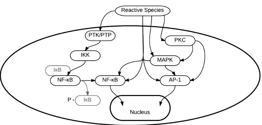
Accumulating ROS have been shown to directly regulate a number of
transcription factors (Kamata and Hirata, 1999); these include the activator protein-1
(AP-1) and nuclear factor kappa-light-chain-enhancer of activated B cells (NF-κB)
(Meyer et al., 1993). This ability to rapidly adapt gene expression to different
environmental conditions is crucial for the growth and survival of every organism.
cysteine residues, leading to either the activation or inactivation of the respective
transcription factor (Meyer et al., 1993). With ROS both activating the AP
NF-κB transcription factors directly and indirectly (Fig. 1
[image:25.595.118.536.252.452.2]activate the inflammatory pathway (Fig. 1
Fig. 1-3. Reactive species directly and indirectly regulate a number of transcription
factors, including NF
inflammatory mechanisms, amongst other pathways.
Animals with higher metabolic rates often have shorter lifespans; however,
animals that produce fewer ROS and RNS from metabolism, such as birds and
primates, tend to live longer than would be predicted by their metabolic rates (Ku et
al., 1993). This has led to the free
age, levels of reactive species increase in many tissues (Drew and Leeuwenburgh,
2002), with levels of antioxidant enzymes, such as SOD, catalase and GPx, showing
different expression levels
teine residues, leading to either the activation or inactivation of the respective
transcription factor (Meyer et al., 1993). With ROS both activating the AP
B transcription factors directly and indirectly (Fig. 1-3), ROS accumulation can
e the inflammatory pathway (Fig. 1-1).
Reactive species directly and indirectly regulate a number of transcription NF-κB and AP-1 which activate genes which upregulate pro inflammatory mechanisms, amongst other pathways.
Animals with higher metabolic rates often have shorter lifespans; however,
animals that produce fewer ROS and RNS from metabolism, such as birds and
primates, tend to live longer than would be predicted by their metabolic rates (Ku et
s led to the free-radical theory of ageing (Harman, 1957). As we
age, levels of reactive species increase in many tissues (Drew and Leeuwenburgh,
2002), with levels of antioxidant enzymes, such as SOD, catalase and GPx, showing
different expression levels between studies and tissues (Rao et al., 1990; Hussain et teine residues, leading to either the activation or inactivation of the respective
transcription factor (Meyer et al., 1993). With ROS both activating the AP-1 and
3), ROS accumulation can
Reactive species directly and indirectly regulate a number of transcription 1 which activate genes which upregulate
pro-Animals with higher metabolic rates often have shorter lifespans; however,
animals that produce fewer ROS and RNS from metabolism, such as birds and
primates, tend to live longer than would be predicted by their metabolic rates (Ku et
radical theory of ageing (Harman, 1957). As we
age, levels of reactive species increase in many tissues (Drew and Leeuwenburgh,
2002), with levels of antioxidant enzymes, such as SOD, catalase and GPx, showing
al., 1995; Ínal et al., 2001). Additionally, transgenic Drosophila overexpressing SOD
and catalase live 34% longer than controls (Orr and Sohal, 1994), whilst conversely,
mice knocked out for genes encoding GPx or SOD do not display a phenotype of
rapid ageing (Reaume et al., 1996; Ho et al., 1997). Although increased antioxidant
enzyme activity can promote longevity, loss of these factors does not appear to be
detrimental to lifespan. Age-related increases in reactive species lead to
over-oxidation and irreversible changes in protein structure and function. It appears that
progressive oxidative damage is a conserved, central mechanism of age-related
functional decline (Muller et al., 2007), with many species showing an
age-dependent upregulation of oxidative stress-response genes (Yanker et al., 2008). It is
the damaging effect of increasing oxidant concentrations that many believe is the
underlying culprit of eukaryotic ageing (Finkel and Holbrook, 2000).
The brain is particularly susceptible to oxidative damage due to its high
oxygen consumption, roughly 20% of the oxygen used by the entire body, and due to
high concentrations of phospholipids which are especially prone to oxidative damage
(Wu et al., 2004). With ageing, there is a significant and progressive increase in the
level of oxidatively damaged DNA and lipids in the brain (Head, 2009), with dietary
antioxidants shown to prevent oxidative damage of the brain in aged rats and reduce
cognitive decline (Liu et al., 2002; Wu et al., 2004).
Hormonal imbalance
The endocrine system experiences an age-related decline in function, with an
imbalance of hormonal production. One hormone that increases in production with
ageing is the catabolic hormone, cortisol, due to over-stimulation of the
hypothalamic-pituitary-adrenal (HPA) axis (Luz et al., 2003). Conversely, the
production of many anabolic hormones decreases with ageing. The anabolic
enhance the proliferation and activity of cellular mediators of immunity, with DHEA
also reducing pro-inflammatory cytokine production (Inserra et al., 1998). With
ageing, reduction in levels of these hormones, and a parallel increase in cortisol
levels, leads to increased chronic inflammation (Buford and Willoughby, 2008). Of
the anabolic hormones, androgens such as oestrogen and testosterone in particular,
greatly decline. These hormones have been shown to modulate the production of
pro-inflammatory cytokines, such as IL-6 (Pottratz et al., 1994), at least partially
through inhibition of NF-κB activity (Keller et al., 1996), and lower levels of these
hormones is associated with increased production of pro-inflammatory cytokines
(Maggio et al., 2006). This interaction occurs in both directions, with the immune
system shown to modulate the endocrine system. Exogenous cytokines have been
used to highlight the marked change that these can have on the HPA axis in rats
(Besedovsky et al., 1977; Besedovsky and del Rey, 1987) and humans (Jablons et
al., 1989; Mastorakos et al., 1993; Späth-Schwalbe et al., 1994).
In the brain, anabolic hormone receptors are distributed throughout and assist
in regulating the transcription of a vast array of genes involved in cognition and
behaviour. Sufficient activation of these receptors is essential for normal brain
functioning; when hormonal imbalances or deficiencies disrupt this activation,
cognitive deficits occur as a result (Sonntag et al., 2005). Studies in rats suggest that
the hormone, oestrogen, also has the ability to function directly as a neurotransmitter
in the CNS (Balthazart and Ball, 2006); with increased levels of androgens linked to
improved cognitive function (Bagger et al., 2005; Newman et al., 2005), and
decreases linked to lower functional capacity (Gray et al., 2005; Daniel, 2006). The
connection between these hormones and cognitive function appears to be partly due
to the role androgens play in maintaining synaptic density; with hippocampal
synaptic maintenance shown to be androgen-dependent (MacLusky et al., 2006).
Lower levels of DHEA, which is particularly active in the CNS, have also been tied
to impaired cognitive performance; a deficit which can be improved with DHEA
Mitochondrial dysfunction
Gene expression studies suggest that a reduction in the expression of
mitochondrial genes with ageing is strongly conserved from C. elegans to humans
(Zahn et al., 2007; Yanker et al., 2008). A decline in mitochondrial function seems to
be an important modulating factor on the ageing process in all species examined, and
it can have positive or negative effects on lifespan, depending on the context
(Sedensky and Morgan, 2006). Reduction in mitochondrial function has been shown
to shorten lifespan in a number of species (Trifunovic et al., 2004; Kujoth et al.,
2005; Rea et al., 2007), while augmentation of mitochondrial function has been
shown to extend lifespan (Lin et al., 2002; Schriner et al., 2005). Mitochondria are
highly dynamic organelles that fuse and divide in response to environmental stimuli,
developmental status, and energy requirements (Seo et al., 2010). A recent wave of
studies demonstrates the pleiotropic role of these mechanisms in many cellular
processes, such as mitochondrial metabolism, redox signalling, maintenance of
mitochondrial DNA (mtDNA), and autophagy (Chan, 2006; Twig et al., 2008; Seo et
al., 2010; Zorzano et al., 2010). Among other functions, they provide energy for
anabolic reactions by securing adenosine-5’-triphosphate (ATP) produced from
catabolic reactions (Fig. 1-4). Hydrolysis of ATP to adenosine-5’-diphosphate
(ADP) or adenosine-5’-monophosphate (AMP) provides energy for most biological
processes making mitochondria essential for normal cell function and maintenance
Fig. 1-4. Mitochondria provide energy, in the form of ATP, for biosynthetic anabolic reactions by securing ATP produced in energy
In quiescent cells, the fission machinery is active and mitochondria are
present as distinct small spheres or short rods. This machinery contributes to the
elimination of irreversibly damaged mitochondria through autophagy. In respons
stresses, such as inflammation, a mitochondrial shift towards fusion favours the
generation of interconnected mitochondria, which contribute to a rapid provision of
energy to the cell by maximising ATP synthesis. An ongoing fusion
allows mitochondrial functional and genetic complementation, generating the
appropriate distribution of new organelles during cell division (Fig. 1
Dysfunctional regulation of these mechanisms is thought to be one of the intrinsic
causes of mitochondrial dys
death during the ageing process (Seo et al., 2010). An imbalance between
mitochondrial fusion, fission, biogenesis and autophagy events may cause substantial
changes in mitochondrial number, biomass,
Mitochondria provide energy, in the form of ATP, for biosynthetic anabolic reactions by securing ATP produced in energy-yielding catabolic reactions.
In quiescent cells, the fission machinery is active and mitochondria are
present as distinct small spheres or short rods. This machinery contributes to the
elimination of irreversibly damaged mitochondria through autophagy. In respons
stresses, such as inflammation, a mitochondrial shift towards fusion favours the
generation of interconnected mitochondria, which contribute to a rapid provision of
energy to the cell by maximising ATP synthesis. An ongoing fusion
mitochondrial functional and genetic complementation, generating the
appropriate distribution of new organelles during cell division (Fig. 1
Dysfunctional regulation of these mechanisms is thought to be one of the intrinsic
causes of mitochondrial dysfunction, which contributes to oxidative stress and cell
death during the ageing process (Seo et al., 2010). An imbalance between
mitochondrial fusion, fission, biogenesis and autophagy events may cause substantial
changes in mitochondrial number, biomass, shape and function.
Mitochondria provide energy, in the form of ATP, for biosynthetic anabolic tabolic reactions.
In quiescent cells, the fission machinery is active and mitochondria are
present as distinct small spheres or short rods. This machinery contributes to the
elimination of irreversibly damaged mitochondria through autophagy. In response to
stresses, such as inflammation, a mitochondrial shift towards fusion favours the
generation of interconnected mitochondria, which contribute to a rapid provision of
energy to the cell by maximising ATP synthesis. An ongoing fusion-fission cycle
mitochondrial functional and genetic complementation, generating the
appropriate distribution of new organelles during cell division (Fig. 1-5).
Dysfunctional regulation of these mechanisms is thought to be one of the intrinsic
function, which contributes to oxidative stress and cell
death during the ageing process (Seo et al., 2010). An imbalance between
Fig. 1-5. The mitochondrial fusion
mitochondrial functional and genetic complementation, generating the appropriate distribution of new organelles during cell division
As key regulators of re
reactive species and, therefore, play a prominent role in oxidative stress, whilst
oxidative damage to mitochondrial DNA (mtDNA) results in dysregulation of cell
and organ function leading to overall sys
al., 2006). Tissues affected by age
organs, ovaries and testes, exhibit loss of function with age (Wei and Lee, 2002).
There is a clear decline in mitochondrial functio
ageing, particularly in postmitotic tissues (Boffoli et al., 1994), with a variety of
mtDNA alterations, such as point mutations, deletions, and oxidative modifications,
increasing with age (Lee et al., 1997). Although, i
mitochondrial dysfunction is the primary cause of ageing, with mutations in genes
that confer longevity slowing the accumulation of mtDNA deletions compared to
wild-type (Melov et al., 1995).
The mitochondrial fusion-fission cycle. This mechanism allows mitochondrial functional and genetic complementation, generating the appropriate distribution of new organelles during cell division
As key regulators of redox homeostasis, mitochondria are the main source of
reactive species and, therefore, play a prominent role in oxidative stress, whilst
oxidative damage to mitochondrial DNA (mtDNA) results in dysregulation of cell
and organ function leading to overall system decline, recognised as ageing (Bua et
al., 2006). Tissues affected by age-related mtDNA damage, such as muscle, adrenal
organs, ovaries and testes, exhibit loss of function with age (Wei and Lee, 2002).
There is a clear decline in mitochondrial function in humans and other animals with
ageing, particularly in postmitotic tissues (Boffoli et al., 1994), with a variety of
mtDNA alterations, such as point mutations, deletions, and oxidative modifications,
increasing with age (Lee et al., 1997). Although, it remains uncertain as to whether
mitochondrial dysfunction is the primary cause of ageing, with mutations in genes
that confer longevity slowing the accumulation of mtDNA deletions compared to
type (Melov et al., 1995).
fission cycle. This mechanism allows mitochondrial functional and genetic complementation, generating the appropriate
dox homeostasis, mitochondria are the main source of
reactive species and, therefore, play a prominent role in oxidative stress, whilst
oxidative damage to mitochondrial DNA (mtDNA) results in dysregulation of cell
tem decline, recognised as ageing (Bua et
related mtDNA damage, such as muscle, adrenal
organs, ovaries and testes, exhibit loss of function with age (Wei and Lee, 2002).
n in humans and other animals with
ageing, particularly in postmitotic tissues (Boffoli et al., 1994), with a variety of
mtDNA alterations, such as point mutations, deletions, and oxidative modifications,
t remains uncertain as to whether
mitochondrial dysfunction is the primary cause of ageing, with mutations in genes
Evidently these key theories of ageing can all be attributable to each other –
autoimmunity, oxidative stress, hormonal imbalance, and mitochondrial dysfunction.
These are four thoroughly entwined mechanisms that raise similar considerations as
the chicken or the egg causality dilemma, and the importance of investigating
upstream factors.
1.2.2 Memory Decline Associated with Ageing
Cognitive decline does not affect all individuals equally; some degree of
cognitivedepreciation as we age is considered normal, but often this decay becomes
debilitating over time. As stated in the previous section, structural and
neurophysiological changes occur in the brain with ageing, along with associated
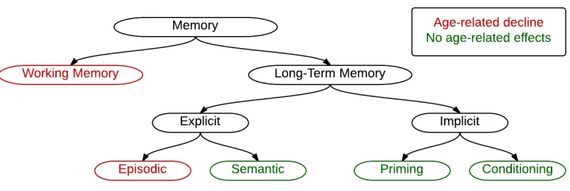
variable decline in cognition. Far from exhibiting uniform depreciation, it appears
that all cognitive faculties do not show the same age-related decline (Fig. 1-6). There
is much evidence to suggest that certain functions are relatively resistant to the
effects of ageing. Knowledge or facts about the world, semantic memory, is
relatively spared in ageing (Light, 1991; Wingfield and Stine-Morrow, 2000; Beier
and Ackerman, 2001), with some evidence to suggest that older adults can, in fact,
outperform younger adults on tests of semantic memory (Wegesin, 2000) and
priming (Laver and Burke, 1993). Skill acquisition also remains intact into old age
(Brown et al., 2009), and it seems that there are no age-related effects on skill
performance, provided the skill is kept in practice (Krampe and Ericsson, 1996).
Contrary to this, episodic and working memories typically show a pronounced
age-related impairment (Salthouse and Babcock, 1991; Mäntylä and Nilsson, 1997; Park
et al., 2002). Decline in episodic memory is a hallmark of dementia onset, but certain
types of episodic memory tasks have been shown to be markedly impaired with
normal ageing also. Memory for associations is typically more affected than memory
for item information (Castel and Craik, 2003), and source and temporal order
judgements also show increased vulnerability to age-related deficits (Cabeza et al.,
2002) with this often being attributed to the inability of older adults to actively
inhibit competing information or that which is no longer relevant, rendering them
[image:32.595.118.520.234.369.2]more susceptible to interference (Hasher and Zacks, 1988).
Fig. 1-6. A diagram showing the sub-types of memory, with those affected by ageing
highlighted in red.
Whilst adaptations in the brain can be observed and compared to young
subjects, it is not always clear whether these alterations have a degenerative effect on
cognition or if they have occurred to compensate for the adverse effects of other
modifications. Different studies help to shed light on which alterations are
debilitating and which are compensatory. It has been observed that with ageing the
interaction between various brain regions associated with higher-order cognitive
functions becomes less co-ordinated (Andrews-Hanna et al., 2007) and that neural
activity becomes less localised in some regions (Cabeza et al., 2002; Park and
Reuter-Lorenz, 2009). Interestingly, aged individuals with delocalised activity
exhibit better cognitive performance than those with more localised activity,
suggesting that this delocalisation is a compensatory response to less co-ordinated
interactions (Cabeza et al., 2002). Often ageing is associated with neurodegenerative
disease. Functional magnetic resonance imaging (fMRI) studies suggest that
measurements of activity in the hippocampus and associated cortical regions can
distinguish normal ageing from pathological ageing (Bishop et al., 2010). Normal
ageing is associated with reduced metabolic activity in the subiculum and dentate
gyrus, whereas reduced activity in the entorhinal cortex is thought to be an early
indicator of Alzheimer’s disease (Small et al., 2002). Neuronal loss is minimal in
most cortical regions with normal ageing (Morrison and Hof, 1997), and it is thought
that the breakdown in these brain systems may be at least partially due to disruption
of myelinated fibres on these connective neurons (Andrews-Hanna et al., 2007). In
Alzheimer’s disease, there is evident neuronal loss, particularly in the entorhinal
cortex and region I cornu ammonis (CA1) of the hippocampus, together with volume
loss in the medial temporal lobe (Braak and Braak, 1991; West et al., 1994;
Gomez-Isla et al., 1996; Price et al., 2001; Rodrigue and Raz, 2004). Focussing on
alterations in the brain regions important for learning and memory, and further
distinguishing healthy ageing from abnormal degeneration, will help us further
understand age-related cognitive decline.
The medial temporal lobes and memory
Initial understanding of the function of the medial temporal lobes came
through studies involving patients with epileptic seizures that underwent medial
temporal lobotomy to remove damaged tissue and prevent further seizures. One such
patient, known as H.M., attracted the attention of Scoville and Milner (1957) when it
was observed that removal of the medial temporal lobes had little effect on
perceptual abilities, intelligence quotient (IQ), or personality, but left H.M. with an
inability to form new memories (Corkin, 2002); H.M. was experiencing anterograde
amnesia. Although exhibiting intact working memory and long-term memory, H.M.
was no longer able to consolidate explicit information from working memory to
long-term memory and, hence, was unable to form new memories after his surgery.
1964), it became apparent that the structures within the temporal lobe were important
for the formation and consolidation of explicit memories.
Subsequent research has confirmed that damage specifically to the
hippocampal formation causes deficits in spatial and learning and memory in rats
(Morris et al., 1982), monkeys (Zola-Morgan and Squire, 1990; Zola et al., 2000),
and humans (Eichenbaum, 2001). Hippocampal lesions impair spatial learning and
highlight an essential involvement of the hippocampal formation in allocentric
spatial tasks. O’Keefe and Nadel (1978) proposed that the hippocampus mediates a
neuronal representation of the physical environment; they termed this “cognitive
mapping”, and it has been found that different types of cells interact extensively to
produce this cognitive map in rats. “Place cells” were found to fire bursts of action
potentials when an animal occupies a particular position in space (O’Keefe and
Dostrovsky, 1971), “head direction cells” increase firing rates when an animal’s
head points in a certain direction (Taube et al., 1990), and “grid cells” fire within
spatial firing fields arranged in a triangular grid thought to encode a cognitive
representation of Euclidean space (Hafting et al., 2005). Dusek and Eichenbaum
(1997) extended the properties of cognitive mapping to non-spatial dimensions of
memory organisation in animals, indicating that the role of the hippocampal region
in explicit memory expression may be as general in rats as it is in humans. It is
believed that these cells are present in humans too, but it is not yet possible to
determine this. More recently, the hippocampus has received increasing attention for
its potential role in energy regulation (Davidson et al., 2007). Hippocampal damage
interferes with energy and body weight regulation, with this thought to be due to
disruption of higher-order learning and memory processes contributing to the control
of appetite and consumptive behaviour.
Broadly speaking the hippocampal formation comprises of the hippocampus
proper, dentate gyrus (DG), subiculum, with some also including the presubiculum,
parasubiculum and entorhinal cortex. The hippocampus has four main histological
divisions: region I cornu ammonis (CA1), region II cornu ammonis (CA2), region III
pyramidal cells have extensive apical and basal dendrites and axons that diverge,
sending projections both anter
mainly project to the septal nuclei, subiculum, as well as other structures. The
subiculum receives afferents mainly from the CA1 of the hippocampus and the
entorhinal cortex. Subicular cells project to
perirhinal cortex, and also to subcortical structures such as mammillary bodies,
hypothalamus, amygdala and nucleus accumbens. The mammillary body projections
to the pons may provide an important link between the hippo
cerebellum, permitting hippocampal influences on motor behaviour. The major
afferents to the hippocampal formation (the dentate gyrus and Ammon’s horn) come
from the entorhinal cortex via the perforant pathway, which in turn receives input
from the entire neocortex. Finally, there are commissural afferents from the
pyramidal cells of contralateral hippocampus and from contralateral entorhinal
cortex. Spatial memory was found to have many sub
particular the dentate gyrus.
[image:35.595.115.525.484.629.2]
Fig. 1-7. A diagram showing the key regions of the hippocampal formation in the rat
brain, at the designated coronal slice. Adapted from Figure 38 in Paxinos and Watson (1998).
pyramidal cells have extensive apical and basal dendrites and axons that diverge,
sending projections both anterior and posterior in the alveus. CA1 pyramidal cells
mainly project to the septal nuclei, subiculum, as well as other structures. The
subiculum receives afferents mainly from the CA1 of the hippocampus and the
entorhinal cortex. Subicular cells project to the entorhinal cortex, deep layers of
perirhinal cortex, and also to subcortical structures such as mammillary bodies,
hypothalamus, amygdala and nucleus accumbens. The mammillary body projections
to the pons may provide an important link between the hippo
cerebellum, permitting hippocampal influences on motor behaviour. The major
afferents to the hippocampal formation (the dentate gyrus and Ammon’s horn) come
from the entorhinal cortex via the perforant pathway, which in turn receives input
rom the entire neocortex. Finally, there are commissural afferents from the
pyramidal cells of contralateral hippocampus and from contralateral entorhinal
cortex. Spatial memory was found to have many sub-regions in the hippocampus, in
te gyrus.
A diagram showing the key regions of the hippocampal formation in the rat brain, at the designated coronal slice. Adapted from Figure 38 in Paxinos and pyramidal cells have extensive apical and basal dendrites and axons that diverge,
ior and posterior in the alveus. CA1 pyramidal cells
mainly project to the septal nuclei, subiculum, as well as other structures. The
subiculum receives afferents mainly from the CA1 of the hippocampus and the
the entorhinal cortex, deep layers of
perirhinal cortex, and also to subcortical structures such as mammillary bodies,
hypothalamus, amygdala and nucleus accumbens. The mammillary body projections
to the pons may provide an important link between the hippocampus and the
cerebellum, permitting hippocampal influences on motor behaviour. The major
afferents to the hippocampal formation (the dentate gyrus and Ammon’s horn) come
from the entorhinal cortex via the perforant pathway, which in turn receives input
rom the entire neocortex. Finally, there are commissural afferents from the
pyramidal cells of contralateral hippocampus and from contralateral entorhinal
regions in the hippocampus, in
Another standard feature linked to classical anterograde a
recognition memory, a role designated to the perirhinal cortex rather than the
hippocampus. Studies involving rats and monkeys all agree that the severity of
impairment in standard tests of recognition memory in these species is greater
following perirhinal lesions than hippocampal lesions. The perirhinal cortex is
involved in discriminating the familiarity and recentness of items (Davachi, 2004). A
lesion to the perirhinal cortex in both monkeys and rats leads to the impairment of
visual recognition memory, disrupting stimulus
recognition abilities (Murray and Mishkin, 1986; Zola
et al., 1993; Meunier et al., 1993; Buckley and Gaffan, 1998).
perirhinal cortex in
suggest that it is part of a larger semantic system that is crucial for endowing objects
with meaning (Murray, 2007).
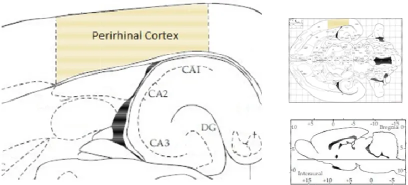
Fig. 1-8. A diagram showing the location of the perirhinal cortex in r
hippocampal formation in the rat brain, at the designated horizontal slice. Adapted from Figure 98 in Paxinos and Watson (1998).
Another standard feature linked to classical anterograde a
recognition memory, a role designated to the perirhinal cortex rather than the
hippocampus. Studies involving rats and monkeys all agree that the severity of
impairment in standard tests of recognition memory in these species is greater
following perirhinal lesions than hippocampal lesions. The perirhinal cortex is
involved in discriminating the familiarity and recentness of items (Davachi, 2004). A
lesion to the perirhinal cortex in both monkeys and rats leads to the impairment of
l recognition memory, disrupting stimulus-stimulus associations and object
recognition abilities (Murray and Mishkin, 1986; Zola-Morgan et al., 1989; Suzuki
et al., 1993; Meunier et al., 1993; Buckley and Gaffan, 1998).
the formation and retrieval of stimulus-stimulus associations
suggest that it is part of a larger semantic system that is crucial for endowing objects
with meaning (Murray, 2007).
A diagram showing the location of the perirhinal cortex in r
hippocampal formation in the rat brain, at the designated horizontal slice. Adapted from Figure 98 in Paxinos and Watson (1998).
Another standard feature linked to classical anterograde amnesia is a loss of
recognition memory, a role designated to the perirhinal cortex rather than the
hippocampus. Studies involving rats and monkeys all agree that the severity of
impairment in standard tests of recognition memory in these species is greater
following perirhinal lesions than hippocampal lesions. The perirhinal cortex is
involved in discriminating the familiarity and recentness of items (Davachi, 2004). A
lesion to the perirhinal cortex in both monkeys and rats leads to the impairment of
stimulus associations and
object-Morgan et al., 1989; Suzuki
et al., 1993; Meunier et al., 1993; Buckley and Gaffan, 1998). The role of the
stimulus associations
suggest that it is part of a larger semantic system that is crucial for endowing objects
[image:36.595.115.523.444.629.2]The perirhinal cortex is comprised of two regions, Brodmann areas 35 and
36, with both divided into three subdivisions; making this a six-layered structure
(Fig. 1-8). Layer IV, in area 35, lacks any cells. The perirhinal cortex projects to
distal CA1 pyramidal cells, overlapping projections from the entorhinal cortex
(Suzuki and Amaral, 1994; van Hoesen and Pandya, 1975). The same CA1 cells
send return projections back to the perirhinal cortex. Inputs from the subiculum
terminate in both superficial and deep layers. The majority of inputs come from
high-level sensory areas that vary between animals: in the monkey these come from
high-level visual areas, whereas, in rat these are primarily from olfactory and, to a
lesser extent, auditory areas.
Neurogenesis and neurotrophins
One of the key features that differentiates the hippocampus from other brain
regions is an ability to constantly produce new neurons. It is thought that this ability
to renew neurons allows this region to carry out its role in learning and memory.
Although the vast majority of neurons in the mammalian brain are formed prenatally,
parts of the adult brain retain the ability to grow new neurons from neural stem cells
in a process known as neurogenesis. Such replication is a normal process for many
cell types, but for many years it was believed that neurons could not replicate (Gage,
2002). This is understandable due to the fact that neurons in most brain regions
cannot replicate, however, it has been discovered that neurogenesis can occur in
adult cells in the hippocampus, particularly in the dentate gyrus and the
subventricular zone. Hippocampal neurogenesis has been shown to occur in birds
(Barnea and Nottebohm, 1994), rodents (Kempermann et al., 1998), monkeys
(Kornack and Rakic, 1999) and humans (Eriksson et al., 1998). Neurogenesis has
been measured by immunohistological detection of the incorporation of
bromodeoxyuridine (BrdU) into the DNA of proliferating cells (Kempermann et al.,
1998) or use of a retroviral vector expressing green fluorescent protein (GFP) to
label dividing cells (van Praag et al., 2002). The latter method also highlighting the
zone, and that the newly generated neurons are functionally similar to mature dentate
granule cells. One of the implications of a role for adult neurogenesis in learning and
memory is that neurogenesis can be regulated by numerous factors associated with
an animal's behavioural and cognitive states (Deng et al., 2010). An animal's
experiences, including hippocampal-dependent learning (Gould et al., 1999),
environmental enrichment (Nilsson et al., 1999) and aerobic exercise (van Praag et
al., 1999), can affect the rate of neurogenesis and enhance cognition. Inhibition of
neurogenesis in the rat brain, using low dose irradiation, has been shown to interfere
with hippocampal-dependent memory function (Winocur et al., 2006). With ageing,
the rate of neurogenesis decreases (von Bohlen und Halbach, 2010), in parallel to
loss of several aspects of cognitive function. Assessment with BrdU labelling shows
that reduced proliferation in the hippocampus is not attributable to a general
aged-related metabolic impairment, because the density of BrdU-pos