ORIGINAL RESEARCH
PEDIATRICS
Characterization of Extensive Microstructural Variations
Associated with Punctate White Matter Lesions in
Preterm Neonates
XX. Li,XJ. Gao,XM. Wang,XJ. Zheng,XY. Li,XE.S. Hui,XM. Wan, andXJ. Yang
ABSTRACT
BACKGROUND AND PURPOSE: Punctate white matter lesions are common in preterm neonates. Neurodevelopmental outcomes of the neonates are related to the degree of extension. This study aimed to characterize the extent of microstructural variations for different punctate white matter lesion grades.
MATERIALS AND METHODS: Preterm neonates with punctate white matter lesions were divided into 3 grades (from mild to severe: grades I–III). DTI-derived fractional anisotropy, axial diffusivity, and radial diffusivity between patients with punctate white matter lesions and controls were compared with Tract-Based Spatial Statistics and tract-quantification methods.
RESULTS:Thirty-three preterm neonates with punctate white matter lesions and 33 matched controls were enrolled. There were 15, 9, and 9 patients, respectively, in grades I, II, and III. Punctate white matter lesions were mainly located in white matter adjacent to the lateral ventricles, especially regions lateral to the trigone, posterior horns, and centrum semiovale and/or corona radiata. Extensive microstruc-tural changes were observed in neonates with grade III punctate white matter lesions, while no significant changes in DTI metrics were found for grades I and II. A pattern of increased axial diffusivity, increased radial diffusivity, and reduced/unchanged fractional anisotropy was found in regions adjacent to punctate white matter lesion sites seen on T1WI and T2WI. Unchanged axial diffusivity, increased radial diffusivity, and reduced/unchanged fractional anisotropy were observed in regions distant from punctate white matter lesion sites.
CONCLUSIONS: White matter microstructural variations were different across punctate white matter lesion grades. Extensive change patterns varied according to the distance to the lesion sites in neonates with severe punctate white matter lesions. These findings may help in determining the outcomes of punctate white matter lesions and selecting treatment strategies.
ABBREVIATIONS:AD⫽axial diffusivity; CST⫽corticospinal tract; FA⫽fractional anisotropy; GCC⫽genu of the corpus callosum; IFO⫽inferior fronto-occipital fasciculus; OR⫽optic radiation; PWML⫽punctate white matter lesion; RD⫽radial diffusivity; SCC⫽splenium of corpus callosum
P
unctate white matter lesions (PWMLs) are common in neo-nates and have been found in ⬎20% of preterm neonates (⬍37 weeks of gestation).1-5These lesions may cause severeneu-rologic disorders, such as cerebral palsy.2,4PWMLs can be
iden-tified on conventional MR imaging as hyperintensity on T1WI and hypointensity on T2WI.1-3,6PWMLs without cystic lesions
can be divided into 3 grades.6The grading scale ascends in severity
on the basis of the number, size, and distribution of cerebral white matter lesions. Extensive microstructural alterations in white matter beyond the PWMLs visible on conventional MR imaging have been observed.3 The neurodevelopmental outcome of
neonates is related to the degree of extension associated with PWMLs.7,8However, little is known about the extent of
micro-structural variations for different PWML grades. More detail is needed regarding the size ranges, shapes, and locations of neona-tal/infantile PWMLs, PWML diffusion characteristics, and the distinction between hemorrhagic and nonhemorrhagic PWMLs.9
DTI could provide quantitative metrics that reveal micro-structural alterations associated with lesions.10,11DTI metrics,
es-pecially directional diffusivities, are sensitive to underlying histo-pathologic processes.12Several methods for analyzing DTI data
Received July 27, 2016; accepted after revision January 26, 2017.
From the Department of Radiology (X.L., J.G., M. Wang, Y.L., J.Y.) and Clinical Re-search Center (J.Z.), the First Affiliated Hospital, Xi’an, Shaanxi, China; Department of Biomedical Engineering (X.L., M. Wan, J.Y.), the Key Laboratory of Biomedical Information Engineering of the Ministry of Education, School of Life Science and Technology, Xi’an Jiaotong University, Xi’an, China; and Department of Diagnostic Radiology (E.S.H.), University of Hong Kong, Hong Kong, China.
Xianjun Li and Jie Gao contributed equally to this work.
This work was supported by grants from the National Natural Science Foundation of China (No.81171317, 81471631), the National Key Research and Development Pro-gram of China (2016YFC0100300), and the 2011 New Century Excellent Talent Sup-port Plan from the Ministry of Education of China (NCET-11– 0438).
Please address correspondence to Jian Yang, PhD, Department of Radiology, The First Affiliated Hospital, Xi’an Jiaotong University, Xi’an, Shaanxi, China; e-mail: [email protected]
Indicates open access to non-subscribers at www.ajnr.org
have been proposed. Tract-Based Spatial Statistics (TBSS; http:// fsl.fmrib.ox.ac.uk/fsl/fslwiki/TBSS) is an automated approach for assessing alterations on major white matter tracts.13The tract
quantification method was proposed to characterize the location of alterations in white matter.14 These automated quantified
methods have been used to detect variations due to brain devel-opment or injury3,13-15and may enable characterization of
alter-ations associated with PWMLs.
The goal of this study was to explore white matter microstruc-tural variations associated with PWMLs of different grades in pre-term neonates and to characterize the change in microstructural patterns along white matter tracts.
MATERIALS AND METHODS
This is a cross-sectional and observational study. It was approved by the institutional review board of the First Affiliated Hospital of Xi’an Jiaotong University. The parents of the neonates were informed of the risks of MR imaging and gave written consent.
Subjects
Preterm neonates were enrolled from the neonatal intensive care unit of the First Affiliated Hospital of Xi’an Jiaotong University, from January 2011 to October 2012. During this period, the care, management, MR imaging scanner, and sequences did not change. The inclusion criterion was evidence of punctate lesions in cerebral white matter, which presented as hyperintensity on T1WI and hypointensity on T2WI. Subjects with a clinical di-agnosis of congenital malformations of the central nervous system, infections, metabolic disorders, hydrocephalus, gray matter lesions, or major destructive white matter lesions such as cystic degeneration and infarction were excluded. Brain MR imaging was also performed on preterm neonates with comor-bid conditions of neonatal asphyxia, hypocalcemia, aspiration pneumonia, and so forth. The preterm neonates without any MR imaging abnormality and matched for sex, gestational age, postnatal age at MR imaging, and birth weight were selected as controls.
MR Imaging Acquisition
The MR imaging datasets used in this study were acquired for clinical examination and diagnosis. To reduce head movement and complete the MR imaging procedure, we sedated patients with a relatively low dose of oral chloral hydrate (25–50 mg/kg).16
Patient selection, monitoring, and management were performed following the “Guidelines for Monitoring and Management of Pediatric Patients during and after Sedation for Diagnostic and Therapeutic Procedures: An Update.”17Neonates were laid in a
supine position and snugly swaddled in blankets. A pediatrician was present during the MR imaging. Micro earplugs were placed bilaterally in the external acoustic meatuses of the subjects to pro-tect their hearing. The subjects’ heads were immobilized by molded foam. Temperature, heart rate, and oxygen saturation were monitored throughout the procedure.
Three-dimensional fast spoiled gradient-recalled echo T1WI, fast spin-echo T2WI, and single-shot echo-planar DTI were per-formed on a 3T scanner (Signa HDXT; GE Healthcare, Milwaukee, Wisconsin) with an 8-channel head coil. The other parameters for
DTI were the following: 35 gradient directions; b-values⫽0 and 1000 s/mm2; TR/TE⫽5500/95–105 ms; section thickness⫽4 mm without a gap; FOV⫽180⫻180 mm2; matrix size⫽128⫻ 128; and voxel size⫽1.41⫻1.41⫻4 mm3.
MR Imaging Interpretation
To provide clues for the etiology of PWMLs, the comorbid con-ditions of neonates were recorded by the clinician from the neo-natal intensive care unit of the institution. Two radiologists, blinded to the clinical history of the neonates, independently an-alyzed the MR imaging. Both of the radiologists had⬎10 years of experience in the interpretation of the neonatal brain MR imag-ing. Neonates with PWML were grouped into grades I, II, and III (from mild to severe) by using the following MR imaging grading method6: grade I: 1 or 2 relatively small lesions (diameter,ⱕ3
mm); grade II: a)ⱖ3 lesions, or b) 1 large lesion (diameter,ⱖ5 mm); and grade III: a)ⱖ3 lesions, and b) multiple large lesions (diameter,ⱖ5 mm). The locations of PWMLs were recorded as follows: anterior region (anterior to the frontal horn of the lateral ventricles), central region (between the frontal horn and the tri-gon of the lateral ventricles), and posterior region (posterior to the trigon of the lateral ventricles).1The lesion load in the
afore-mentioned regions was calculated separately. Lesions longer than 5 mm were segmented into subsections in units of 5 mm. Every subsection was counted while we calculated the lesion load.
Preprocessing of DTI Data
DTI data were processed by using the FMRIB software library (FSL; http://www.fmrib.ox.ac.uk/fsl).18 First, the eddy current
correction was performed. Then, brain regions were extracted by using the FSL Brain Extraction Tool (http://fsl.fmrib.ox.ac.uk/fsl/ fslwiki/BET). To exclude the influence of artifacts, we rejected artifact-corrupted volumes (directions) automatically before the tensor estimation.19 The number of rejected volumes varied
across subjects (median⫽2; range, 0⬃11). DTI metrics of frac-tional anisotropy (FA), axial diffusivity (AD), and radial diffusiv-ity (RD) were calculated by using the FMRIB Diffusion Toolbox (http://fsl.fmrib.ox.ac.uk/fsl/fslwiki/FDT).
Image registration was performed by using an optimized pro-tocol.20,21First, the group mean FA image in native space was
created from the subjects in this study. This method was de-scribed in a previous atlas creation study.22Second, images of
all the subjects were registered to the group mean image. The single-subject FA image with the minimum mean displace-ment score was selected as the final target.13,20Finally, all
in-dividual FA images were registered to the target by using a combination of linear and nonlinear registration methods.20
Other metrics were normalized into the target space by using the deformation parameters of FA.
Voxelwise Analysis of TBSS
The normalized individual FA images were up-sampled to a voxel size of 1⫻1⫻1 mm3and then were averaged to create the mean FA.13A mean FA skeleton was extracted from the mean FA to
represent the center of white matter tracts.13The threshold of
Tract-Quantification Analysis
To characterize the change patterns along white matter tracts, we quantified the DTI metrics on the representative tracts: projection fibers of the corticospinal tract (CST) and optic radiation (OR); commissural fibers of the splenium of the corpus callosum (SCC) and genu of the corpus callosum (GCC); and association fibers of the inferior fronto-occipital fasciculus (IFO). First, images of all
sub-jects were normalized to the neonatal template.22Second, measurement planes
were equally spaced14on the tract
proba-bilistic map (cmrm.med.jhmi.edu) for the neonatal template.22 Measurements
were then averaged on each plane.14
Fi-nally, DTI metrics were measured at 100 equivalent levels.15The combination of
changes in DTI metrics was used to char-acterize damage types.12
Statistical Analysis
Each case was matched with a control by sex (the same sex in PWML and control groups), gestational age (differences,⬍1 week), postnatal age at scanning (differ-ences,⬍5 days), and birth weight (differ-ences,⬍0.5 kg). Because case and control groups were dependent after matching, Wilcoxon signed rank tests were used for the group difference in demographics between each grade of PWML and its matched control group due to non-normal distributions of variables. tests and intraclass correlation coefficients were used to deter-mine intrarater and interrater agreement for the PWML grading and the lesion number respectively.P⬍.05 was significant for the above analyses.
The lesion number was counted repeatedly in anterior, cen-tral, and posterior regions for each PWML subject. The Friedman test was used to assess the variation of lesion number across re-gions. Then pair-wise comparisons among the 3 regions were per-formed by using Wilcoxon signed rank tests.P⬍.017 (.05/3) was considered significant after the Bonferroni correction. The anal-yses above were performed by using SPSS (Version 17.0; IBM, Armonk, New York).
The FSL Randomize tool (http://fsl.fmrib.ox.ac.uk/fsl/fslwiki/ Randomise/UserGuide) was used for the voxelwise analysis to com-pare PWML groups and controls. The number of permutations was 10,000. Tests in TBSS were considered significant atP⬍.05 after the threshold-free cluster enhancement and family-wise error rate correction.
[image:3.594.53.537.56.159.2]In tract-quantification analysis, the Wilcoxon signed rank test in MATLAB (Version 7.11; MathWorks, Natick, Massachusetts) was used to evaluate differences in regional values of DTI metrics between PWML groups and controls.P ⬍.05 was considered significant for this analysis.
Table 1: Demographics of preterm neonates with PWMLs and controlsa
PWML Grade I (n= 15)
Controls (n= 15)
P
Value
PWML Grade II (n= 9)
Controls (n= 9)
P
Value
PWML Grade III (n= 9)
Controls (n= 9)
P
Value
GA (wk) 34⫹2 34⫹1 .21 33⫹6 33⫹6 .76 33⫹0 33⫹2 .37
(32⫹0⬃35⫹6) (32⫹6⬃36⫹2) (30⫹5⬃34⫹5) (30⫹1⬃35⫹0) (32⫹3⬃35⫹4) (32⫹1⬃35⫹6) Postnatal age at
scan (days)
8 8 .53 10 10 .67 10 9 .80
(1⬃13) (1⬃13) (6⬃14) (8⬃14) (4⬃13) (1⬃14)
PMA (wk) 35⫹5 36⫹0 .32 35⫹3 35⫹1 .23 34⫹5 35⫹2 .40
(33⫹1⬃37⫹2) (33⫹3⬃37⫹1) (32⫹1⬃36⫹0) (32⫹1⬃36⫹4) (33⫹1⬃36⫹3) (32⫹5⬃36⫹5)
Birth weight (g) 1780 1760 .22 1950 1750 .26 1920 1700 .51
(1470⬃2360) (1410⬃2400) (1480⬃2700) (1460⬃2360) (1650⬃2760) (1460⬃2570)
Sex (male/female) 10:5 10:5 – 4:5 4:5 – 3:6 3:6 –
Note:—GA indicates gestational age; PMA, postmenstrual age.
a
[image:3.594.51.297.216.602.2]GA, PMA, and birth weight values are medians (ranges). Values in the row of sex are subject numbers. Wilcoxon signed rank tests were used to test the group difference between each grade of PWML and its matched control group due to non-normal distributions of variables. The sex ratios were not tested because they were the same in PWML and the corresponding control groups.
Table 2: Clinical history of subjects
No. of Subjects (Column-Based Percentage)
Controls (n= 33) All Grades
(n= 33)
Grade I (n= 15)
Grade II (n= 9)
Grade III (n= 9)
Hypoxic-ischemic encephalopathy 20 (61%) 7 (47%) 5 (56%) 8 (89%) 0 (0%) Neonatal asphyxia 17 (52%) 7 (47%) 3 (33%) 7 (78%) 7 (21%) Neonatal respiratory distress
syndrome
13 (40%) 4 (27%) 3 (33%) 6 (67%) 6 (18%)
Neonatal pneumonia 11 (33%) 6 (40%) 2 (22%) 3 (33%) 2 (6%) Electrolyte disturbances 10 (30%) 4 (27%) 4 (44%) 2 (22%) 0 (0%)
Neonatal anemia 9 (27%) 4 (27%) 3 (33%) 2 (22%) 4 (12%)
Metabolic acidosis 8 (24%) 3 (20%) 2 (22%) 3 (33%) 7 (21%) Congenital heart disease 6 (18%) 3 (20%) 0 (0%) 3 (33%) 3 (9%) Neonatal hypoglycemia 4 (12%) 0 (0%) 2 (22%) 2 (22%) 1 (3%) Neonatal intracranial hemorrhage 4 (12%) 0 (0%) 2 (22%) 2 (22%) 0 (0%) Neonatal polycythemia 2 (6%) 0 (0%) 1 (11%) 1 (11%) 0 (0%)
Hyperbilirubinemia 2 (6%) 1 (7%) 1 (11%) 0 (0%) 6 (18%)
Agenesis of bronchus 1 (3%) 0 (0%) 1 (11%) 0 (0%) 0 (0%)
Neonatal hypocalcemia 0 (0%) 0 (0%) 0 (0%) 0 (0%) 9 (27%)
Aspiration pneumonia 0 (0%) 0 (0%) 0 (0%) 0 (0%) 7 (21%)
[image:3.594.54.369.216.585.2]RESULTS
Demographics
Thirty-three preterm neonates with PWMLs and 33 matched controls were enrolled. The intrarater and interrater agreement for PWML grading was 97% (value⫽0.953; standard error⫽ 0.906) and 93.3% (value ⫽0.906; standard error⫽0.063), respectively. Fifteen, 9, and 9 neonates with PWMLs were classi-fied into grades I, II, and III, respectively. No significant differ-ences in gestational age, postnatal age at MR imaging, postmen-strual age, or birth weight were found between neonates with PWMLs and controls (Table 1). In this study, PWMLs were as-sociated with many comorbid conditions (Table 2). More than
half of the neonates with PWMLs had a clinical history of hypoxic-ischemic en-cephalopathy (61%) and neonatal as-phyxia (52%).
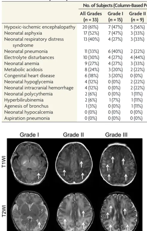
PWML Location
PWMLs were mainly located in the white matter adjacent to lateral ventricles, espe-cially the regions lateral to the trigone, posterior horns, and the centrum semi-ovale and/or corona radiata (Fig 1). The intrarater and interrater correlation coef-ficients for the lesion number counting were 0.994 (95% confidence interval, 0.989⬃0.997) and 0.974 (95% confidence interval, 0. 948⬃0. 987), respectively. For grades I and II, most of the lesions were located in the posterior region (Table 3). For grade III, more lesions were located in the central and posterior regions than in the anterior region (Table 3).
Microstructural Alterations in Different PWML Grades
The extent of microstructural alterations was different across PWML grades. There were no significant changes in grades I and II compared with controls (Pⱖ.05). For neonates with PWML grade III, re-duced FA, increased AD, and increased RD were observed in the centrum semi-ovale and/or corona radiata, white matter near the trigone of the lateral ventricles, SCC, and OR (Fig 2). These regions were near PWML sites seen on T1WI and T2WI (Fig 1). Reduced FA, unchanged AD, and increased RD were observed in re-gions distant from the lesion sites, including the CST in the posterior limb of the internal capsule, more extensive areas in the OR, and the central part of the SCC, GCC, IFO, and the external capsule (Fig 2).
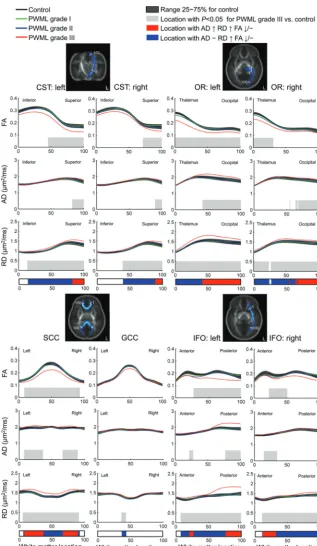
Microstructural Alterations along Tracts
Different patterns of microstructural changes associated with severe PWMLs (grade III) were found along white matter tracts (Fig 3). Increased AD, increased RD, and reduced/unchanged FA were found in the superior part of the CST, the occipital proximal region of the OR, the peripheral re-gions of the SCC near the lateral ventricles, the anterior and pos-terior regions of the left IFO, and the anpos-terior region of the right IFO. Unchanged AD, increased RD, and unchanged/re-duced FA were observed in the posterior limb of the internal capsule part of the CST, the region of the OR proximal to the thalamus, central regions of the SCC and GCC, the central region of the left IFO, and the central and posterior regions of the right IFO.
Table 3: Lesion load of PWMLs in the anterior, central, and posterior regionsa
Grade
Lesion No. (Median and Range) PValue
Anterior Central Posterior
Anterior vs Central
Central vs Posterior
Anterior vs Posterior I 0 (0⬃0) 0 (0⬃1) 1 (1⬃2) .32 ⬍.001b,c ⬍.001b,c
II 0 (0⬃6) 1 (0⬃9) 5 (3⬃12) .17 .02b ⬍.01b,c
III 8 (2⬃18) 24 (5⬃38) 16 (12⬃29) ⬍.01b,c .37 .01b,c aThe inter-region comparisons were performed with the Wilcoxon signed rank test after the Friedman test. The
anterior region is anterior to the frontal horn of the lateral ventricles. The central region is between the frontal horn and the trigon of the lateral ventricles. The posterior region is posterior to the lateral ventricles.
b
P⬍.05.
cP⬍.017 (.05/3), significant after the Bonferroni correction.
DISCUSSION
The results demonstrated different levels of microstructural alter-ations across the 3 PWML grades. Extensive white matter altera-tions were revealed by DTI metrics in neonates with PWML grade III. There were 2 main patterns of alterations found with DTI. The distribution of alterations was related to the distance to lesions visible on the conventional MR imaging.
The etiology of PWMLs is nonspecific.8PWMLs were
associ-ated with various comorbid conditions in this study (Table 2).
Knowledge of the pathogenesis of PW-MLs is limited.7Evidence has revealed that
PWMLs may correspond to vascular con-gestion with infiltration of activated micro-glia.8Late myelination defects indicate that
gliosis and/or loss of oligodendrocytes is possible.7Although the etiology and
patho-genesis are nonspecific and complex, the lo-cation of PWMLs is regular.
PWML Location
PWMLs were common along the corona radiata, in the posterior periventricular white matter, and along the OR. This finding is consistent with previous ones.7,8,23The distribution of injury is in
agreement with areas of high microglia density and deep medullary venous anat-omy.7,23 Microglia accumulate in
re-stricted laminar bands, most notably around 19 –30 gestational weeks, at the axonal crossroads in the centrum semi-ovale, extending caudally in the immature white matter to the OR.24The most
vul-nerable area is the region of the terminal veins, which is a collection of medullary veins in the posterior frontal, central, and parietal regions.23In summary, metabolic
demand and regional cerebral blood flow contribute to the distribution of the white matter lesions.25
Extensive Microstructural Alterations in Different PWML Grades
The developmental outcome of PWMLs is related to the degree of extension.7,8
The results in this study reveal that exten-sive microstructural changes were differ-ent across PWML grades. This finding may be due to different pathophysiologic mechanisms2,6 or different effects on
brain development.2 Rare isolated
PW-MLs can disappear during brain develop-ment and leave no residual abnormal-ity.3,7,26,27We found that there were no
extensive microstructural alterations as-sociated with PWML grades I and II. Sev-eral studies have shown that PWML-re-lated alterations extend beyond the immediate area of injury.3,28
Extensive microstructural alterations were revealed here in neo-nates with severe PWMLs. Relatively larger lesions leave areas of hypomyelination, which may relate to focal gliosis, oligodendro-glial injury, or axonal swelling in lesion sites.2,6,7A cross-talk
ex-ists between axons and oligodendrocytes during development.29
This cross-talk maintains proper metabolic function of axons, trophic support, cytoskeletal arrangement, ion channel organiza-FIG 3. Changes in DTI metrics (FA, AD, and RD) along white matter tracts in preterm neonates
[image:5.594.53.371.43.590.2]tion, and axonal transport.29Damage to axons or glial cells in
PWML sites would affect healthy cross-talk between the axons and the oligodendrocytes along the tract. This may influence the proliferation of oligodendrocyte precursor cells and further my-elination, even in areas distant from the lesion sites.
In this study, severe PWML-related alterations were wide-spread along various white matter tracts, including the CST, OR, SCC, GCC, and IFO. The corticospinal tracts are the major pro-jectional motor fibers of the brain. It has been found that altered structural integrity in the CST may result in delayed psychomotor development,30motor impairment,8and cerebral palsy.30,31High
rates of motor impairment or cerebral palsy have been found in subjects with PWMLs.2,8Visual function in preterm neonates at
term-equivalent age is directly related to the development of white matter in the OR.32In agreement with previous findings,
PWMLs are often present in the OR and may be associated with impaired visual function.4,8The corpus callosum is the primary
center for the interhemispheric integration of information.33
Pic-togram test performance,34Psychomotor Developmental Index
scores,30and the speed of bimanual motor coordination33are
related to the structural integrity of the SCC. The development of the GCC is involved in the maturation of semantic coding,34
in-telligence,35and visual learning, possibly through a higher level
integration of visual information relayed to the frontal lobes by the IFO.36The IFO connects the occipital lobe with the frontal
cortex, playing a critical role in neurocognitive maturation of pro-cessing speed, visual learning,36and semantic processing.37The
widespread alterations in projection, commissural, and associa-tion fibers associated with PWMLs may result in motor, sensory, and cognitive disorders.4,7,8
Regional Microstructural Alteration Patterns
Changes in DTI metrics demonstrated a PWML site-related spa-tial distribution: significantly increased AD, increased RD, and decreased/unchanged FA in regions adjacent to the PWML sites, with unchanged AD, increased RD, and decreased/unchanged FA in regions distant from the PWML sites. These 2 patterns may be related to different mechanisms underlying the effects of PWMLs on brain development, including the delayed oligodendrocyte proliferation or the death of oligodendrocyte progenitors, and the disturbed maturation of oligodendrocytes. During normal brain development, the proliferation of glial cell bodies is linked to de-creases in diffusivity indices in all directions. This process would lead to unchanged or increased FA.38The disrupted cross-talk
between an axon and oligodendrocytes would delay the early pro-cess of the oligodendrocyte proliferation or lead to the death of oligodendrocyte progenitors.29,39Increases of AD and RD in
re-gions adjacent to PWMLs may reveal this process. The increase in RD without changes in AD could reflect demyelination or dysmy-elination.40,41In the regions distant from the lesion sites, the
change in AD was not significant. This finding suggested that maturation of oligodendrocytes was disturbed without loss of oli-godendrocytes in these regions.39
According to previous studies7,8and one of our ongoing
co-hort studies, outcomes during infancy and/or preschool age were relevant to the PWML grading and extensive microstructural al-terations observed on MR imaging during the neonatal period.
Early intervention is associated with improved outcomes.42
Con-sidering that extensive microstructural alterations were different across PWML grades, patients with different PWML grades should be treated with different approaches. Widespread micro-structural changes were observed in neonates with PWML grade III. This finding suggests that early multifaceted treatment strat-egies (including rehabilitation and interventions associated with motor and cognitive competence, and so forth) might be benefi-cial to patients.
This study had several limitations. Widespread variations were observed in neonates with severe PWMLs. However, only several representative tracts were selected during the tract-quantification analysis. Besides the selected tracts, other structures (superior fronto-occipital fascicle, tapetum, and so forth) would also be vulnerable. The follow-up work is not finished. We will try to verify outcomes of the enrolled subjects in the future work. The sample size of neonates with PWMLs in each group is relatively small. Furthermore, this work is an in vivo study on the human brain. Pathologic experiments are needed to reveal the exact mi-crostructural changes associated with PWMLs.
CONCLUSIONS
White matter microstructural variations were different across PWML grades. Extensive change patterns varied according to the distance to lesion sites in neonates with severe PWMLs. These findings may help in determining outcomes of PWMLs and se-lecting appropriate early treatment strategies.
ACKNOWLEDGMENTS
The authors are grateful to Drs Li Liu, Xihui Zhou, and Xiaoquan Li in the neonatology department for preparing and monitoring the neonates before and during imaging.
Disclosures: Jian Yang—RELATED:Grant: National Natural Science Foundation of China, Ministry of Education of China, Ministry of Science and Technology of China,
Comments: This work was supported by grants from the National Natural Science Foundation of China (No.81171317, 81471631), the National Key Research and Develop-ment Program of China (2016YFC0100300), and the 2011 New Century Excellent Tal-ent Support Plan from the Ministry of Education of China (NCET-11-0438). * *Money paid to the institution.
REFERENCES
1. Tortora D, Panara V, Mattei PA, et al.Comparing 3T T1-weighted sequences in identifying hyperintense punctate lesions in preterm neonates.AJNR Am J Neuroradiol2015;36:581– 86CrossRef Medline
2. Kersbergen KJ.Different patterns of punctate white matter lesions in serially scanned preterm infants. PLoS One 2014;9:e108904
CrossRef Medline
3. Bassi L, Chew A, Merchant N, et al.Diffusion tensor imaging in preterm infants with punctate white matter lesions.Pediatr Res 2011;69:561– 66CrossRef Medline
4. de Bruïne FT, van den Berg-Huysmans AA, Leijser LM, et al.Clinical implications of MR imaging findings in the white matter in very preterm infants: a 2-year follow-up study.Radiology2011;261:899 – 906CrossRef Medline
5. Dyet LE, Kennea N, Counsell SJ, et al.Natural history of brain le-sions in extremely preterm infants studied with serial magnetic res-onance imaging from birth and neurodevelopmental assessment. Pediatrics2006;118:536 – 48CrossRef Medline
abnormalities in newborn infants. Clin Radiol 2001;56:647–55
CrossRef Medline
7. Raybaud C, Ahmad T, Rastegar N, et al.The premature brain: devel-opmental and lesional anatomy. Neuroradiology 2013;55:23– 40
CrossRef Medline
8. Rutherford MA, Supramaniam V, Ederies A, et al.Magnetic reso-nance imaging of white matter diseases of prematurity. Neuroradi-ology2010;52:505–21CrossRef Medline
9. Niwa T, de Vries LS, Benders MJ, et al.Punctate white matter lesions in infants: new insights using susceptibility-weighted imaging. Neuroradiology2011;53:669 –79CrossRef Medline
10. Le Bihan D, Iima M.Diffusion magnetic resonance imaging: what water tells us about biological tissues.PLoS Biol2015;13:e1002203
CrossRef Medline
11. Hu¨ppi PS, Murphy B, Maier SE, et al.Microstructural brain devel-opment after perinatal cerebral white matter injury assessed by dif-fusion tensor magnetic resonance imaging. Pediatrics2001;107: 455– 60CrossRef Medline
12. Wang S, Wu EX, Tam CN, et al.Characterization of white matter injury in a hypoxic-ischemic neonatal rat model by diffusion tensor MRI.Stroke2008;39:2348 –53CrossRef Medline
13. Smith SM, Jenkinson M, Johansen-Berg H, et al.Tract-based spatial statistics: voxelwise analysis of multi-subject diffusion data. Neuro-image2006;31:1487–505CrossRef Medline
14. Groeschel S, Tournier JD, Northam GB, et al.Identification and in-terpretation of microstructural abnormalities in motor pathways in adolescents born preterm.Neuroimage2014;87:209 –19CrossRef Medline
15. Yeatman JD, Dougherty RF, Myall NJ, et al.Tract profiles of white matter properties: automating fiber-tract quantification.PLoS One 2012;7:e49790CrossRef Medline
16. Bracken J, Heaslip I, Ryan S.Chloral hydrate sedation in radiology: retrospective audit of reduced dose.Pediatr Radiol2012;42:349 –54
CrossRef Medline
17. Cote´ CJ, Wilson S; American Academy of Pediatrics, American Acad-emy of Pediatric Dentistry, Work Group on Sedation.Guidelines for monitoring and management of pediatric patients during and after sedation for diagnostic and therapeutic procedures: an update. Pe-diatrics2006;118:2587– 602CrossRef Medline
18. Smith SM, Jenkinson M, Woolrich MW, et al.Advances in functional and structural MR image analysis and implementation as FSL. Neu-roimage2004;23(suppl 1):S208 –19CrossRef Medline
19. Li X, Yang J, Gao J, et al.A robust post-processing workflow for datasets with motion artifacts in diffusion kurtosis imaging.PLoS One2014;9:e94592CrossRef Medline
20. Ball G, Counsell SJ, Anjari M, et al.An optimised tract-based spatial statistics protocol for neonates: applications to prematurity and chronic lung disease.Neuroimage2010;53:94 –102CrossRef Medline
21. Li X, Gao J, Wang M, et al.Rapid and reliable tract-based spatial statistics pipeline for diffusion tensor imaging in the neonatal brain: applications to the white matter development and lesions. Magn Reson Imaging2016;34:1314 –21CrossRef Medline
22. Oishi K, Mori S, Donohue PK, et al.Multi-contrast human neonatal brain atlas: application to normal neonate development analysis. Neuroimage2011;56:8 –20CrossRef Medline
23. Raets M, Dudink J, Raybaud C, et al.Brain vein disorders in new-born infants. Dev Med Child Neurol 2015;57:229 – 40 CrossRef Medline
24. Verney C, Monier A, Fallet-Bianco C, et al.Early microglial coloni-zation of the human forebrain and possible involvement in periventricular white-matter injury of preterm infants.J Anat2010; 217:436 – 48CrossRef Medline
25. Takashima S, Itoh M, Oka A.A history of our understanding of cerebral vascular development and pathogenesis of perinatal brain damage over the past 30 years.Semin Pediatr Neurol2009;16:226 –36
CrossRef Medline
26. Ramenghi LA, Fumagalli M, Righini A, et al.Magnetic resonance imaging assessment of brain maturation in preterm neonates with punctate white matter lesions. Neuroradiology 2007;49:161– 67
CrossRef Medline
27. Cornette L, Tanner S, Ramenghi L, et al.Magnetic resonance imag-ing of the infant brain: anatomical characteristics and clinical sig-nificance of punctate lesions.Arch Dis Child Fetal Neonatal Ed2002; 86:F171–77CrossRef Medline
28. Miller SP, Vigneron DB, Henry RG, et al.Serial quantitative diffu-sion tensor MRI of the premature brain: development in newborns with and without injury.J Magn Reson Imaging2002;16:621–32
CrossRef Medline
29. Alizadeh A, Dyck SM, Karimi-Abdolrezaee S.Myelin damage and repair in pathologic CNS: challenges and prospects.Front Mol Neu-rosci2015;8:35CrossRef Medline
30. Pannek K, Scheck SM, Colditz PB, et al.Magnetic resonance diffu-sion tractography of the preterm infant brain: a systematic review. Dev Med Child Neurol2014;56:113–24CrossRef Medline
31. Jaspers E, Byblow WD, Feys H, et al.The corticospinal tract: a bio-marker to categorize upper limb functional potential in unilateral cerebral palsy.Front Pediatr2015;3:112CrossRef Medline
32. Bassi L, Ricci D, Volzone A, et al.Probabilistic diffusion tractogra-phy of the optic radiations and visual function in preterm infants at term equivalent age.Brain2008;131:573– 82CrossRef Medline
33. Muetzel RL, Collins PF, Mueller BA, et al.The development of cor-pus callosum microstructure and associations with bimanual task performance in healthy adolescents.Neuroimage2008;39:1918 –25
CrossRef Medline
34. Kozlovskiy SA, Vartanov AV, Pyasik MM, et al.Functional role of corpus callosum regions in human memory functioning.Int J Psy-chophysiol2012;85:396 –97CrossRef
35. Kontis D, Catani M, Cuddy M, et al.Diffusion tensor MRI of the corpus callosum and cognitive function in adults born preterm. Neuroreport2009;20:424 –28CrossRef Medline
36. Peters BD, Ikuta T, DeRosse P, et al.Age-related differences in white matter tract microstructure are associated with cognitive perfor-mance from childhood to adulthood. Biol Psychiatry 2014;75: 248 –56CrossRef Medline
37. Ivanova MV, Isaev DY, Dragoy OV, et al.Diffusion-tensor imaging of major white matter tracts and their role in language processing in aphasia.Cortex2016;85:165– 81CrossRef Medline
38. Dubois J, Dehaene-Lambertz G, Kulikova S, et al.The early devel-opment of brain white matter: a review of imaging studies in fetuses, newborns and infants. Neuroscience 2014;276:48 –71
CrossRef Medline
39. Back SA, Riddle A, McClure MM.Maturation-dependent vulnera-bility of perinatal white matter in premature birth.Stroke2007;38: 724 –30CrossRef Medline
40. Song SK, Yoshino J, Le TQ, et al.Demyelination increases radial diffusivity in corpus callosum of mouse brain.Neuroimage2005;26: 132– 40CrossRef Medline
41. Song SK, Sun SW, Ramsbottom MJ, et al.Dysmyelination revealed through MRI as increased radial (but unchanged axial) diffusion of water.Neuroimage2002;17:1429 –36CrossRef Medline