Glanzmann thrombasthenia. Cooperation
between sequence variants in cis during splice
site selection.
Y Jin, … , B Coller, P F Bray
J Clin Invest.
1996;
98(8)
:1745-1754.
https://doi.org/10.1172/JCI118973
.
Glanzmann thrombasthenia (GT), an autosomal recessive bleeding disorder, results from
abnormalities in the platelet fibrinogen receptor, GP(IIb)-IIIa (integrin alpha(IIb)beta3). A
patient with GT was identified as homozygous for a G-->A mutation 6 bp upstream of the
GP(IIIa) exon 9 splice donor site. Patient platelet GP(IIIa) transcripts lacked exon 9 despite
normal DNA sequence in all of the cis-acting sequences known to regulate splice site
selection. In vitro analysis of transcripts generated from mini-gene constructs demonstrated
that exon skipping occurred only when the G-->A mutation was cis to a polymorphism 116
bp upstream, providing precedence that two sequence variations in the same exon which
do not alter consensus splice sites and do not generate missense or nonsense mutations,
can affect splice site selection. The mutant transcript resulted from utilization of a cryptic
splice acceptor site and returned the open reading frame. These data support the
hypothesis that pre-mRNA secondary structure and allelic sequence variants can influence
splicing and provide new insight into the regulated control of RNA processing. In addition,
haplotype analysis suggested that the patient has two identical copies of chromosome 17.
Markers studied on three other chromosomes suggested this finding was not due to
consanguinity. The restricted phenotype in this patient may provide information regarding
the expression of potentially imprinted genes on chromosome 17.
Research Article
Find the latest version:
J. Clin. Invest.
© The American Society for Clinical Investigation, Inc. 0021-9738/96/10/1745/10 $2.00
Volume 98, Number 8, October 1996, 1745–1754
Glanzmann Thrombasthenia
Cooperation between Sequence Variants in
Cis
during Splice Site Selection
Ying Jin, Harry C. Dietz,* Robert A. Montgomery,‡ William R. Bell, Iain McIntosh, Barry Coller,§ and Paul F. Bray
Departments of Medicine, *Pediatrics, and ‡Surgery, Johns Hopkins University School of Medicine, Baltimore, Maryland 21205; and the §Department of Medicine, Mt. Sinai Hospital, New York 10029
Abstract
Glanzmann thrombasthenia (GT), an autosomal recessive bleeding disorder, results from abnormalities in the platelet fibrinogen receptor, GPIIb-IIIa (integrin aIIbb3). A patient
with GT was identified as homozygous for a G→A muta-tion 6 bp upstream of the GPIIIa exon 9 splice donor site. Patient platelet GPIIIa transcripts lacked exon 9 despite normal DNA sequence in all of the cis-acting sequences
known to regulate splice site selection. In vitro analysis of transcripts generated from mini-gene constructs demon-strated that exon skipping occurred only when the G→A mutation was cis to a polymorphism 116 bp upstream,
pro-viding precedence that two sequence variations in the same exon which do not alter consensus splice sites and do not generate missense or nonsense mutations, can affect splice site selection. The mutant transcript resulted from utiliza-tion of a cryptic splice acceptor site and returned the open reading frame. These data support the hypothesis that pre-mRNA secondary structure and allelic sequence variants can influence splicing and provide new insight into the reg-ulated control of RNA processing. In addition, haplotype analysis suggested that the patient has two identical copies of chromosome 17. Markers studied on three other chromo-somes suggested this finding was not due to consanguinity. The restricted phenotype in this patient may provide infor-mation regarding the expression of potentially imprinted genes on chromosome 17. (J. Clin. Invest. 1996. 98:1745–
1754.) Key words: RNA splicing • exon skipping •platelet membrane glycoprotein complex IIb-IIIa • molecular genet-ics • Glanzmann thrombasthenia
Introduction
Glanzmann thrombasthenia (GT)1 is an autosomal recessive
bleeding disorder due to quantitative or qualitative abnormali-ties in the platelet fibrinogen receptor (1, 2). The disease was
first described in 1918 by Glanzmann, a Swiss pediatrician (3). In 1974 Nurden and Caen demonstrated the absence of plate-let membrane glycoproteins IIb-IIIa (GPIIb-IIIa) in three pa-tients with this disorder (4). Subsequent studies characterized, isolated, and demonstrated that GPIIb-IIIa functioned as the fibrinogen receptor in platelets (5–12), thus providing the bio-chemical explanation for the bleeding phenotype seen in GT. With the isolation of cDNA and genomic clones for GPIIb and GPIIIa (13–19), a number of mutations in the genes have been identified which result in GT. The first molecular defect in thrombasthenia was described in 1990 (20) and since then . 20 different mutations have been characterized (for review see reference 21). These studies have provided a wealth of infor-mation about platelet physiology, as well as biochemical, im-munologic, and molecular biologic information about GPIIb-IIIa and other molecules which comprise the integrin family of adhesive molecules (22, 23).
GT is categorized into three types according to the amount of immunologically detectable GPIIb-IIIa: type I, , 5%; type II, 10–20%; and variant thrombasthenia with nearly normal levels of GPIIb-IIIa (1). Four mutant GPIIIa alleles have been described in five kindreds with variant thrombasthenia (24– 28): three of these mutations affect two amino acids (Asp119 and Arg214) and demonstrate a necessary region for fibrino-gen binding (24–26, 28). A third mutation alters the cytoplas-mic Ser752 and impairs the signaling required for GPIIb-IIIa function (27, 29). However, the patients with variant throm-basthenia represent only 8% of all thrombasthenics (1), and the majority of mutations described to date have been in type I patients. Most of these are caused by small mutations (30–40). Of the 10 point mutations published in full form, five have re-sulted in mRNA splicing abnormalities (30–33, 38). These splicing abnormalities usually generate unstable mRNAs yielding no translated protein products, or in a few cases, trun-cated and dysfunctional protein products. Several point muta-tions have resulted in mRNA splicing defects in which entire exons have been deleted (30, 31, 33). Although alternate exon splicing has been thoroughly investigated (see reviews 41, 42), many mechanistic details are still poorly understood. Most mu-tations which affect splicing are located within consensus splice donor, acceptor, and branch sites and cause exon skipping as opposed to cryptic splice site utilization (43, 44). Nakai et al. scanned 209 mammalian genetic disease splicing mutations
and observed that . 90% create or destroy consensus
se-quences and that most occurred at the 59 splice site region; none were upstream of the 21 position (45).
We previously reported the preliminary characterization of a patient with type I GT who had an unusual mutation result-ing in abnormal mRNA splicresult-ing manifested by the skippresult-ing of exon 9 in the GPIIIa gene (46). We now report the full charac-terization of this genetic defect and the inheritance of the mu-tant allele. Our data indicate that a mutation 6 bp upstream of the splice donor site of exon 9 acts in concert with an
addi-Address correspondence to Paul F. Bray, Ross 1015, Johns Hopkins University School of Medicine, 720 Rutland Avenue, Baltimore, MD 21205. Phone: 410-955-7417; FAX: 410-955-0185.
Received for publication 30 May 1996 and accepted in revised form 6 August 1996.
tional exonic sequence variation to cause exon skipping with the subsequent utilization of a cryptic splice site, and that the phenotype in this patient was apparently due to disomy of chromosome 17. We discuss the possible reasons for these splicing abnormalities and the genetic implications of chromo-some 17 disomy.
Methods
Subjects. Subject RS is a 24-yr-old African American female with a lifelong bleeding history. Her bleeding is primarily from her nasal and gingival mucosa and with her menses. She required platelet transfu-sions at age 10 for epistaxis. Her clinical features are typical for Glanz-mann thrombasthenia and careful history and physical examination revealed no unusual or additional clinical findings. Her platelets do not aggregate in response to physiologic stimuli, but do aggregate to ristocetin. Platelets from the patient had trace amounts of GPIIb, but lacked detectable GPIIIa by Western immunoblotting and have been previously shown not to bind soluble fibrinogen (47). No history of consanguinity could be elicited despite intense questioning. The fa-ther is unavailable for study. The mofa-ther of the patient was asymp-tomatic and had normal platelet aggregation. Blood from normal in-dividuals was used to prepare genomic DNA for control experiments. All of these studies have been conducted according to the principles expressed in the Declaration of Helsinki.
Reagents. Taq polymerase was from Perkin Elmer Cetus Corp. (Norwalk, CT), isotopes were from Amersham Corp. (Arlington Heights, IL), ds Cycle Sequencing System from Gibco BRL (Gaith-ersburg, MD).
Quantitation of vitronectin receptors. Monoclonal antibody LM609 (the generous gift of Dr. David Cheresh, Scripps Research Institute, La Jolla, CA) specific for avb3 was used to quantitate the platelet
vitro-nectin receptor as previously described (48).
Single strand conformation polymorphism (SSCP) analysis. Pre-paration of genomic DNA and screening for mutations was per-formed by SSCP analysis as previously described (31). Radiolabeled PCR products were denatured in 95% formamide and electropho-resed through 6% acrylamide, 10% glycerol, and 1 3 TBE (89 mM Tris-borate pH 5 8.3, 2 mM EDTA) nondenaturing gels. The gels were run for 16 h at 10 W at room temperature, dried, and exposed to film for 48 h.
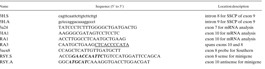
Oligonucleotide synthesis. Oligonucleotides for SSCP were as previously described (31). Primers for RNA analysis were designed from GPIIIa cDNA sequence (16) and are listed in Table I. Nucle-otide sequence numbering is according to Zimrin et al., (18); exon numbering, according to Villa-Garcia et al., (19) and Zimrin et al. (18).
RNA preparation. Total platelet RNA was prepared and reverse transcribed into cDNA with random primers as previously described (49). Using oligonucleotides designed from exonic sequence, the platelet cDNA was amplified and analyzed on 2% agarose gels. Ab-normal fragments were gel purified and the nucleotide sequence de-termined as described below.
Nucleotide sequence determination. Sequence information was obtained from eluted DNA from SSCP gels (31), directly from ampli-fied genomic DNA, or from reverse transcribed platelet mRNA. 2 ml of the PCR products were used in a sequencing reaction which in-cluded a [g-33]P-labeled primer and 2.5 units Taq polymerase (ds
Cy-cle Sequencing System; Gibco BRL). 20–30 cyCy-cles of amplification were performed as previously described (31). Extension products were separated on 6% acrylamide/7 M urea gels and assayed by auto-radiography.
Southern blot analysis. Genomic DNA preparation for Southern blot analysis, as well as the blotting procedure were performed as pre-viously described (20). A 2.0-kb cDNA probe 59 to the internal EcoRI site in the GPIIIa cDNA (50) and a 345-bp probe containing exons 1–3 of the fibrillin cDNA (51) were used for genomic DNA hy-bridizations. Equivalency of loading between lanes was determined by using the ImageQuaNT version 4.1 software on a phosphorimager, (445SI-486; Molecular Dynamics, Sunnyvale, CA).
Reverse transcriptional PCR (RT-PCR) products derived from platelet RNA were separated on a 2% agarose gel, transferred to ny-lon filters, and hybridized with an internal oligonucleotide as de-scribed (52). Relative abundance of RNA transcripts was determined by carefully aligning the autoradiogram with the filter, cutting out that portion of the filter corresponding to a particular band, and de-termining the signal strength with a scintillation counter.
Minigene transcript analysis of mutant and wild-type alleles. Ap-proximately 2 kb of genomic DNA was PCR amplified from the pa-tient and her mother using PCR primers RSY.S and RSY.A con-tained in exons 8 and 10, respectively. These fragments were cloned in frame into the ornithine d-aminotransferase (OAT) cDNA con-tained in pGEM-4Z (Promega Corp., Madison, WI) as described (52). The EcoRI fragment from these plasmids were isolated and cloned into the pcDNAI/Amp expression plasmid (Invitrogen, San Diego, CA), and mapped for the proper orientation. The nucleotide sequence of the entire exon 9 and the flanking 50 bp was determined, and demonstrated that we had isolated the wild-type maternal allele (pcDNA.E1), the mutant maternal allele (pcDNA.E2), and the single patient allele (pcDNA.RS).
[image:3.612.58.562.580.712.2]Point mutations were introduced into pcDNA.E1 and pcDNA.RS using the Altered Sites II in vitro mutagenesis kit (Promega Corp.). Briefly, the EcoRI fragments of pcDNA.E1 and pcDNA.RS were cloned into pALTER-1 and single-strand DNA prepared. The de-sired mutation was generated using a synthetic oligonucleotide and
Table I. GPIIIa Oligonucleotide Primers
Name Sequence (59 to 39) Location:description
3H.S cagttcaattcttgtcttcttgt intron 8 for SSCP of exon 9
3H.A gctccaggacaaaggccct intron 9 for SSCP of exon 9
3a24 TATCCCTCTTTGGGGCTGATGACTG exon 7 for mRNA analysis
3IA1 AAGGGCGATAGTCCTCCTC exon 10 for mRNA analysis
RA1 ACCTTGGCCTCAATGCTGAAG exon 10 for mRNA analysis
RA3 CAATGCTGAAGCTCACCCCATA spans exons 10 and 8
3aex8 CCAGCTCATTGTTGATGCTT exon 8 probe for Southern
RSY.S ACCGGAACCAATTCTGTCCATGGATTCCAGCA exon 8 sense for minigene RSY.A GGCATGCATCAAAGGTGACCTGGACGAT exon 10 antisense for minigene
T4 DNA polymerase, subcloned back into pcDNAI/Amp, and the plasmid was sequenced to confirm the mutation had been properly introduced.
Plasmid DNA was purified using Nucleic Acid purification col-umns (Qiagen Inc., Chatsworth, CA) and electroporated into K562 cells as described (19). 16 h later total RNA was isolated, reversed transcribed into cDNA, and analyzed by PCR amplification with OAT and GPIIIa specific primers. To confirm the identity of these PCR products, they were cloned into the pCRII plasmid (Invitrogen) and sequenced.
Microsatellite analysis. Oligonucleotides were used to PCR am-plify the region of genomic DNA known to contain polymorphic di-nucleotide repeats. These primers have been described previously (53–55) and were identified by searching the official Genome Data Base (GDB) (56, 57) source node at Johns Hopkins University in Baltimore on the GDB main computer. PCR conditions were as above except the sense primer was end-labeled with 32P-gATP and
the annealing temperatures varied between 55–628C. Products were separated in 6% acrylamide/7-M urea gels, and assayed by autorad-iography.
RNA secondary structure prediction. RNA secondary structure was analyzed by the algorithm of Zuker and Stieger (58) using the computer program RNAFOLD in the PC/GENE software from In-telliGenetics (Mountain View, CA) on a Computer Ergonomics (Co-lumbia, MD) 486 computer. The input sequence contained all of exon 9 (four combinations of sequence variants) and the last and first 10 bp of introns 8 and 9, respectively. A graphic display was generated from this prediction using the program MOLECULE written by Dr. J. Ryan Thompson (Carnegie-Mellon University, Pittsburgh, PA) ac-cording to the algorithm of Lapalme et al. (59).
Results
Because the molecular defect in GT may be in either of two genes, we performed preliminary studies to assess GPIIIa ex-pression. Platelets from patient RS and a normal control were studied for their vitronectin receptor (avb35av/GPIIIa)
con-tent (48). The patient’s platelets did not bind an antibody to the vitronectin receptor (LM609), suggesting a GPIIIa defect (data not shown). Genomic Southern blotting of patient DNA using a series of restriction enzymes was normal (see below), suggesting normal gene dosage and the lack of a large rear-rangement. We used the SSCP technique to analyze all GPIIIa exons from patient RS. Fig. 1 A shows such an analysis of exon 9 from a group of normal individuals and patient RS. Despite the presence of additional bands due to the known silent C/A polymorphism at position 20507 in this exon (18), a clearly ab-normal pattern of migration was seen for the sample from RS (Fig. 1, lane 13). The abnormal 209-bp fragment was isolated and sequenced, and a G to A substitution was found 6 bp
up-stream of the splice donor site (ACGGTGAGgt→
ACAGT-GAGgt) at position 20624 of the GPIIIa gene (Fig. 1 B). There were no other nucleotide differences between the sequence from the patient DNA fragment and that of the normal con-trol. This G to A substitution represents a synonymous change and has not previously been reported. To address whether this sequence change represented a polymorphism, we performed SSCP analysis of exon 9 on DNA samples from 45 normal indi-viduals and 4 additional patients with GT (90 and 8 chromo-somes, respectively). In no case did we find the SSCP pattern corresponding to this G to A transition (data not shown). Be-cause the patient was African American, we also studied DNA from 58 African Americans using an HphI restriction digest of the PCR fragments (the HphI recognition sequence is
specifi-cally encoded by the G variant) and again found no alleles containing the A at position 20624. Thus, the A was not present in 206 chromosomes from normal individuals lessening the possibility that the sequence variation at position 20624 represents a polymorphism.
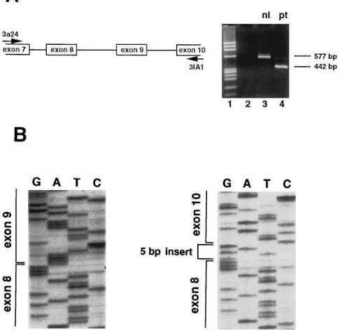
[image:4.612.315.555.63.381.2]Direct sequencing of genomic DNA revealed the patient was homozygous for the G to A substitution, suggesting this may be responsible for the nonexpression of GPIIIa. How-ever, because this nucleotide substitution does not change an amino acid, we sought to determine whether it alters pre-mRNA splicing. The major PCR product identified when pa-tient platelet RNA was amplified with sense and antisense primers complementary to regions of exons 7 and 10, respec-tively, was smaller than normal (Fig. 2 A, lane 3). There were also two less abundant products of approximately normal size observed in the patient sample which were only consistently seen upon overloading of PCR products (see below). Isolation and sequencing of the major 442-bp fragment from the patient sample revealed the absence of exon 9 but the insertion of 5 bp between the sequences corresponding to exons 8 and 10. (Fig. 2 B). These 5 bp match precisely the 5 bp preceding the exon 10 spice acceptor site and are preceded in genomic DNA by an
Figure 1. SSCP analysis of exon 9 of the GPIIIa gene. (A) PCR amplified genomic DNA from 11 different normal individuals (lanes
AG dinucleotide. This suggested that the majority of the tran-scripts manifest the skipping of exon 9 and the utilization of a cryptic splice acceptor site 5 bp upstream of exon 10. Note that the isolated skipping of exon 9 would result in a frameshift and consequent premature termination codon, and mutant tran-script instability would be predicted (52). However, some pro-portion of such transcripts utilize a cryptic splice site, restoring the open reading frame (ORF) and hence stability of those molecules. We have determined the sequence of the two faint
upper bands seen in Fig. 2 A, lane 4. The lower of these two is normal in size, contains exon 9, the G to A substitution, and otherwise has normal sequence (data not shown). The slightly larger than normal fragment has been difficult to separate from the normal fragment, but the sequence we have obtained indicates it is a heteroduplex comprised of both the 577 and 442 bp cDNA species. Fig. 3 summarizes the abnormal splicing from the mutant allele and indicates the position of the cryptic splice site.
We considered several additional issues with respect to the molecular genetics of this mutation. First, is the G to A substi-tution in exon 9 necessary to cause the abnormal splicing phe-notype? Second, is the G to A substitution in exon 9 sufficient to cause the abnormal splicing phenotype? Third, how was the abnormal allele inherited? To address the first question we de-termined the nucleotide sequence corresponding to all of the
cis-acting elements known to influence splice site selection. These include the splice donor, branch point, and splice accep-tor for introns 8 and 9 (Fig. 3). No sequence alteration was identified in these regions, and we conclude that the G to A substitution in exon 9 is associated with, and perhaps necessary for the abnormal splicing.
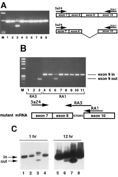
Since the mother is heterozygous for the G→A substitution (see below), if the G→A substitution causes exon skipping we would predict that some portion of her platelet GPIIIa tran-scripts would demonstrate exon 9 skipping. To determine whether exon 9 skipping did not occur at low levels in normal individuals and to study maternal transcripts, we analyzed platelet RNA from the mother, the patient, and a series of nor-mal individuals (Fig. 4 A). Using primers flanking exon 9, we observed a faint band in the mother’s sample (Fig. 4 A, lane 2) which comigrated with the fragment manifesting exon skip-ping from RS. The abnormal fragment was not seen in platelet RNA from normal individuals (Fig. 4 A, lanes 1, 4–9). Al-though we did not rigorously attempt to perform a quantita-tive RT-PCR, these data nevertheless suggested that a small percent of the maternal platelet mRNA contained GPIIIa transcripts lacking exon 9. We confirmed this by utilizing an antisense PCR primer which would only anneal to cDNA de-rived from mRNA that had skipped exon 9 and utilized the cryptic splice site in intron 9 during splicing. Fig. 4 B demon-strates that platelet total RNA from the patient (Fig. 4 B, lane
[image:5.612.56.299.69.305.2]3) and the mother (Fig. 4 B, lane 2), but not a normal control
Figure 2. Platelet GPIIIa RNA analysis. (A) The cartoon on the left indicates the relative positions of the PCR primers in exons 7 and 10 of the GPIIIa gene, and does not reflect a specific sized PCR product. To the right is shown an ethidium-stained agarose gel containing products from RT-PCR reactions using primers 3a24 and 3IA1 with no input template (lane 2), and platelet cDNA from a normal individ-ual (lane 3), and patient RS (lane 4). Size markers are fx174 DNA di-gested with HaeIII (lane 1). (B) Nucleotide sequence of the frag-ments shown in A. (Left) GPIIIa mRNA from normal platelets; (right) platelet GPIIIa mRNA from RS.
[image:5.612.58.444.565.739.2](Fig. 4 B, lane 1), contain such transcripts. Shifting the anti-sense primer 10 bp in the 39 direction, such that it is entirely complementary to exon 10 sequence, allows the amplification of the “exon in” species in normal individuals (Fig. 4 B, lanes
4, 7–11) and both the “exon in” and “exon out” species in the
mother (Fig. 4 B, lane 5) and patient (Fig. 4 B, lane 6). We used Southern blot analysis to estimate the relative abundance of the two species of GPIIIa mRNA in the mother’s platelets and to confirm the identity of the smaller cDNA fragment (Fig. 4 C). Prolonged exposures revealed the exon out form in the maternal amplified cDNA (Fig. 4 C, lane 6) but not in nor-mal controls (Fig. 4 C, lanes 5 and 8). Within the limits of this RT-PCR amplification, we estimate that the exon 9 deleted form represents z 3% of the maternal platelet GPIIIa tran-scripts. These comprehensive RNA analyses indicated that (a) the maternal and patient 442-bp amplified fragment (Fig. 2 A
and Fig. 4 A) was indeed the result of exon 9 skipping, and (b) this abnormal splicing did not appear to occur in normal indi-viduals. These data also suggest the G→A substitution was necessary for exon 9 skipping.
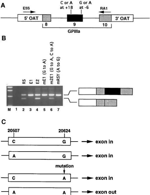
[image:6.612.57.298.56.417.2]To prove whether the G to A transition is necessary and sufficient to dictate abnormal splicing, we analyzed the splicing of this portion of the GPIIIa gene transfected into K562 cells. Fig. 5 A schematically displays the minigene constructs used in these experiments. As shown in Fig. 5 B, the patient’s allele and the mother’s mutant allele cause exon 9 skipping (Fig. 5 B, lanes 2 and 4), whereas the mother’s wild-type allele splices normally (Fig. 5 B, lane 3). Note that in the context of these ar-tificial minigene constructs this abnormal splicing process is not maximally efficient and a substantial portion of the pro-cessed transcripts contain exon 9. The key finding, however, is that exon 9 is deleted only in the presence of the G to A substi-tution (Fig. 5, lanes 2 and 4). To confirm the identity of the PCR products in lanes 2 and 3 we subcloned and sequenced these fragments (data not shown). The larger fragments in lanes 2 and 3 contained sequence for OAT and GPIIIa exon
8–10. The smaller fragment in lane 2 contained OAT and
GPIIIa exon 8 and 10 sequence, with the previously identified 5 bp of intronic sequence between exons 8 and 10.
We next analyzed splicing of the patient and wild-type ma-ternal alleles in which the 26 position from the splice donor site of intron 9 had been switched; i.e., to a wild-type G in the patient allele and a mutant A in the maternal allele. The muta-tion in patient exon 9 is necessary for exon skipping, since re-placing it with the normal G corrects the splicing abnormality (Fig. 5 B, lane 7). However, introducing the mutant A into the maternal wild-type allele was not sufficient to cause exon skip-ping (Fig. 5 B, lane 5). Because the G to A substitution in exon 9 could not be solely responsible for the splicing defect, and because the mother was heterozygous for the C/A polymor-phism at the 59 end of exon 9 (nucleotide 20507), while the pa-tient was homozygous A at this position, we hypothesized that the G to A mutation at the 39 end of exon 9 (position 20624) would result in abnormal splicing only when allelic with the 59 polymorphic A. This hypothesis is summarized in Fig. 5 C. To exclude the possible contribution to splicing of any other se-quence variations in the maternal mutant allele, we introduced both sequence substitutions into the maternal wild-type allele. We observed that the 39 A caused abnormal splicing only in the context of the 59 A (compare Fig. 5 B, lane 6 with lane 5).
We were puzzled by the fact that the patient was homozy-gous for the G to A substitution, since this appeared to be an exceedingly rare sequence variant and there was no history of consanguinity. One possibility consistent with this finding was haploinsufficiency due to a large gene deletion. The quantita-tive Southern blot data shown in Fig. 6 discount this explana-tion. Fig. 6 A indicates that equivalent amounts of normal,
ma-Figure 4. Analysis of GPIIIa exon 9 splicing. (A) Primers 3a24 and 3IA1 were used to amplify cDNA from normal (lanes 1, 4–9), mater-nal (lane 2), and patient (lane 3) platelets. Size markers (M) are fx174 DNA digested with HaeIII. Position of primers on each script is displayed to the right. The 5-bp insertion in the smaller tran-script is not shown. (B) Primers 3a24 and RA3 were used to amplify cDNA from normal (lane 1), maternal (lane 2), and patient (lane 3) platelets. Primer RA3 is specific for mRNA lacking exon 9 that has utilized the cryptic splice site. Different antisense primers RA3 and RA1 were used for products shown on this gel, and are indicated be-low. Primers 3a24 and RA1 were used to amplify cDNA from normal (lanes 4, 7–11), maternal (lane 5), and patient (lane 6) platelets. Primer RA1 will amplify mRNA independent of the presence of exon 9. Size markers (M) are fx174 DNA digested with HaeIII. Note that antisense primers RA1 and RA3 generate smaller products than seen in A. (C) Southern analysis of PCR products indicating relative abun-dance of GPIIIa transcripts with or without exon 9. Normal (lanes 1
and 4), maternal (lane 2), and patient (lane 3) platelet RNA was re-verse transcribed, PCR amplified with primers 3a24 and 3IA1, sepa-rated in 1.5% agarose, transferred to nylon membranes, and hybrid-ized with 32P-labeled primer 3aex8. The same filter is shown twice,
ternal, and patient genomic DNA were loaded (Fig. 6 A, lanes
1–6). In Fig. 6 B, note the equivalent intensity of the z 3.3 and z 5.2 kb TaqI fragments between the control (Fig. 6 B, lane 1) and the patient (Fig. 6 B, lane 3), as well as all BamHI
frag-ments (Fig. 6 B, lanes 4–6), indicating that the patient has two copies of the GPIIIa gene. Essentially equivalent DNA load-ing of all lanes was confirmed by reprobload-ing this filter with a cDNA probe from the fibrillin gene (Fig. 6 B, lanes 7–12).
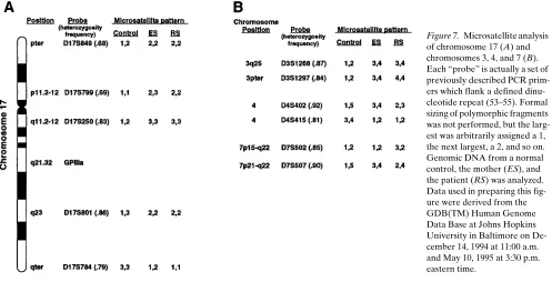
Because the patient was homozygous for a total of three GPIIIa polymorphisms, including the TaqI polymorphism at position 20507 of exon 9 (as shown in Fig. 6 B), we considered the possibility that she had two identical or nearly identical copies of chromosome 17. To address this issue, we examined five highly informative microsatellite polymorphisms distrib-uted throughout chromosome 17. We found that the patient was homozygous for all five of these markers (Fig. 7 A). Note the normal control is heterozygous for D17S849, D17S250, and D17S801 and homozygous for D17S799, and D17S784. The mother is heterozygous for D17S799 and D17S784, and ho-mozygous for D17S250, D17S801, and D17S849. Using the cal-culated population frequencies for the different marker alleles, there is a 1 in 2,000 chance that an individual would be ho-mozygous for all five markers. The frequency of the GPIIIa polymorphisms are not known, but we have analyzed 40 unre-lated individuals to determine the allelic frequencies at posi-tion 20507 (data not shown). The frequency of the homozy-gous “A” state is 0.39. By including this GPIIIa marker, there would be only a 1 in 5,000 chance that an individual would be homozygous for these 6 chromosome 17 polymorphisms, strongly suggesting two copies of the same chromosome 17 in this patient. This idea becomes virtually certain if one consid-ers that none of the 206 normal chromosomes studied had the A at position 20624.
Two identical copies of a chromosome may result when both copies were inherited from one parent (uniparental isodi-somy), or as a product of a consanguinous union. To address these possibilities, we analyzed chromosomes 3, 4, and 7 to de-termine if the patient had the expected frequency of heterozy-gosity elsewhere in the genome. We studied two polymorphic markers on each chromosome and found that similar to the control, the patient was heterozygous for four of the six mark-ers (Fig. 7 B). Because paternal DNA was not available for study, we cannot eliminate the possibility of consanguinity with 100% certainty. However, the studies on chromosomes 3, 4, and 7 are inconsistent with a consanguineous inheritance.
Discussion
We have performed a detailed molecular genetic analysis of the GPIIIa gene in a patient with GT and identified a novel mechanism for producing the thrombasthenic phenotype as well as a novel cause of exon skipping. The most significant findings from our studies are (a) that two sequence variations within a single exonic which do not alter consensus splice se-quences and which do not generate nonsense or missense mu-tations, can produce an RNA splicing abnormality. The pro-found effect on splicing of such nucleotide changes supports a hypothesis whereby exon secondary structure effects mRNA splicing; (b) we believe this to be the first example of chromo-some 17 disomy in any human disease; (c) Assuming this pa-tient inherited two copies of maternal chromosome 17, the lack of any additional clinical phenotype beyond that of typical GT has implications regarding the possibility of imprinted genes on chromosome 17.
The spectrum of mutations identified to date in type I thrombasthenia includes nonsense mutations, frameshift
[image:7.612.57.297.59.371.2]tations, and mutations that interfere with pre-mRNA splicing. The unifying factor appears to be a deficiency of functional protein, either due to unstable transcripts or the production of unstable or truncated and dysfunctional protein products. Iwa-moto et al. described a patient with a nonsense mutation in exon 28 of the GPIIb gene which resulted in exon 28 skipping (33). One of the mutant alleles described by Kato et al. pro-duced a GPIIb transcript lacking exon 26 secondary to a C→G mutation at the 23 position of the exon 26 splice acceptor site (30). And we have described exon 5 skipping in the GPIIIa gene due to a G to A mutation at position 11 of the splice do-nor site (31).
The mutation described in this report results in the skip-ping of exon 9 of the GPIIIa gene. This mutation is unusual in several respects. First, the G→A substitution is 6 bp upstream
of the exon 9 splice donor site, a position that shows no appar-ent sequence preference nor conservation in mammalian genes (61). None of the 24 single base mutations affecting exon splicing identified by Chen and Chasin were located in the 26 position from the splice donor site (44). To consider alternate mutations as the cause for exon 9 skipping, we sequenced the patient’s entire exon 9 and flanking regions known to affect splice site selection, but the only abnormality was the G→A substitution. Because none of 206 chromosomes from 103 nor-mal individuals contained this nucleotide change, it does not satisfy the definition of a polymorphism.
[image:8.612.55.558.58.241.2]The G→A mutation was necessary but not sufficient to ef-fect exon 9 skipping in our patient’s GPIIIa gene, and required the presence of an additional distal allelic variation. The pa-tient allele is polymorphic for a known substitution at the 59 Figure 6. Southern blot demon-strating patient RS has two GPIIIa genes. (A) Ethidium stained 0.8% agarose gel con-taining 7.5 mg of genomic DNA digested with TaqI (lanes 1–3) or BamHI (lanes 4–6) from a normal control (lanes 1 and 4), the patient’s mother (lanes 2
and 5), and the patient (lanes 3
and 6). Size markers (M) are lDNA digested with HindIII and fx174 DNA digested with HaeIII. (B) The gel in A was transferred to a nylon mem-brane and hybridized with a 32
P-labeled 2.0 kb GPIIIa cDNA (lanes 1–6) or 345 bp fibrillin cDNA probe (lanes 7–12). Note the patient is homozygous for the uncommon form of the known TaqI polymorphism. This results in the absence of the z 4.6- and z 2.9-kb
frag-ments, and the double intensity of the z 7.5-kb fragment. PhosphorImager analysis of the fibrillin hybridization indicated that relative to the
[image:8.612.58.555.492.746.2]normal control (lanes 7 and 10), an essentially equivalent amount (68%) of RS genomic DNA was loaded (lanes 9 and 12).
end (position 20507) of exon 9 and at several positions in the flanking introns (data not shown). Rather than characterize the effect of each nucleotide change, we introduced the two exon 9 sequence alterations of the mutant allele into the con-text of the maternal wild-type allele, and showed that the pres-ence of both was required for abnormal splicing. Such cooper-ativity between widely spaced sequence variants suggested that a perturbation of pre-mRNA secondary structure might be central to the exon skipping phenotype. We used the com-puter program RNAFOLD to predict the most energy-effi-cient RNA secondary structure for the four possible combina-tions of sequence variants in exon 9 (data not shown). An identical structure was predicted when either a C or an A was present at position 20507 (the 59 polymorphism), indicating that not all nucleotide changes alter structure as predicted by the input parameters of our model. However, the mutant A at position 20624 produced a dramatic alteration in the predicted RNA secondary structure. Interestingly, with the A at position 20624, different structures were obtained for a C or an A at the 59 end of the exon, with the latter variant predicting a long in-traexonic hairpin loop. We are unaware of any naturally occur-ring alternative exon splicing which results from cooperation between widely spaced sequence variations at the 59 and 39 re-gions of a single exon and which do not involve the consensus splice sequences. It is possible that the A variant at position 20624 alters the GPIIIa pre-mRNA conformation, but only in combination with the A variant at position 20507 does the con-formation preclude efficient splicing of exon 9, perhaps by in-terfering with exon scanning or by reducing the affinity of small nuclear ribonuclear proteins for their cis-acting ele-ments. We realize that such modeling may not accurately re-flect the situation in vivo, but at least it is consistent with the data shown in Fig. 5. Such effects of secondary exon structure on splicing have been postulated by others (62, 63). Matsuo et al. observed exon skipping despite normal consensus splice se-quences when the exon contained a 52-bp deletion, and showed a significant alteration in the predicted secondary structure of the pre-mRNA (64). Carothers et al. found that intraexonic sequence changes could counter the effects of al-lelic mutations within the splice site consensus (65). Such re-vertants restored normal splicing. Interestingly, the position of these revertant mutations clustered between 5 and 8 bp up-stream of the splice donor site.
We are also intrigued by the utilization of the cryptic splice site (AGGT) 5 bp upstream of the exon 10 splice acceptor site in all transcripts containing the exon 9 deletion. Scrutiny of the intron upstream of GPIIIa exon 10 (Fig. 3) reveals that the cryptic splice site has a preceding polypyrimidine tract that better conforms to the mammalian consensus (61) than does the constitutively utilized splice site. Of note, use of the normal splice acceptor site violates the accepted rule in which the first AG downstream of the branch point is utilized (66, 67). We were unable to detect any transcripts from normal individuals lacking exon 9 or which contained exon 9 and used the cryptic splice site. We have previously presented data indicating that maintenance of an ORF can act as an additional level of scru-tiny during splice site selection, based upon the skipping of ex-ons containing nex-onsense mutatiex-ons (52) and the restoration of the normal splicing phenotype by revertant mutations that re-store the ORF (68). In the majority of these cases, simple skip-ping of the exon was sufficient to restore the ORF. In the mu-tant GPIIIa allele characterized in the current study, the
majority of detectable GPIIIa transcripts have maintained an ORF after exon 9 skipping by a more complex mechanism: that of utilizing the cryptic splice acceptor 5 bp upstream of exon 10. While far reaching consequences of secondary struc-ture changes in exon 9 could be invoked to account for the si-multaneous occurrence of exon skipping and cryptic splice site utilization, in light of our prior work, it is tempting to speculate that the selection of an alternative splice site acceptor for in-tron 10 is a consequence of the premature termination codon that would result upon exon 9 skipping if the normal acceptor were utilized.
Because the mutant GPIIIa transcript maintained an open reading frame, it would be predicted to translate into protein. GPIIIa stability (and plasma membrane insertion) requires as-sembly with GPIIb (69). However, no GPIIIa was detected by Western blot analysis. Presumably, the loss of 45 and/or the addition of two amino acids from exon 9 skipping or the 5-bp insertion, respectively, results in a GPIIIa molecule unable to pair with GPIIb. Exon 9 contains residues 350–395 of mature GPIIIa, including an NH2-linked glycosylation site and 2
cys-teine residues which are disulfide linked to one another (70); the 5-bp insertion would produce a Val-Ser insertion. Al-though expression studies are beyond the scope of this work, we speculate that residues GPIIIa 350–395 are important for GPIIb-IIIa assembly and/or stability. This would be consistent with data of Calvete et al. who analyzed proteolytic fragments of the isolated receptor and identified GPIIIa residues 324–366 as one of three interfaces between GPIIb and GPIIIa (71).
Unfortunately, the patient’s father is not available for study so we cannot definitively eliminate the possibility of consan-guinity in this case. Nevertheless, there is strong evidence that the patient has two identical copies of chromosome 17: (a) there are two copies of the GPIIIa gene (Fig. 6); (b) based on calculated and determined allelic frequencies, we estimate that there is only a 1 in 5,000 chance that an individual would be homozygous for the six polymorphisms we have studied. These chances become extraordinarily small when considering the patient is also homozygous for the G→A mutation. Many lines of evidence suggest that the chromosome 17 disomy in this pa-tient is uniparental and not the result of consanguinity. First, repeated questioning of the patient’s mother consistently re-vealed no history of consanguinity. Second, the patient was ho-mozygous at all six chromosome 17 markers, but at just one of six markers on chromosomes 3, 4, and 7. There is no reason why, in consanguinity, one chromosome would be selectively affected. Third, crossovers during the first meiotic division make it quite unlikely that related individuals would have identical copies of chromosome 17. Even the two consan-guinous unions most likely to cause the inheritance of two cop-ies of the same chromosome 17 (father–daughter or brother– sister) would require an extremely rare series of meioses in which either double crossovers in the intervals of markers we have assayed and/or nonrecombination took place. Taken to-gether, and in the context of the biology of meiotic crossovers, our haplotype studies are not compatible with consanguinity and are most consistent with uniparental isodisomy.
genes on this chromosome. Genomic, or gametic, imprinting refers to the phenomenon in which a gamete-specific modifica-tion in the parental generamodifica-tion can sometimes lead to func-tional differences between maternal and paternal genomes in diploid cells of the offspring (73). Genes become altered in some as yet undefined fashion during gametogenesis such that they are either expressed or not expressed during embryogen-esis and in adult life. For any given imprinted gene, it is pre-dictable that only the allele inherited from a parent of a spe-cific gender will be expressed. Human genetic disorders and cancers have been described in which the phenotype results from a mutation in only the expressed parental allele (74, 75). Only a handful of imprinted genes have been described; none are on human chromosome 17 (74). Assuming maternal isodi-somy in patient RS, the fact that her phenotype is restricted to Glanzmann thrombasthenia implies chromosome 17 must not harbor genes expressed exclusively from the paternal allele whose loss leads to significant disease. Likewise, chromosome 17 must not harbor genes expressed exclusively from the ma-ternal allele that lead to significant disease when expressed from two copies.
In summary, we have described a patient with Glanzmann thrombasthenia and an abnormally spliced GPIIIa mRNA due to the concerted effect of allelic sequence variants. Because the nucleotide substitutions neither created premature termi-nation codons nor altered known splice site sequences, our data support the hypothesis that pre-mRNA secondary struc-ture influences splicing. The choice of a cryptic splice site is consistent with cellular mechanisms favoring transcripts con-taining an ORF. Finally, we have presented evidence that the thrombasthenic phenotype in this patient may result from uni-parental isodisomy for a maternal chromosome containing a mutant GPIIIa gene, a finding which carries certain implica-tions regarding imprinted genes on chromosome 17. Such ob-servations highlight the potential secondary gain that can be afforded by the comprehensive analysis of mutant alleles asso-ciated with human disease.
Acknowledgments
We wish to thank Dr. Andrew Feinberg for helpful discussions re-garding genomic imprinting, Dr. Garry Cutting for normal African-American DNA samples and assistance with the RNAFOLD pro-gram, and Ray Kendzior, Jr. for technical assistance.
Supported by grants 43020, 02815, 36260, and HL-19278 from the National Institutes of Health, by the Rogers-Wilbur Foundation, and by the Smilow Family Foundation. Dr. Bray is an Established Investigator of the American Heart Association. Dr. Dietz is a Richard Starr Ross Research Scholar. Dr. Bell is the Edythe Harris Lucas-Clara Lucas Lynn Professor of Hematology.
References
1. George, J.N., J.P. Caen, and A.T. Nurden. 1990. Glanzmann’s thrombas-thenia: the spectrum of clinical disease. Blood. 75:1383–1395.
2. George, J.N., and A.T. Nurden. 1994. Inherited disorders of the platelet membrane: Glanzmann thrombasthenia, Bernard-Soulier syndrome and other disorders. In Hemostasis and Thrombosis. Basic Principles and Clinical Prac-tice. R.W. Coleman, J. Hirsh, V.J. Marder, and E.W. Salzman, editors. J.B. Lip-pincott Co., Philadelphia. 652–672.
3. Glanzmann, E. 1918. Hereditare hamorrhagische thrombasthenie: ein beitrag zur pathologie der blut plattchen. J. Kinderkr. 88:1–13.
4. Nurden, A.T., and J.P. Caen. 1974. An abnormal platelet glycoprotein pattern in three cases of Glanzmann’s thrombasthenia. Br. J. Haematol. 28:253– 260.
5. Marguerie, G.A., E.F. Plow, and T.S. Edgington. 1979. Human platelets possess an inducible and saturable receptor specific for fibrinogen. J. Biol.
Chem. 254:5357–5363.
6. Bennett, J.S., and G. Vilaire. 1979. Exposure of platelet fibrinogen recep-tors by ADP and Epinephrine. J. Clin. Invest. 64:1393–1401.
7. McEver, R.P., N.L. Baenziger, and P.W. Majerus. 1980. Isolation and quantitation of the platelet membrane glycoprotein deficient in thrombasthenia using a monoclonal hybridoma antibody. J. Clin. Invest. 66:1311–1318.
8. Nachman, R.L., L.K. Lawrence, and L. Leung. 1982. Complex formation of platelet membrane glycoproteins IIb and IIIa with fibrinogen. J. Clin. Invest.
69:263–269.
9. Gogstad, G.O., F. Brosstad, M.-B. Krutnes, I. Hagen, and N.O. Solum. 1982. Fibrinogen-binding properties of the human platelet glycoprotein IIb-IIIa complex: a study using crossed-radioimmunoelectrophoresis. Blood. 60:663– 671.
10. Bennett, J.S., G. Vilaire, and D.B. Cines. 1982. Identification of the fi-brinogen receptor on human platelets by photoaffinity labeling. J. Biol. Chem.
257:8049–8054.
11. Coller, B.S., E.I. Peerschke, L.E. Scudder, and C.A. Sullivan. 1983. A murine monoclonal antibody that completely blocks the binding of fibrinogen to platelets produces a thrombasthenic-like state in normal platelets and binds to glycoproteins IIb and/or IIIa. J. Clin. Invest. 72:325–338.
12. Bennett, J.S., J.A. Hoxie, S.F. Leitman, G. Vilaire, and D. Cines. 1983. Inhibition of fibrinogen binding to stimulated human platelets by a monoclonal antibody. Proc. Natl. Acad. Sci. USA. 80:2417–2421.
13. Poncz, M., R. Eisman, R. Heidenreich, S.M. Silver, G. Vilaire, S. Surrey, E. Schwartz, and J.S. Bennett. 1987. Structure of the platelet membrane glyco-protein IIb. J. Biol. Chem. 262:8476–8482.
14. Bray, P.F., J.-P. Rosa, G.I. Johnston, D.T. Shiu, R.G. Cook, C. Lau, Y.W. Kan, R.P. McEver, and M.A. Shuman. 1987. Platelet glycoprotein IIb: chromosomal location and tissue expression. J. Clin. Invest. 80:1812–1817.
15. Fitzgerald, L.A., B. Steiner, S.C. Rall, Jr., S.-S. Lo, and D.R. Phillips. 1987. Protein sequence of endothelial glycoprotein IIIa derived from a cDNA clone. J. Biol. Chem. 262:3936–3949.
16. Rosa, J.-P., P.F. Bray, O. Gayet, G.I. Johnston, R.G. Cook, K.W. Jack-son, M.A. Shuman, and R.P. McEver. 1988. Cloning of glycoprotein IIIa cDNA from human erythroleukemia cells and localization of the gene to chromosome
17. Blood. 72:593–600.
17. Heidenreich, R., R. Eisman, S. Surrey, K. Delgrosso, J.S. Bennett, E. Schwartz, and M. Poncz. 1990. Organization of the gene for platelet glycopro-tein IIb. Biochemistry. 29:1232–1244.
18. Zimrin, A.B., S. Gidwitz, A. Lord, E. Schwartz, J.S. Bennett, G.C. White III, and M. Poncz. 1990. The genomic organization of platelet glycopro-tein IIIa. J. Biol. Chem. 265:8590–8595.
19. Villa-Garcia, M., L. Li, G. Riely, and P.F. Bray. 1994. Isolation and characterization of a TATA-less promoter for the human b3 integrin gene.
Blood. 83:668–676.
20. Bray, P.F., and M.A. Shuman. 1990. Identification of an abnormal gene for the GPIIIa subunit of the platelet fibrinogen receptor resulting in Glanz-mann thrombasthenia. Blood. 75:881–888.
21. Bray, P.F. 1994. Inherited disease of platelet glycoproteins: consider-ations for rapid molecular characterization. Thromb. Haemostasis. 72:492–502.
22. Plow, E.F., and M.H. Ginsberg. 1989. Cellular adhesion: GPIIb-IIIa as a prototypic adhesion receptor. Prog. Hemostasis Thromb. 9:117–156.
23. Hynes, R.O. 1992. Integrins: modulation, and signaling in cell adhesion.
Cell. 69:11–25.
24. Loftus, J.C., T.E. O’Toole, E.F. Plow, A. Glass, A.L. Frelinger, and M.H. Ginsberg. 1990. A b3 integrin mutation abolishes ligand binding and alters divalent cation-dependent conformation. Science (Wash. DC). 249:915–918.
25. Bajt, M.L., M.H. Ginsberg, A.L. Frelinger III, M.C. Berndt, and J.C. Loftus. 1992. A spontaneous mutation of integrin aIIbb3 (platelet glycoprotein IIb-IIIa) helps define a ligand binding site. J. Biol. Chem. 267:3789–3794.
26. Lanza, F., A. Stierl, D. Fournier, M. Morales, G. André, A.T. Nurden, and J.-P. Cazenave. 1992. A new variant of Glanzmann’s thrombasthenia (Strasbourg I). Platelets with functionally defective glycoprotein IIb-IIIa com-plexes and a glycoprotein III 214Arg→214Trp mutation. J. Clin. Invest. 89:1995– 2004.
27. Chen, Y.-P., I. Djaffar, I. Pidard, B. Steiner, A.-M. Cieutat, J.P. Caen, and J.-P. Rosa. 1992. Ser752→Pro mutation in the cytoplasmic domain of inte-grin b3 subunit and defective activation of platelet integrin aIIbb3 (glycoprotein IIb-IIIa) in a variant of Glanzmann thrombasthenia. Proc. Natl. Acad. Sci. USA. 89:10169–10173.
28. Djaffar, I., and J.-P. Rosa. 1993. A second case of variant Glanzmann’s thrombasthenia due to substitution of platelet GPIIIa (integrin b3) Arg214 by Trp. Hum. Mol. Genet. 2:2179–2180.
29. Chen, Y.-P., T.E. O’Toole, J. Ylanne, J.-P. Rosa, and M.H. Ginsberg. 1994. A point mutation in the integrin b3 cytoplasmic domain (S752→ P) impairs bidirectional signaling through aIIbb3 (platelet glycoprotein IIb-IIIa). Blood. 84: 1857–1865.
on the biosynthetic pathway of glycoprotein IIb-IIIa complex. Blood. 79:3212– 3218.
31. Jin, Y., H. Dietz, A.T. Nurden, and P.F. Bray. 1993. SSCP is a rapid and effective method for the identification of mutations and polymorphisms in the gene for GPIIIa. Blood. 82:2281–2288.
32. Simsek, S., H. Heyboer, L.G. de Bruihne, R. Goldschmeding, H.T.M. Cuijpers, and A.E.G., von dem Borne. 1993. Glanzmann’s thrombasthenia caused by homozygosity for a splice defect that leads to deletion of the first cod-ing exon of the glycoprotein IIIa mRNA. Blood. 81:2044–2049.
33. Iwamoto, S., E. Nishiumi, E. Kajii, and S. Ikemoto. 1994. An exon 28 mutation resulting in alternative splicing of the glycoprotein IIb transcript and Glanzmann’s thrombasthenia. Blood. 83:1017–1023.
34. Poncz, M., S. Rifat, B.S. Coller, P.J. Newman, S.J. Shattil, T. Parrella, P. Fortina, and J.S. Bennett. 1994. Glanzmann thrombasthenia secondary to a Gly273→Asp mutation adjacent to the first calcium-binding domain of platelet glycoprotein IIb. J. Clin. Invest. 93:172–179.
35. Wilcox, D.A., J.L. Wautier, D. Pidard, and P.J. Newman. 1994. A single amino acid substitution flanking the fourth calcium binding domain of aIIb pre-vents maturation of the aIIbb3 integrin complex. J. Biol. Chem. 269:4450–4457.
36. Chen, F., B.S. Coller, and D.L. French. 1993. Homozygous mutation of platelet glycoprotein IIIa (b3) Cys374→Tyr in a Chinese patient with Glanzmann thrombasthenia. Blood. 82:163a (Abstr.).
37. Wilcox, D.A., C.M. Paddock, J. Gill, and P.J. Newman. 1995. Glanz-mann thrombasthenia resulting from a single amino acid substitution between the second and third calcium-binding domains of GPIIb. Role of the GPIIb amino terminus in integrin subunit association. J. Clin. Invest. 95:1553–1560.
38. Schlegel, N., O. Gayet, M.C. Morel-Kopp, B. Wyler, M.F. Hurtaud-Roux, C. Kaplan, and J. McGregor. 1995. The molecular genetic basis of Glanz-mann’s thrombasthenia in a gypsy population in France: identification of a new mutation on the aIIb gene. Blood. 86:977–982.
39. Tomiyama, Y., H. Kashiwagi, S. Kosugi, M. Shiraga, Y. Kanayama, Y. Kurata, and Y. Matsuzawa. 1995. Abnormal processing of the glycoprotein IIb transcript due to a nonsense mutation in exon 17 associated with Glanzmann’s thrombasthenia. Thromb. Haemostasis. 73:756–762.
40. Peretz, H., N. Rosenberg, S. Usher, E. Graff, P.J. Newman, B.S. Coller, and U. Seligsohn. 1995. Glanzmann’s thrombasthenia associated with deletion-insertion and alternative splicing in the glycoprotein IIb gene. Blood. 85:414– 420.
41. Sharp, P.A. 1987. Splicing of messenger RNA precursors. Science
(Wash. DC). 235:766–771.
42. Wu, J.Y., and T. Maniatis. 1993. Specific interactions between proteins implicated in splice site selection and regulated alternative splicing. Cell. 75: 1061–1070.
43. Talerico, M., and S.M. Berget. 1990. Effect of 59 splice site mutations on splicing of the preceding intron. Mol. Cell. Biol. 10:6299–6305.
44. Chen, I.-T, and L.A. Chasin. 1993. Direct selection for mutations affect-ing specific splice sites in a hamster dihydrofolate reductase minigene. Mol.
Cell. Biol. 13:289–300.
45. Nakai, K., and H. Sakamoto. 1994. Construction of a novel database containing aberrant splicing mutations of mammalian genes. Gene. 141:171– 177.
46. Jin, Y., H.C. Dietz, and P.F. Bray. 1993. A mutation in the GPIIIa gene results in an mRNA splicing abnormality and suggests maintenance of reading frame is a deliberate process. Blood. 82:210a (Abstr.).
47. Faraday, N., P. Goldschmidt-Clermont, K. Dise, and P.F. Bray. 1994. Quantitation of soluble fibrinogen binding to platelets by fluorescence acti-vated flow cytometry. J. Lab. Clin. 123:728–740.
48. Coller, B.S., D.A. Cheresh, E. Asch, and E. Seligsohn. 1991. Platelet vit-ronectin receptor expression differentiates Iraqi–Jewish from Arab patients with Glanzmann thrombasthenia in Israel. Blood. 77:75–83.
49. Djaffar, I., D. Vilette, P.F. Bray, and J.-P. Rosa. 1991. Quantitative iso-lation of RNA from human platelets. Thromb. Res. 62:127–135.
50. Bray, P.F., G. Barsh, J.P. Rosa, X.Y. Luo, E. Magenis, and M.A. Shu-man. 1988. Physical linkage of the genes for platelet membrane glycoprotein IIb and IIIa. Proc. Natl. Acad. Sci. USA. 85:8683–8687.
51. Eldadah, Z.A., T. Brenn, H. Furthmayr, and H.C. Dietz. 1995. Expres-sion of a mutant human fibrillin allele upon a normal human or murine genetic background recapitulates a Marfan cellular phenotype. J. Clin. Invest. 95:874– 880.
52. Dietz, H.C., and R.J. Kendizior, Jr. 1994. Maintenance of an open read-ing frame as an additional level of scrutiny durread-ing splice site selection. Nature
Genet. 8:183–188.
53. Weber, J.L., A.E. Kwitek, P.E. May, M.R. Wallace, F.S. Collins, and D.H. Ledbetter. 1990. Dinucleotide repeat polymorphisms at the D17S250 and D17S261 loci. Nucleic Acids Res. 18:4640a (Abstr.).
54. Weissenbach, J., G. Gyapay, C. Dib, A. Vignal, J. Morissette, P. Millas-seau, G. Vaysseix, and M. Lathrop. 1992. A second-generation linkage map of the human genome. Nature (Lond.). 359:794–801.
55. Gyapay, G., J. Morissette, A. Vignal, C. Dib, C. Fizames, P. Millasseau, S. Marc, G. Bernardi, M. Lathrop, and J. Weissenbach. 1994. The 1993-94 Généthon human genetic linkage map. Nature Genet. 7:246–339.
56. Fasman, K.H., A.J. Cuticchia, and D.T. Kinsbury. 1994. The GDB(TM) human genome data base anno 1994. Nucleic Acids Res. 22:3462–3469.
57. Cuticchia, A.J., K.H. Fasman, D.T. Kinsbury, R.J. Robbins, and P.L. Pearson. 1993. The GDB(TM) human genome data base anno 1993. Nucleic
Acids Res. 21:3003–3006.
58. Zucker, M., and P. Stiegler. 1981. Optimal computer folding of large RNA sequences using thermodynamics and auxiliary information. Nucleic
Ac-ids Res. 9:133–148.
59. Lapalme, G., R.J. Cedergren, and D. Sankoff. 1982. An algorithm for the display of nucleic acid secondary structure. Nucleic Acids Res. 10:8351– 8356.
60. Mitchell, G.A., J.E. Looney, L.C. Brody, G. Steel, M. Suchanek, J.F. Engelhardt, H.F. Willard, and D. Valle. 1988. Human ornithine-d -aminotrans-ferase. cDNA cloning and analysis of the structural gene. J. Biol. Chem. 263: 14288–14295.
61. Padgett, R.A., P.J. Grabowski, M.M. Konarska, S. Seiler, and P.A. Sharp. 1986. Splicing of messenger RNA precursors. Annu. Rev. Biochem. 55: 1119–1150.
62. Steingrimsdottir, H., G. Rowley, G. Dorado, J. Cole, and A.R. Leh-mann. 1992. Mutations which alter splicing in the human hypoxanthine guanine phosphoribosyltransferase gene. Nucleic Acids Res. 20:1201–1208.
63. Menichini, P., A. Inga, G. Fronza, R. Iannone, P. Degan, P. Cam-pomenosi, and A. Abbondandolo. 1994. Defective splicing induced by 4NQO in the hamster hprt gene. Mutat. Res. 323:159–165.
64. Matsuo, M., H. Nishio, Y. Kitoh, U. Francke, and H. Nakamura. 1992. Partial deletion of a dystrophin gene leads to exon skipping and to loss of an in-tra-exon hairpin structure from the predicted mRNA precursor. Biochem.
Bio-phys. Res. Commun. 182:495–500.
65. Carothers, A.M., G. Urlaub, D. Grunberger, and L.A. Chasin. 1993. Splicing mutants and their second-site suppressors at the dihydrofolate reduc-tase locus in Chinese hamster ovary cells. Mol. Cell. Biol. 13:5085–5098.
66. Smith, C.W., E.B. Porro, J.G. Patton, and B. Nadal-Ginard. 1989. Scan-ning from an independently specified branch point defines the 39 splice site of mammalian introns. Nature (Lond.). 342:243–247.
67. Smith, C.W., T.T. Chu, and B. Nadal-Ginard. 1993. Scanning and com-petition between AGs are involved in 39 splice site selection in mammalian in-trons. Mol. Cell. Biol. 13:4939–4952.
68. Dietz, H.C., D. Valle, C.A. Francomano, R.J. Kendzior, R.E. Pyeritz, and G.R. Cutting. 1993. The skipping of constitutive exons in vivo induced by nonsense mutations. Science (Wash. DC). 259:680–683.
69. O’Toole, T.E., J.C. Loftus, E.F. Plow, A.A. Glass, J.R. Harper, and M.H. Ginsberg. 1989. Efficient surface expression of platelet GPIIb-IIIa re-quires both subunits. Blood. 74:14–18.
70. Calvete, J.J., A. Henschen, and J. González-Rodríguez. 1991. Assign-ment of disulphide bonds in human platelet GPIIIa. A disulphide pattern of the
b-subunits of the integrin family. Biochem. J. 274:63–71.
71. Calvete, J.J., K. Mann, M.V. Alvarez, M.M. López, and J. González-Rodríguez. 1992. Proteolytic dissection of the isolated platelet fibrinogen recep-tor, integrin GPIIb/IIIa: localization of GPIIb and GPIIIa sequences putatively involved in the subunit interface and in intrasubunit and intrachain contacts.
Biochem. J. 282:523–532.
72. Ramirez-Duenas, M.L., C. Medina, R. Ocampo-Campos, and H. Rivera. 1992. Severe Silver-Russell syndrome and translocation. Clin. Genet. 41:51–53.
73. Barlow, D.P. Imprinting: a gamete’s point of view. 1994. Trends Genet.
10:194–199.
74. Nicholls, R.D. 1994. New insights reveal complex mechanisms involved in genomic imprinting. Am. J. Hum. Genet. 54:733–740.
75. Rainier, S., L.A. Johnson, C.J. Dobry, A.J. Ping, P.E. Grundy, and A.P. Feinberg. 1993. Relaxation of imprinted genes in human cancer. Nature