Inducible nitric oxide synthase: what difference
does it make?
C Nathan
J Clin Invest.
1997;
100(10)
:2417-2423.
https://doi.org/10.1172/JCI119782
.
Perspective
Find the latest version:
J. Clin. Invest.
© The American Society for Clinical Investigation, Inc. 0021-9738/97/11/2417/07 $2.00
Volume 100, Number 10, November 1997, 2417–2423 http://www.jci.org
Perspectives Series: Nitric Oxide and Nitric Oxide Synthases
Inducible Nitric Oxide Synthase: What Difference Does It Make?
Carl Nathan
Seaver Laboratory, Division of Hematology-Oncology, Department of Medicine, Cornell University Medical College, New York 10021
Fire sweeps through the brush. In its aftermath, dormant seeds of chaparral, savanna, heath, and scrub begin to germinate in response to a “go” signal in the smoke. Even though smoke-soaked water kills the seeds, in diluted form it triggers their de-velopment. The chemical cues are nitrogen oxides (1). This les-son of death and life in the field mirrors comparable events within us, where reactive nitrogen intermediates (RNI)1
de-liver both death- and life-promoting messages. As described in Michel and Feron’s introduction to this series (2), RNI include not only nitric oxide (NO), the primary reactive product of ni-tric oxide synthases (NOSs), but also those species resulting from NO’s rapid oxidation, reduction, or adduction in physio-logic milieus, such as NO2, NO22, N2O3, N2O4, S-nitrosothiols,
and peroxynitrite (OONO2). In mammals, there is a rough correspondence between toxic and homeostatic functions of NO and its production in large and small quantities, respec-tively.
The high-output path of NO production is the hallmark of the second isoform of NOS to be cloned, NOS2. NOS2 was named “iNOS” (3) to connote its independence of elevated in-tracellular Ca21, the distinguishing biochemical feature prima-rily responsible for conferring the capacity of this isoform for more sustained catalysis than typically exercised either by nNOS (NOS1) or eNOS (NOS3) (4). Because iNOS is ex-pressed in most cells only after induction by immunologic and inflammatory stimuli, the “i” doubles for “inducible.” 5 yr af-ter mouse iNOS cDNA was cloned (3, 5, 6), and 2 yr afaf-ter the NOS2 gene was disrupted in mice through homologous recom-bination (7–9), it is timely to take stock: What does iNOS con-tribute to mammalian pathophysiology? The complexity of this question has elicited multiple responses addressed to dif-ferent facets of an answer (e.g., references 10–15). The ap-proach of this Perspective is to focus on lessons emerging from
iNOS “knock-out” mice. The compound phenotype of these mice (Table I) invites prediction, the limitations of patho-physiologic analysis through gene disruption deserve reflec-tion, and the bottom line demands inspection: In what light does this new knowledge cast iNOS as a potential therapeutic target?
Role of iNOS in control of infection
Fang’s masterful review in this space 5 mo ago (15) summa-rized evidence based on tissue expression and pharmacologic intervention to the effect that mice use RNI to help control a variety of infections. Fang also recounted genetic evidence, based on experiments using iNOS-deficient mice. The latter theme is updated here. Evidence from iNOS-deficient mice paints precisely the picture one would predict for a major path-way of host defense: depending on the infection, the contribu-tion of iNOS to host proteccontribu-tion is critical, ancillary, deleteri-ous, or imperceptible. The same can be said of every major weapon in the arsenal of the immune system, reflecting the di-versity of infectious agents’ metabolic, invasive, and evasive pathways, and the host’s need to deploy a variety of weapons in response.
Infections in which iNOS is critical for host survival.By most measures, including proportion of the population infected, du-ration of infection, and number of resulting deaths, Mycobac-terium tuberculosis is one of the most successful pathogens of humankind. Nonetheless, the vast majority of infected individ-uals remain disease-free. Thus, it is of great interest to under-stand what biochemical mechanisms are used by most immu-nocompetent individuals to hold the organism in check, or stated differently, to learn what the pathogen must overcome to escape from death or dormancy. Many strains of genetically manipulated mice have increased susceptibility to death from M. tuberculosis, including those with disrupted genes affecting CD8 T cell development or encoding T cell receptors, IFN-g, IFN-g receptor, or tumor necrosis factor receptor-1. Com-pared with these, the susceptibility of iNOS-deficient mice ap-pears to be at least as great (16). Since most of the other im-mune pathways whose role in antituberculous defense has been tested by genetic disruption lead (among other things) to the induction of iNOS, these results suggest that failure to in-duce iNOS may be sufficient to explain the sensitivity of such mice to infection with M. tuberculosis. The most immunodefi-cient mice previously studied in this setting, those with severe combined immunodeficiency, still display residual resistance as revealed by the further sensitivity manifest upon treatment with glucocorticoids (17). In contrast, iNOS-deficient mice are not rendered any more susceptible when steroid treated; they are already as susceptible as steroid-suppressed wild-type mice (16). Since the tuberculosis-exacerbating effect of corticoster-oids is quantitatively indistinguishable from the effect of iNOS deficiency, and corticosteroids suppress iNOS, suppression of
Address correspondence to Carl Nathan, Box 57, Cornell Univer-sity Medical College, 1300 York Avenue, New York, NY 10021. Phone: 212-746-2985; FAX: 212-746-8536; E-mail: cnathan@mail. med.cornell.edu
Received for publication 8 October 1997.
1. Abbreviations used in this paper: EAE, experimental allergic en-cephalomyelitis; iNOS, nitric oxide synthase type 2, whose activity is independent of elevated intracellular Ca21 and whose expression is
iNOS may be an important mechanism for the tuberculosis-promoting effects of corticosteroids. In the study cited (16), the host’s dependence on iNOS was manifest in all three sites examined—lung, liver, and spleen—as well as in shortened time to death. In similar work from another lab, dependence on iNOS was manifest in liver and spleen but much less so in lung, as assessed by colony counts (18). The reason for the dis-crepancy is unknown.
Another chronic intracellular pathogen whose control is ef-fected predominantly by macrophages is the protozoan leish-mania. The marked susceptibility of iNOS-deficient mice to Leishmania major infection was initially believed to be mani-fest only in the later stages of infection (8). It now appears on closer examination to reflect two distinct roles of iNOS in the wild-type host, which operate at very early and at later time points, respectively (Bogdan, C., personal communication). The more familiar mechanism starts to come into play after several days of infection, when the acquired immune response marshals CD41 T cells that secrete IFN-g to activate macro-phages. Expression of iNOS by the activated macrophages ap-pears to play an antimicrobial role that is direct as well as in-dispensable. A fundamentally different role of iNOS was discerned in the first day after inoculation, by which time iNOS has already been induced in an IFN-a/b–dependent manner. Expression of iNOS at day 1 is not sufficiently widespread to kill a substantial proportion of the parasites, but is nonetheless essential for four key elements of the innate immune response: (1) prevention of the dissemination of iNOS-negative cells bearing parasites; (2) responsiveness on the part of natural killer (NK) cells to the NK cell activating factor, IL-12; (3) re-lease by NK cells of IFN-g; and (4) suppression by IFN-g of the production of TGF-b, a potent iNOS-suppressing cytokine (Bogdan, C., personal communication). Thus, iNOS appears to play both regulatory and effector roles.
One of the most thoroughly documented antiinfectious roles of iNOS, before the advent of iNOS-deficient mice, was
against ectromelia virus, the agent of mousepox (19). Follow-up studies have confirmed that iNOS-deficient mice are sub-stantially more susceptible to the virus in vivo than are wild-type mice (Karupiah, G., personal communication).
Infections in which iNOS plays a beneficial but not a domi-nant role. The first report of iNOS-deficient mice documented their increased susceptibility to Listeria monocytogenes (7). Nonetheless, considerable resistance still remained, as subse-quently dramatized by the more profound susceptibility of mice deficient in the transcription factors IRF-2 or ICSBP (20). The latter mice appear to express iNOS normally.
Murine toxoplasmosis provides a fascinating example of a different sort of partially protective function of iNOS, one in which its role depends on the anatomic compartment (21). Pro-liferation of the protozoan Toxoplasma gondii is considerably greater in the brains of iNOS-deficient mice than in wild-type mice, contributing to earlier death. In contrast, in other body compartments, the infection is controlled to the same extent regardless of the presence or absence of iNOS. Although ex-planted peritoneal exudate macrophages infected in vitro are strictly dependent upon iNOS to kill T. gondii, some other mechanism protects the peritoneal cavity from which the mac-rophages are collected (21).
Infection in which the expression of iNOS is detrimental to the host. Genetic deficiency of iNOS substantially protects mice from death caused by intranasal inoculation with influ-enza A virus (Karupiah, G., personal communication). This seemingly paradoxical result is fully consonant with an earlier pharmacologic study (22). In this infection in mice, the inflam-matory response appears to be a more important cause of mor-tality than the cytopathic effects of the virus, and iNOS ap-pears to contribute substantially to the inflammation.
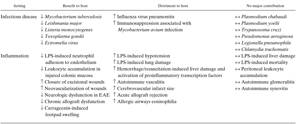
[image:3.612.56.563.73.282.2]During infection with Mycobacterium avium, iNOS-defi-cient mice suffer no greater replication of bacteria in liver and spleen than in control mice, but their splenic lymphocytes are relieved of the inhibition of mitogen responses characteristic Table I. Roles of iNOS Inferred from Studies in iNOS-deficient Mice
Setting Benefit to host Detriment to host No major contribution
Infectious disease ↓ Mycobacterium tuberculosis ↑ Influenza virus pneumonitis ↔ Plasmodium chabaudi
↓ Leishmania major ↑ Immunosuppression associated with ↔ Plasmodium yoelii
↓ Listeria monocytogenes Mycobacterium avium infection ↔ Trypanosoma cruzi
↓ Toxoplasma gondii ↔ Pseudomonas aeruginosa
↓ Ectromelia virus ↔ Legionella pneumophila
↔ Chlamydia trachomatis
Inflammation ↓ LPS-induced neutrophil ↑ LPS-induced hypotension ↔ LPS-induced liver damage adhesion to endothelium ↑ LPS-induced lung damage ↔ LPS-induced mortality
↓ Leukocyte accumulation in ↑ Hemorrhage/resuscitation-induced liver damage and ↔ Peritoneal leukocyte injured colonic mucosa activation of proinflammatory transcription factors accumulation
↑ Closure of excisional wounds ↑ Autoimmune vasculitis ↔ Autoimmune glomerulitis
↑ Neovascularization of wounds ↑ Cerebrovascular infarct size ↔ Autoimmune synovitis
↓ Neurologic dysfunction in EAE ↑ Acute allograft rejection
↓ Chronic allograft dysfunction ↑ Allergic airways eosinophilia
↓ Carrageenin-induced footpad swelling
↑, ↓, ↔, increase, decrease, or no major change, respectively, in one or more of the following: microbial titer, host mortality, or tissue response. In
of infected wild-type mice (23). Thus, the predominant action of iNOS in this setting is to cause immunosuppression.
Infections in which no significant effect of iNOS deficiency has been established. Notwithstanding exacerbatory effects re-ported when infected mice were treated with NOS inhibitors, genetic deficiency of iNOS has had no discernible impact on Chagasic trypanosomiasis (Tanowitz, H., personal communi-cation), or on malaria, as judged by parasitemia after infection with Plasmodium chabaudi (Stevenson, M.M., personal com-munication), or by IFN-g–mediated protection against liver stage Plasmodium yoelii (Tsuji, M., and F. Zavala, personal communication). In several other infections unaffected by iNOS deficiency, there had been little or no preceding phar-macologic basis for expecting the enzyme to be involved. Pathogens in the latter category include Pseudomonas aerugi-nosa (Gros, P., personal communication), Legionella pneumo-phila (Gros, P., personal communication), and Chlamydia tra-chomatis (Perry, L.L., K. Feilzer, and H. Caldwell, personal communication).
Potential relevance of murine studies to human diseases in-volving infections controlled by macrophages. Because iNOS has been difficult to demonstrate in human macrophages de-rived in vitro from normal donors’ monocytes, the question arises whether there is any clinical significance in the demon-stration that tissue macrophages from rodents use RNI to re-sist certain infections. With hindsight, the controversy seems confined chiefly to the expression of iNOS in healthy donors’ mononuclear phagocytes after attempts to activate the cells in vitro. In monocytes or macrophages from patients with a wide range of infectious or inflammatory diseases, iNOS has been more readily detected or induced (for review see reference 14). Human macrophages express iNOS, for example, when collected from the lungs of patients with tuberculosis (24). In-flammatory (but not normal) human alveolar macrophages could be induced in vitro by mycobacterial infection to express iNOS, and they appeared to use iNOS to control the replica-tion of mycobacteria (25). Other clinical settings that have pre-sented with iNOS-positive monocytes or macrophages include alcoholic hepatitis, endemic malaria, rheumatoid arthritis, os-teoarthritis, giant cell arteritis, and multiple sclerosis (for re-view see reference 14), as well hepatitis A under treatment with IFN-a (26). In sum, it is difficult to recreate reproducibly in vitro the macrophage-priming or -activating environments that arise in infected or inflamed human hosts. Given that hu-man macrophages often express iNOS when activated in vivo, the problems encountered in vitro are more appropriately viewed as a deficiency of our culture techniques and immuno-logic knowledge than as an inadequacy of the cell. Further work is required to determine what contribution iNOS makes in human macrophages when it is fully expressed; most func-tional studies have addressed the contribution that iNOS does not make, when it is not fully expressed.
Role of iNOS in inflammation
The foregoing findings in infections are mirrored by studies of inflammation induced by nonreplicative stimuli in iNOS-defi-cient mice. Depending on the setting, the role of iNOS has ranged from enhancing inflammation to retarding it. In fact, the multifaceted nature of some inflammatory syndromes across organs or through time has allowed more than one of these roles of iNOS to be manifest in a single model. As a re-sult, responses to endotoxic bacterial LPS, systemic
autoimmu-nity, and allografts are each discussed under more than one heading below.
Inflammatory settings in which the capacity to express iNOS appears to have a predominantly deleterious effect. The first phe-notype associated with iNOS deficiency was resistance to the hypotension induced by injection of LPS in anesthetized mice (7). Similarly, lung damage after LPS injection is markedly re-duced in iNOS-deficient mice compared with wild-type mice, as gauged by the lung wet/dry ratio and content of lactate de-hydrogenase in bronchoalveolar lavage fluid (Hussain, S.N.A., personal communication). Another form of shock, that caused by hemorrhage and resuscitation, is often followed by severe inflammation in the lungs associated with induction of neutro-phil-mobilizing cytokines. In mice and rats, this response is preceded by and probably dependent upon activation of tran-scription factors NF-kB and Stat-3. Activation of these tran-scription factors is markedly diminished in iNOS-deficient mice compared with wild-type mice after hemorrhage and re-suscitation (Billiar, T.R., personal communication). Thus, in shock states, iNOS is not merely an effector of organ dysfunc-tion, but also a regulator of other effectors. This echoes the role of iNOS in marshaling the innate immune response in leishmaniasis, as described above.
Given the prominence of iNOS in human macrophages in-filtrating the intima in giant cell arteritis (27), it is of interest to gauge whether iNOS has the potential to contribute to the de-velopment of vasculitis. While there is no reported mouse model of giant cell arteritis, the systemic autoimmune disease that develops in the MRL-lpr/lpr mouse includes vasculitis. This vasculitis is markedly ameliorated by iNOS deficiency (28). Occlusion of the middle cerebral artery induces iNOS in the postischemic brain of wild-type mice beginning after 24 h and peaking at 96 h after occlusion (29). By 96 h, the resulting infarcts are 28% smaller in iNOS-deficient mice than in wild-type mice (29). This is an important extension of the observa-tion that genetic deficiency of nNOS reduces infarct volume measured at 24 and 72 h after occlusion of the middle cerebral artery (30).
In untreated mice, acute rejection of major histocompati-bility complex–mismatched, wild-type cardiac allografts is ameliorated in iNOS-deficient recipients compared with wild-type recipients of the same strain (Koeglin, J., and M.E. Rus-sell, personal communication). However, the role of iNOS in acute graft rejection is reversed in chronic rejection, as dis-cussed in the next section.
In a model of allergic airways disease in which immunized mice are challenged with aerosolized ovalbumin, many fewer eosinophils are recovered from the lungs of iNOS-deficient mice (Xiong, Y., and A. Ramsay, personal communication). Levels of IL-4 and -5 are unchanged. It is unclear by what mechanism iNOS promotes eosinophil accumulation in wild-type mice (Xiong, Y., and A. Ramsay, personal communica-tion).
Footpad swelling 24 h after injection of carrageenin is di-minished in iNOS-deficient mice compared with wild-type mice (8). It is not known whether the accumulation of fluid, fibrin, or cells is preferentially affected.
puri-fied E-selectin. These responses are markedly exaggerated in mice lacking iNOS (31). Thus, expression of iNOS during sep-sis may help retard neutrophil margination, sequestration and activation. Likewise, leukocyte accumulation in the colonic mucosa is more prolonged in iNOS-deficient mice than in wild-type mice after injury by intrarectal instillation of acetic acid (32). Considering that colonic mucosal injury may involve host responses to LPS arising from colonic flora, this finding may be another manifestation of the ability of iNOS to de-crease inflammatory neutrophil–endothelial interactions trig-gered by LPS.
Aseptic wounding induces iNOS. Closure of excisional wounds is delayed by 31% in iNOS-deficient mice compared with wild-type mice (Billiar, T., personal communication). The defect in healing of excisional wounds is quantitatively cor-rected by a single topical administration of an adenoviral vec-tor containing iNOS cDNA (Billiar, T., personal communica-tion). Likewise, iNOS deficiency markedly interferes with the angiogenesis necessary to sustain survival of a skin flap (Frau-lin, F., A. Kane, G. Mitchell, R. Romeo, W. Morrison, and A. Stewart, personal communication). These observations are among the few to demonstrate the requirement for a specific enzyme in wound healing.
SJL mice suitably immunized with myelin basic protein un-dergo a T lymphocyte–dependent demyelinating syndrome termed “experimental allergic encephalomyelitis” (EAE), widely considered a model of multiple sclerosis. Contrary to expectations based on acute pharmacologic inhibition of NOS in wild-type mice and rats, iNOS-deficient mice backcrossed to the SJL background suffer EAE that is more severe and pro-longed (32a). Perhaps this observation reflects the loss of the immunosuppressive action of iNOS at the time of immuniza-tion.
In cardiac allografts in immunosuppressed mice, expression of iNOS in parenchymal cells in the grafted heart decreases the severity of chronic rejection, apparently by inhibiting inflam-matory cell accumulation and blunting neointimal smooth mus-cle cell proliferation and the associated graft arteriosmus-clerosis (Koeglin, J., and M.E. Russell, personal communication).
Inflammatory settings in which the host’s ability to express iNOS appears inconsequential. Injection of LPS into iNOS-deficient mice primed with heat-killed Propionobacterium ac-nes causes just as much liver damage and margination of neu-trophils in the pulmonary vasculature as in wild-type mice (7). Despite the role of iNOS in causing hypotension (7), echocar-diographic analysis suggests that the cardiac dilatation in this model is iNOS-independent (33). Most important, iNOS defi-ciency does not consistently alleviate LPS-induced mortality in conscious mice compared with genetically matched controls (7, 9). In another study, LPS-induced mortality was less in iNOS-deficient mice, but the comparison was made to wild-type mice of a different genetic background (8). Thus, in septic shock, harmful and protective effects of iNOS may contend against each other. Even if the deleterious effects of iNOS predomi-nate, the existence of multiple derangements, each capable of causing death, may obscure the benefit of inhibiting any one such pathway in isolation (7).
Despite the ability of iNOS to interfere with neutrophil– endothelial interaction (31), iNOS deficiency has no impact on the mobilization of leukocytes into the peritoneal cavity after injection of several inflammatory irritants (thioglycollate broth, sodium periodate, and IFN-g plus LPS) (7).
In the same MRL-lpr/lpr mouse model of multisystem au-toimmune disease in which iNOS deficiency decreases vasculi-tis, iNOS deficiency exerts no protective effect against glomer-ulitis and synovitis (28).
Potential roles of iNOS in homeostasis
The veil of normalcy. As much as iNOS-deficient mice bring home the message that iNOS is sometimes important in shap-ing the host’s response to infection or inflammation, the same mice appear to teach us that the enzyme has nothing to con-tribute to homeostasis in the unchallenged host. The mice are born to heterozygous parents with the expected Mendelian frequency, indicating the absence of fetal wastage. They gain weight in step with their wild-type littermates, offer no distin-guishing features to the pathologist or clinical chemist, and re-produce normally with homozygous-deficient mates (7). This is hardly surprising, since so little iNOS is expressed in the un-perturbed host.
Nonetheless, a conclusion that iNOS has no role in homeo-stasis would be premature, nor should such a conclusion auto-matically be extrapolated to humans. Within the confines of the vivarium, the mice are spared important physiologic chal-lenges, such as vigorous exercise and changes in climate. Their apparent normalcy may obscure roles played by iNOS in the wild-type host for which alternate mechanisms are called into play when iNOS is congenitally deficient. Finally, more sophis-ticated studies may reveal subtle phenotypes in iNOS-deficient mice even though they are uninfected and uninflamed. A case in point is described below.
Regulation of transcytosis in pulmonary capillary endothe-lium. The pulmonary capillary endothelium in iNOS-deficient mice displays a markedly greater number of transcytotic vesi-cles than in wild-type mice. At rest, the rate of albumin trans-port out of the pulmonary vasculature in iNOS-deficient mice matches the high level induced in wild-type mice by the activa-tion of complement, and is not further responsive to comple-ment activation (Doerschuk, C.M., personal communication). Apparently, expression of iNOS in the normal, unperturbed mouse exerts a tonic suppressive effect on pulmonary capillary transport function. Two questions arise from these observa-tions: First, by what mechanism do products of iNOS regulate endothelial transcytosis? Second, from what source in normal mice do iNOS-derived products reach pulmonary capillaries? That otherwise normal pulmonary alveolar capillaries register the lack of iNOS suggests that iNOS, rather than nNOS or eNOS, provides a major portion of the RNI to which these cells are constitutively exposed. This surprising notion leads to the following speculation.
Possible contribution of iNOS to the S-nitrosylation of he-moglobin. One of the few settings for seemingly constitutive expression of iNOS in humans is in the respiratory epithelium, especially in large airways (34), as well as in occasional alveo-lar macrophages (35). The appearance of iNOS in these cells probably reflects their response to inhalation of microbes and irritants. Ozone, for example, induces iNOS (36), and ex-planted airway epithelial cells lose iNOS, but maintain or re-gain it in response to substances produced in response to IFN-g
and IL-4 (37). Through the inspired air, NO produced by con-tinuously expressed iNOS in the larger airways could reach al-veoli, there to dissolve in the lining fluid, where it is likely to be stored as S-nitrosothiols.
disposed reservoir of iNOS-derived RNI. First, this pool may mediate the constitutive, iNOS-dependent regulation of pul-monary endothelial fluid transport discussed above. Second, some measure of microbial stasis may be achieved by bathing the respiratory mucosa in RNI. A third speculation is inspired by the recent demonstration that when hemoglobin passes through the lung, cysteine 93 in the b chain is charged with a nitroso group, whose discharge in arterioles regulates their di-ameter in response to the need for flow, as sensed by oxygen tension (38). The source of NO in this homeostatic circuit has not been defined. The hypothesis put forth here is that evolu-tion of mammals in microbiologically active environments has led to reliance upon the continuous expression of iNOS at the portal of the largest, thinnest interface between the outside world and the interior. The resulting accumulation of iNOS-derived RNI in the bronchoalveolar fluid is envisioned as being harnessed for homeostatic functions at distant sites through the allosteric intermediacy of circulating hemoglobin. This hypothesis predicts that tissue pO2-dependent regulation
of arteriolar flow may be blunted in genetically iNOS-deficient individuals, or in genetically normal individuals under field conditions after prolonged, profound pharmacologic inhibition of iNOS.
Possible role of iNOS in uterine physiology. Propagation of the species requires that the gravid uterus relax extensively without stretch-induced activation, and yet commence forceful contractions at term. How is this physiology engineered in hu-mans? In the nongravid uterus, iNOS is undetectable (39). During pregnancy, iNOS is expressed in myometrial myocytes; at the onset of labor, iNOS expression declines precipitously (39). These and related findings noted by Bansal et al. (39) suggest that iNOS may play a role in regulation of uterine con-tractions in human pregnancy. Although uncomplicated preg-nancy is a cytokine-rich, allografted, non–steady state, it is not a disease. Thus the contribution of iNOS envisioned by Bansal et al. (39) would represent a role for iNOS in normal physiol-ogy. This speculation regarding a facilitatory role of iNOS in gestation does not dismiss that iNOS may be destructive in the same organ when expressed in other cells, times, or amounts. For example, decidual macrophages express iNOS at the im-plantation sites of resorbing embryos in mice with high rates of fetal wastage. Administration of an NOS inhibitor forestalls fe-tal loss, suggesting that iNOS may mediate fefe-tal rejection (40).
The therapeutic horizon
Inhibitors of iNOS. Is iNOS a therapeutic target? The follow-ing discussion does not answer the question, but considers cri-teria that bear on it.
Any molecule whose expression is induced by signals asso-ciated with inflammation is likely to be detected in a wide vari-ety of disease states. It is not surprising, then, that iNOS has been detected in people at sites involved by the following con-ditions: Alzheimer’s disease, multiple sclerosis, AIDS-associ-ated dementia, viral uveitis, pulmonary tuberculosis, asthma, lung cancer, pulmonary sarcoidosis, bacterial pneumonia, Crohn’s disease, ulcerative colitis, rheumatoid arthritis, os-teoarthritis, renal allografts, aortic aneurysms, and psoriasis; in blood monocytes from patients with malaria, rheumatoid ar-thritis, and alcoholic hepatitis; and in neutrophils from infected urine (for review see reference 14). Nonetheless, expression at the time and place of disease meets only the simplest of criteria that iNOS might constitute a therapeutic target.
Another criterion is the plausibility of iNOS’s mechanistic involvement. Here, the cytotoxic and proinflammatory poten-tial of iNOS advances the case for its therapeutic inhibition in those of the diseases discussed above that are not thought to be infectious in etiology, such as Alzheimer’s disease, hemor-rhagic shock/resuscitation, late-phase vasoocclusive stroke, in-flammatory bowel disease, and rheumatoid and osteoarthritis, or in those infectious diseases where the inflammatory effect of iNOS appears to outweigh its antimicrobial effect, such as influenza pneumonia. However, the antiinflammatory role of iNOS emphasizes the possibility of adverse consequences at-tendant on its inhibition.
The next step in evaluating iNOS as a therapeutic target is the demonstration that genetic disruption of NOS2 amelio-rates a disease that has met the first two criteria. Unfortu-nately, this forthright standard is problematic. The most seri-ous impediment is the dearth of faithful models of human inflammatory diseases in animals whose genes can be experi-mentally disrupted. In rodents, how closely does the enteritis caused by gavage with dextran sodium sulfate mimic Crohn’s disease, or the rectal instillation of acetic acid recreate ulcer-ative colitis? To what extent is rheumatoid arthritis produced by intraperitoneal injection of streptococcal cell walls or intra-muscular injection of collagen in adjuvant?
Another problem with using unconditional gene disruption to evaluate a therapeutic target is that a gene product may play different roles at different stages in pathogenesis. Conven-tional knock-out of an enzyme produces a life-long deficiency that differs from pharmacologic inhibition after the onset of disease. In multiple sclerosis, for example, expression of iNOS during the development of autoimmunity may help restrain the expansion of autoreactive T cell clones, while its expres-sion during the destruction of brain may accelerate damage. That mice deficient in iNOS from birth get more persistent EAE than wild-type mice (32a) need not conflict with sugges-tions of benefit from administration of NOS inhibitors given after onset of disease.
The third criterion is to seek proof of principle in humans by characterizing the phenotype of subjects who are geneti-cally deficient in the target. As yet, no primary state of human iNOS deficiency has been reported. A search for primary iNOS deficiency might profitably begin among well nourished, immunocompetent patients with miliary tuberculosis and a positive family history.
Fourth, one needs to anticipate that mechanism-based tox-icity should not be prohibitive. Chronic administration of iNOS inhibitors, for example, might be associated with recru-descence of latent tuberculosis (16) or leishmaniasis (41). Thus, in patients treated with NOS inhibitors, PPD status should be ascertained and the patient monitored in the same manner as is customary with corticosteroid therapy, an iNOS-suppressive, tuberculosis-predisposing modality.
Bearing these obstacles and cautions in mind, it is hoped that the opportunity will arise to test iNOS inhibitors with pharmacologically favorable properties in patients with neuro-degenerative disorders, cerebrovascular ischemia/reperfusion, hemorrhagic shock/resuscitation, rheumatoid and osteoarthri-tis, inflammatory bowel diseases, progressive pancreatic b cell dysfunction (42), and fulminant influenza pneumonitis (22).
suggest the possibility of therapeutic benefit for delivering iNOS by “gene therapy” in some settings (Billiar, T., personal communication). Such approaches have also been contem-plated to reduce postangioplasty restenosis (43).
Inhibitors of RNI resistance genes. Now that the role of RNI in some infections in mice has been established, and the relevance to humans judged a possibility, another question arises: How do pathogens catabolize RNI or otherwise defend themselves against their toxic actions? Several new RNI resis-tance genes have been uncovered recently (15, 44), and RNI resistance properties ascribed to previously reported genes (45). These findings suggest that therapeutic manipulation of the high-output system of NO production need not target only iNOS. To the extent that microbial RNI resistance pathways play a critical role in the host–pathogen relationship, their in-hibition might sensitize the pathogen to the host’s armamen-tarium, or to therapeutic agents, such as NO donors, designed to mimic this component of the host’s attack. Likewise, when mammalian RNI resistance genes are better understood, their inhibition may enhance the efficacy of tumor immunotherapy.
Conclusion
Over the last 2 yr, more than 120 laboratories have set up colo-nies of iNOS-deficient mice. Only a handful have had time to complete their studies. Nonetheless, it is already clear that ex-pression of iNOS sometimes makes a profound difference to the course of infection or inflammation in mice. There is as yet no sound experimental basis on which to reject the presump-tion that iNOS may play a similar role in humans. In both in-fection and inflammation, iNOS appears to act both as a direct effector and as a regulator of other effectors. The impact of iNOS is potentially dichotomous, and the dichotomy is some-times manifest at different some-times or sites in the same experi-mental setting. These complexities do not preclude experimen-tal therapeutic intervention, but demand caution, whether trials be with iNOS inhibitors, iNOS cDNAs, NO, NO donors, or inhibitors of RNI resistance pathways.
Acknowledgments
Thanks are due the investigators who generously shared their unpub-lished results; Drs. John Mudgett and John MacMicking for generat-ing the mice on whose progeny so many of the investigations relied; Dr. Philip Davies for his indispensable support in that endeavor; and Drs. John Mudgett and Qiao-wen Xie for their helpful comments on the manuscript. I apologize to investigators working with iNOS-defi-cient mice whom I was not able to identify for help in preparation of this article, as well as those whose primary papers could only be cited indirectly by reference to reviews.
This work was supported by National Institutes of Health grants HL-51967, AI-34543, and GM-53921.
References
1. Keeley, J.E., and C.J. Fotheringham. 1997. Trace gas emissions and smoke-induced seed germination. Science. 276:1248–1250.
2. Michel, T., and O. Feron. 1997. Nitric oxide synthases: which, where, how, and why? J. Clin. Invest. 100:2146–2152.
3. Xie, Q.-w., H. Cho, J. Calaycay, R.A. Mumford, K.M. Swiderek, T.D. Lee, A. Ding, T. Troso, and C. Nathan. 1992. Cloning and characterization of inducible nitric oxide synthase from mouse macrophages. Science. 256:225–228. 4. Ruan, J., Q.-w. Xie, N. Hutchinson, H. Cho, G.C. Wolfe, and C. Nathan. 1996. The putative calmodulin-binding region of murine inducible nitric oxide synthase is necessary but not sufficient to sustain calmodulin binding and nitric oxide production at trace levels of free Ca21. J. Biol. Chem. 271:22679–22686.
5. Lyons, C.R., G.J. Orloff, and J.M. Cunningham. 1992. Molecular cloning and functional expression of an inducible nitric oxide synthase from a murine macrophage cell line. J. Biol. Chem. 267:6370–6374.
6. Lowenstein, C.J., C.S. Glatt, D.S. Bredt, and S.H. Snyder. 1992. Cloned and expressed macrophage nitric oxide synthase contrasts with the brain en-zyme. Proc. Natl. Acad. Sci. USA. 89:6711–6715.
7. MacMicking, J.D., C. Nathan, G. Hom, N. Chartrain, M. Trumbauer, K. Stevens, Q.-w. Xie, K. Sokol, D.S. Fletcher, N. Hutchinson, H. Chen, and J.S. Mudgett. 1995. Altered responses to bacterial infection and endotoxic shock in mice lacking inducible nitric oxide synthase. Cell. 81:641–650.
8. Wei, X.-q., I.G. Charles, A. Smith, J. Ure, G.-j. Feng, F.-p. Huang, D. Xu, W. Muller, S. Moncada, and F.Y. Liew. 1995. Altered immune responses in mice lacking inducible nitric oxide synthase. Nature. 375:408–411.
9. Laubach, V.W., E.G. Shesely, O. Smithies, and P.A. Sherman. 1995. Mice lacking inducible nitric oxide synthase are not resistant to lipopolysaccharide-induced death. Proc. Natl. Acad. Sci. USA. 92:10688–10692.
10. Nathan, C., and Q.-w. Xie. 1994. Minireview: regulation of biosynthesis of nitric oxide. J. Biol. Chem. 269:13725–13728.
11. Wong, J.M., and T.R. Billiar. 1995. Regulation and function of inducible nitric oxide synthase during sepsis and acute inflammation. Adv. Pharmacol. 34: 155–170.
12. James, S.L. 1995. Role of nitric oxide in parasitic infections. Microbiol. Rev. 59:533–547.
13. Kroncke, K.-D., K. Fehsel, and V. Kolb-Bachofen. 1995. Inducible nitric oxide synthase and its product nitric oxide, a small molecule with complex bio-logical activities. Biol. Chem. Hoppe-Seyler. 376:327–343.
14. MacMicking, J., Q.-w. Xie, and C. Nathan. 1997. Nitric oxide and mac-rophage function. Annu. Rev. Immunol. 15:323–350.
15. Fang, F.C. 1997. Mechanisms of nitric oxide–related antimicrobial activ-ity. J. Clin. Invest. 99:2818–2825.
16. MacMicking, J.D., R.J. North, R. LaCourse, J.S. Mudgett, S.K. Shah, and C.F. Nathan. 1997. Identification of NOS2 as a protective locus against tu-berculosis. Proc. Natl. Acad. Sci. USA. 94:5243–5248.
17. North, R.J., and A.A. Izzo. 1993. Mycobacterial virulence. Virulent strains of Mycobacterium tuberculosis have faster in vivo doubling times and are better equipped to resist growth-inhibiting functions of macrophages in the presence and absence of specific immunity. J. Exp. Med. 177:1723–1733.
18. Adams, L.B., M.C. Dinauer, D. Morganstern, and J.L. Krahenbuhl. 1997. The role of reactive oxygen and nitrogen intermediates in the host re-sponse to Mycobacterium tuberculosis. Abstract 29, 32nd Annual US–Japan Cooperative Medical Science Program’s Tuberculosis and Leprosy Conference, Cleveland, OH, July 21–23, 1997.
19. Karupiah, G., Q.-w. Xie, R.M.L. Buller, C. Nathan, C. Duarte, and J. MacMicking. 1993. Inhibition of viral replication by interferon-g-induced nitric oxide synthase. Science. 261:1445–1448.
20. Fehr, T., G. Schoedon, B. Odermatt, T. Holtschke, M. Schneemann, M.F. Bachmann, T.W. Mak, I. Horak, and R.M. Zinkernagel. 1997. Crucial role of interferon consensus sequence binding protein, but neither of interferon reg-ulatory factor 1 nor of nitric oxide synthesis for protection against murine liste-riosis. J. Exp. Med. 185:921–931.
21. Scharton-Kerstein, T.M., G. Yap, J. Magram, and A. Sher. 1997. Induc-ible nitric oxide synthase is essential for host control of persistent but not acute infection with the intracellular pathogen Toxoplasma gondii.J. Exp. Med. 185: 1261–1273.
22. Akaike, T., Y. Noguchi, S. Ijiri, K. Setoguchi, M. Suga, Y.M. Zheng, B. Dietschold, and H. Maeda. 1996. Pathogenesis of influenza virus-induced pneu-monia: involvement of both nitric oxide and oxygen radicals. Proc. Natl. Acad. Sci. USA. 93:2448–2453.
23. Doherty, T.M., and A. Sher. 1997. Defects in cell-mediated immunity af-fect chronic, but not innate, resistance of mice to Mycobacterium avium infec-tion. J. Immunol. 158:4822–4831.
24. Nicholson, S., M. da G. Bonecini-Almeida, J.R. Lapa e Silva, C. Nathan, Q.-w. Xie, R. Mumford, J.R. Weidner, J. Calaycay, J. Geng, N. Boechat, C. Lin-hares, W. Rom, and J.L. Ho. 1996. Inducible nitric oxide synthase in pulmonary alveolar macrophages from patients with tuberculosis. J. Exp. Med. 183:2293– 2302.
25. Nozaki, Y., Y. Hasegawa, S. Ichiyama, I. Nakashima, and K. Shimokata. 1997. Mechanism of nitric oxide-dependent killing of Mycobacterium BCG in human alveolar macrophages. Infect. Immun. 65:3644–3647.
26. Sharara, A.K., D.J. Perkins, M.A. Misukonis, S.U. Chan, J.A. Dorninitz, and J.B. Weinberg. 1997. IFN-a activation of human mononuclear cells in vitro and in vivo for nitric oxide synthase type 2 mRNA and protein expression. Pos-sible relationship of induced NOS2 to the anti–hepatitis C effects of IFN-a in vivo. J. Exp. Med. In press.
27. Weyand, C.M., A.D. Wagner, J. Bjornsson, and J.J. Goronzy. 1996. Cor-relation of the topographical arrangement and the functional pattern of tissue-infiltrating macrophages in giant cell arteritis. J. Clin. Invest. 98:1642–1649.
28. Gilkeson, G.S., J.S. Mudgett, M.F. Seldin, P. Ruiz, A.A. Alexander, M.A. Misukonis, D.S. Pisetsky, and J.B. Weinberg. 1997. Clinical and serologic manifestations of autoimmune disease in MRL-lpr/lpr mice lacking nitric oxide synthase type 2. J. Exp. Med. 186:365–373.
Delayed reduction of ischemic brain injury and neurological deficits in mice lacking the inducible nitric oxide synthase gene. J. Neurosci. 17:9157–9164.
30. Huang, Z., P.L. Huang, N. Panahian, T. Dalkara, M.C. Fishman, and M.A. Moskowitz. 1994. Effects of cerebral ischemia in mice deficient in neu-ronal nitric oxide synthase. Science. 265:1883–1885.
31. Hickey, M.J., K.A. Sharkey, E.G. Sihota, P.H. Reinhardt, J.D. Mac-Micking, C. Nathan, and P. Kubes. 1997. Inducible nitric oxide synthase-defi-cient mice have enhanced leukocyte-endothelium interactions in endotoxemia. FASEB (Fed. Am. Soc. Exp. Biol.) J. 11:955–964.
32. McCafferty, D.-M., J.S. Mudgett, M.G. Swain, and P. Kubes. 1997. In-ducible nitric oxide synthase plays a critical role in resolving intestinal inflam-mation. Gastroenterology. 112:1022–1027.
32a. Fenyk-Melody, J., A. Garrison, S. Brunnert, J. Weidner, F. Shen, B. Shelton, and J.S. Mudgett. 1997. Experimental autoimmune encephalomyelitis is exacerbated in mice lacking the NOS2 gene. J. Immunol. In press.
33. Hahn, R.T., J. Brause, R.B. Devereux, C.F. Nathan, and S.C. Nicholson. 1997. Cardiac function in septic shock in inducible nitric oxide synthase knock-out mice. J. Am. Coll. Cardiol. In press.
34. Guo, F.H., H.R. De Raeve, T.W. Rice, D.J. Stuehr, F.B. Thunnissen, and S.C. Erzurum. 1995. Continuous nitric oxide synthesis by inducible nitric oxide synthase in normal human airway epithelium in vivo. Proc. Natl. Acad. Sci. USA. 92:7809–7813.
35. Kobzik, L., D.S. Bredt, C.J. Lowenstein, J. Drazen, B. Gaston, D. Sug-arbaker, and J.S. Stamler. 1993. Nitric oxide synthesis by inducible nitric oxide synthase in human and rat lung: immunocytochemical and histochemical local-ization. Am. J. Respir. Cell Mol. Biol. 9:371–377.
36. Pendino, K.J., J.D. Laskin, R.L. Shuler, C.J. Punjabi, and D.L. Laskin. 1993. Enhanced production of nitric oxide by rat alveolar macrophages after in-halation of a pulmonary irritant is associated with increased expression of nitric oxide synthase. J. Immunol. 151:7196–7205.
37. Guo, F.H., K. Uetani, S.J. Haque, B.R.G. Williams, R.A. Dweik, F.B.J.M. Thunnissen, W. Calhoun, and S.C. Erzurum. 1997. Interferon g and in-terleukin 4 stimulate prolonged expression of inducible nitric oxide synthase in human airway epithelium through synthesis of soluble mediators. J. Clin. Invest. 100:829–838.
38. Jia, L., C. Bonaventura, and J.S. Stamler. 1996. S-nitrosohaemoglobin: a dynamic activity of blood involved in vascular control. Nature. 380:221–226.
39. Bansal, R.K., P.C. Goldsmith, Y. He, C.J. Zaloudek, J.L. Ecker, and R.K. Riemer. 1997. A decline in myometrial nitric oxide synthase expression is associated with labor and delivery. J. Clin. Invest. 99:2502–2508.
40. Haddad, E.K., A.J. Duclos, and M.G. Baines. 1995. Early embryo loss is associated with local production of nitric oxide by decidual mononuclear cells. J. Exp. Med. 182:1143–1151.
41. Stenger, S., N. Donhauser, H. Thüring, M. Röllinghoff, and C. Bogdan. 1996. Reactivation of latent leishmaniasis by inhibition of inducible nitric oxide synthase. J. Exp. Med. 180:783–793.
42. Shimabukuro, M., M. Ohneda, Y. Lee, and R.H. Unger. 1997. Role of nitric oxide in obesity-induced b cell disease. J. Clin. Invest. 100:290–295.
43. Tzeng, E., L.L. Shears II, P.D. Robbins, B.R. Pitt, D.A. Geller, S.C. Watkins, R.L. Simmons, and T.R. Billiar. 1996. Vascular gene transfer of the human inducible nitric oxide synthase: characterization of activity and effects on myointimal hyperplasia. Mol. Med. 2:211–225.
44. Ehrt, S., M.U. Shiloh, J. Ruan, M. Choi, S. Gunzburg, C. Nathan, Q.-w. Xie, and L.W. Riley. 1997. A novel antioxidant gene from Mycobacterium tu-berculosis. J. Exp. Med. In press.