RESEARCH ARTICLE STEM CELLS AND REGENERATION
The majority of early primordial germ cells acquire pluripotency by
AKT activation
Yasuhisa Matsui1,2,*, Asuka Takehara1, Yuko Tokitake1,2, Makiko Ikeda1, Yuka Obara1, Yuiko Morita-Fujimura3, Tohru Kimura4and Toru Nakano2,5
ABSTRACT
Primordial germ cells (PGCs) are undifferentiated germ cells in embryos, the fate of which is to become gametes; however, mouse PGCs can easily be reprogrammed into pluripotent embryonic germ cells (EGCs) in culture in the presence of particular extracellular factors, such as combinations of Steel factor (KITL), LIF and bFGF (FGF2). Early PGCs form EGCs more readily than do later PGCs, and PGCs lose the ability to form EGCs by embryonic day (E) 15.5. Here, we examined the effects of activation of the serine/threonine kinase AKT in PGCs during EGC formation; notably, AKT activation, in combination with LIF and bFGF, enhanced EGC formation and caused∼60% of E10.5 PGCs to become EGCs. The results indicate that the majority of PGCs at E10.5 could acquire pluripotency with an activated AKT signaling pathway. Importantly, AKT activation did not fully substitute for bFGF and LIF, and AKT activation without both LIF and bFGF did not result in EGC formation. These findings indicate that AKT signal enhances and/or collaborates with signaling pathways of bFGF and of LIF in PGCs for the acquisition of pluripotency.
KEY WORDS: EGCs, PGCs, LIF, bFGF, AKT
INTRODUCTION
Mammalian germ cells have potential to acquire totipotency through fertilization, and fertilized eggs subsequently generate naïve pluripotent stem cells (Nichols and Smith, 2009) and trophoectoderm cells after zygote cleavage. After implantation, a population of naïve pluripotent stem cells, designated primitive ectoderm or early epiblast, develop into primed pluripotent stem cells or late epiblast, the developmental potential of which is restricted compared with that of naïve pluripotent stem cells. Primordial germ cell (PGC) emergence from the late epiblast results from the functions of extracellular factors such as BMP4 (Lawson et al., 1999) and those of intrinsic transcription regulators including BLIMP1 (PRDM1 – Mouse Genome Informatics) (Ohinata et al., 2005), PRDM14 (Yamaji et al., 2008), TFAP2C (Weber et al., 2010) and OCT4 (POU5F1 –Mouse Genome Informatics) (Okamura et al., 2008). PGCs then undergo a complex differentiation process that includes epigenetic reprogramming and meiosis (Sasaki and Matsui, 2008); ultimately, functional gametes are formed in adult gonads.
Although mammalian PGCs normally progress through this segment of the mammalian germ cell cycle in embryos, they also
have the potential to progress through an alternative shortcut cycle and acquire naïve pluripotency in culture orin vivo. PGCs (Matsui et al., 1992; Resnick et al., 1992) and spermatogonial stem cells (Kanatsu-Shinohara et al., 2004) from mice are converted into naïve pluripotent cells, designated EGCs (embryonic germ cells) or mGSCs (multipotent germline stem cells), respectively, in culture in the presence of particular extracellular factors. Additionally, PGCs can differentiate into teratomas that contain pluripotent cells in embryos in the presence of mutations of genes such as Dnd1 (Youngren et al., 2005) orPten(Kimura et al., 2003). Although PGCs express a number of pluripotency-associated genes including Oct4,NanogandSox2, PGCs themselves are monopotent and can normally differentiate only into gametes (Leitch et al., 2014). Therefore, conversion of PGCs to pluripotent stem cells is considered a reprogramming process.
EGCs were initially established from PGCs cultured in the presence of a combination of Steel factor (KITL–Mouse Genome Informatics), LIF and bFGF (FGF2–Mouse Genome Informatics) (Matsui et al., 1992; Resnick et al., 1992), but subsequent studies have demonstrated that other factors also stimulate EGC formation; these include retinoic acid (RA), forskolin (FK) (Koshimizu et al., 1996), trichostatin A (TSA), which is an inhibitor of histone deacetylase (Durcova-Hills et al., 2008), and inhibitors of mitogen-activated protein kinase (MAPK) signaling and of glycogen synthase kinase 3 (GSK3B), which as a group are designated 2i (Leitch et al., 2010). In addition, we previously reported that activation of the serine/threonine kinase AKT (Ak strain transforming) in PGCs enhances EGC formation (Kimura et al., 2008). However, even in the presence of those factors, only a portion of PGCs could undergo reprogramming into EGCs.
PGCs at earlier embryonic stages show relatively high efficiency of EGC formation, but PGCs gradually lose the potential to be converted into EGCs, and EGC formation is not observed after embryonic day (E)15.5 (Matsui and Tokitake, 2009). Recent findings show that∼20% of E7.5 PGCs are converted into EGCs in the presence of Steel factor, LIF, bFGF, RA, FK and 2i (Leitch et al., 2013) and, based on this high efficiency of reprogramming, it has been suggested that PGCs have latent pluripotency (Leitch and Smith, 2013). Here, we examined in detail the effects of AKT activation on PGC conversion to EGCs. We found that∼60% of E10.5 PGCs were converted into EGCs with combinations of LIF, bFGF and AKT activation, and the results revealed that early PGCs indeed closely associate with pluripotency.
RESULTS
Effects of culture conditions on the efficiency of EGC formation from PGCs
To obtain higher efficiencies of PGC to EGC conversion, we tested several culture conditions. Fluorescence activated cell sorting (FACS) was used to isolate GFP-expressing PGCs from IFITM3
Received 3 June 2014; Accepted 30 September 2014 1
Cell Resource Center for Biomedical Research, Institute of Development, Aging and Cancer, Tohoku University, Sendai, Miyagi 980-8575, Japan.2CREST, JST, Kawaguchi, Saitama 332-0012, Japan.3Frontier Research Institute of Interdisciplinary Sciences (FRIS), Tohoku University, Sendai, Miyagi 980-8578, Japan.4School of Science, Kitasato University, Sagamihara, Kanagawa 252-0373, Japan.5Graduate School of Frontier Biosciences, Osaka University, Suita, Osaka 565-0871, Japan. *Author for correspondence ([email protected])
DEVEL
O
(mil1)-GFP (3.0G) transgenic E12.5 embryos (Tanaka et al., 2004); these PGCs were plated into 24-well culture dishes with Sl/Sl4-m220 feeder cells expressing membrane-bound Steel factor (Matsui et al., 1991). At the end of the culture period, colonies with alkaline phosphatase (ALP)-positive EGCs were counted, and the efficiency of EGC formation was determined as a ratio of the number of EGC colonies to every 100 sorted PGCs that were seeded in a culture well. We found that the efficiency of EGC formation was higher in cultures with knockout serum replacement (KSR) than those with fetal bovine serum (FBS). In the presence of 10% KSR, the efficiency of EGC formation was ∼4%, which was about 5-fold
higher than that with 10% FBS (Fig. 1A). KSR had a similar effect on formation of EGCs from E10.5 PGCs (Fig. 3A).
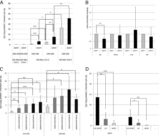
[image:2.612.51.561.200.636.2]Activation of AKT significantly stimulates EGC formation Activation of AKT enhances conversion of PGCs into EGCs (Kimura et al., 2008). Here, we quantitatively reassessed this effect of AKT activation on EGC formation. Specifically, we mated AKT-MER transgenic mice, in which AKT-MER is ubiquitously expressed, with mil1-GFP (3.0G) transgenic mice that specifically express GFP in PGCs. FACS-purified GFP-positive PGCs at different embryonic stages were cultured with or without 4-hydroxy tamoxifen (4OHT),
Fig. 1. The effects of AKT activation on reprogramming of PGCs into EGCs.(A) PGCs purified from AKT-MER×mil1-GFP (3.0G) transgenic embryos at E10.5, E12.5, E14.5 or E15.5 were cultured with or without 4OHT in the presence of LIF and bFGF and either with FBS or KSR. The efficiency of reprogramming is represented by ratios of EGC colony number to initially plated PGC number. (B) The effects of AKT activation on the expression of crucial genes. PGCs of AKT-MER×Oct4-deltaPE transgenic embryos at E12.5 were cultured with or without 4OHT for 2 days and GFP-positive PGCs were purified by cell sorting for quantitative RT-PCR analysis.Arbpwas used as an internal control. The expression levels of each gene in PGCs cultured without 4OHT was set as 1.0. The experiments were performed using three independently cultured PGC samples. (C) PGCs purified from AKT-MER×mil1-GFP (3.0G) transgenic embryos at E12.5 were cultured with or without 100 nM or 1000 nM 4OHT either with FBS or KSR. In some cases, 4OHT was added only for the first day in culture (d0-1), but otherwise 4OHT was present continuously in the culture medium (d0-9). (D) The effects of LIF and bFGF were examined by using medium either with or without 4OHT that contained LIF and/or bFGF or that lacked both LIF and bFGF. PGCs purified from AKT-MER×mil1-GFP (3.0G) transgenic embryos at E12.5 were cultured. Number of independent experiments (n)=3-13 (A), 3 (B), 4-13 (C), 3-11 (D). Data show the mean±s.e.m.; *P<0.05; **P<0.005; ***P<0.001.
DEVEL
O
which activates AKT-MER protein. We used a mixture of male and female embryos because previous studies have indicated that the efficiency of EGC formation from PGCs was equal for male and female PGCs (Kimura et al., 2008; Matsui and Tokitake, 2009).
EGC formation from E12.5 PGCs cultured in 10% FBS or 10% KSR was significantly stimulated by addition of 4OHT (Fig. 1A). Efficiency of EGC formation from E10.5 PGCs was also increased by addition of 4OHT and reached∼50% (Fig. 1A). It is likely that AKT activation results in induction or repression of critical genes for the reprogramming of PGCs into EGCs, and we found that the expression of BAX, a member of the BCL2 gene family that promotes apoptosis in various kinds of cells including PGCs, was downregulated by AKT activation in cultured PGCs (Fig. 1B), supporting the notion that AKT activation prevents apoptosis of PGCs (Kimura et al., 2008). By contrast, the expression of BLIMP1, a key regulator of PGC development, and of c-MYC and KLF4, critical reprogramming factors, the expression of which was downregulated and upregulated, respectively, during conversion of PGCs into EGCs (Durcova-Hills et al., 2008), were not significantly changed by AKT activation (Fig. 1B).
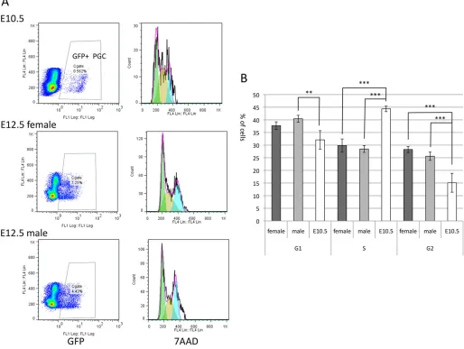
In contrast to E12.5 PGCs, PGCs from E14.5 or E15.5 embryos rarely formed EGC colonies even in the presence of 4OHT (Fig. 1A). All of these finding were consistent with previous findings (Kimura et al., 2008). Developmental stage-dependent decrease of EGC formation likely correlates with changes in cell cycle status. After E13.5, PGCs undergo mitotic arrest, which might be associated with the inability of PGCs to undergo reprogramming. Although PGCs still proliferate at E12.5, we found that a cell cycle profile of E12.5 PGCs was significantly different from that of E10.5 PGCs (Fig. 2). At E10.5, the ratio of PGCs in S phase was relatively high at∼44%, whereas that at E12.5 significantly decreased and was ∼29% (Fig. 2B), indicating that E10.5 PGCs are more proliferative than E12.5 PGCs. This result suggests that the cell cycle change might affect the efficiency of conversion of PGCs into EGCs, and that proliferative PGCs are susceptible to reprogramming.
[image:3.612.49.563.301.688.2]Notably, the presence of 4OHT for only the first day of culture significantly enhanced EGC formation, and the efficiency was slightly increased by continuous stimulation by 4OHT throughout the culture period (Fig. 1C). Enhancement of PGC to EGC conversion with 1000 nM 4OHT did not differ significantly from
Fig. 2. Cell cycle analysis of E10.5 and E12.5 PGCs.(A) Cells containing PGCs obtained from the Oct4-deltaPE-GFP transgenic embryos at E10.5 and E12.5 were stained with 7-amino-actinomycin D (7AAD) for estimating the amount of DNA in the cells, and the cell cycle of GFP-positive PGCs was examined by flow cytometry (right panels). The GFP-positive gates are shown on the left. In the case of E12.5 PGCs, female and male PGCs were separately examined. (B) The percentage of cells in G1/G0 (G1), S and G2/M (G2) phases in E10.5 and E12.5 PGCs are indicated. The data represents three independently prepared PGC samples of each developmental stage. Data show the mean±s.e.m.; **P<0.01; ***P<0.001.
DEVEL
O
that with 100 nM, indicating that 100 nM was enough to stimulate PGCs (Fig. 1C).
AKT activation can substitute for bFGF with regard to PGC to EGC conversion (Kimura et al., 2008). We re-examined the effects of AKT activation with or without bFGF and/or LIF. EGCs were formed in the absence of bFGF, and the efficiency of PGC to EGC conversion in the presence of LIF and 4OHT was slightly higher than that with LIF alone (Fig. 1D). However, in our culture system, the efficiency of EGC formation was much higher in the presence of 4OHT, LIF and bFGF than under any other conditions used. EGCs were also formed without LIF, but the efficiency was lower than that without bFGF. In addition, AKT activation alone did not result in EGC formation (Fig. 1D). Taken together, these results indicate that AKT activation did not fully substitute for bFGF or for LIF, but AKT activation worked synergistically with bFGF and LIF to stimulate EGC formation.
2i does not further enhance EGC formation
[image:4.612.105.509.56.437.2]We next tested the effect of 2i, because a previous study indicated that this condition resulted in an increased efficiency of EGC formation (Leitch et al., 2010). We cultured FACS-purified E10.5 PGCs with medium containing LIF, bFGF and either FBS or KSR for 3 days; medium in some wells was then replaced with serum free medium containing LIF+2i as reported previously (Leitch et al., 2010). The results showed that the efficiency of EGC formation in wells containing LIF+2i medium was∼11-12%, which was similar to or less than that in wells that contained LIF+bFGF medium continuously (Fig. 3A). We then tested the effects of PD0325901 or CHIR99021 alone with KSR, and found that the efficiency was also decreased in the presence of PD0325901 or CHIR99021 (Fig. 3B). Notably, CHIR99021 but not PD0325901 resulted in densely packed and strongly ALP-positive undifferentiated colonies similar to those formed following 2i treatment (Fig. 3C), suggesting Fig. 3. The effects of 2i on the reprogramming of PGCs into EGCs.(A) PGCs purified from MCH×mil1-GFP (3.0G) embryos at E10.5 were cultured with medium containing LIF, bFGF and either FBS or KSR (L, F→L, F). In some cases, the culture medium was changed to N2B27 medium containing 2i+LIF after 3 days in culture (L, F→L, 2i). (B) PGCs purified from MCH×mil1-GFP (3.0G) embryos at E10.5 were cultured with medium containing LIF, bFGF and KSR or FBS with or without Y27632 (Y) or BMP4. In some cases, the culture medium was changed to N2B27 medium containing PD0325901 (PD), CHIR99021 (CH) or both (2i)+LIF after 3 days in culture (L, F→L, PD, CH or 2i). (C) The appearance of EGC colonies derived from PGCs of MCH×mil1-GFP (3.0G) embryos at E10.5. PGCs were cultured with medium containing KSR, LIF and bFGF (a), medium was changed to N2B27 medium containing PD0325901+LIF (b), CHIR99021+LIF (c) or 2i+LIF (d) after 3 days in culture. Images of ALP staining are shown. Scale bar: 100μm. (D) PGCs purified from AKT-MER×mil1-GFP (3.0G) transgenic embryos at E12.5 were cultured with KSR and with or without 100 nM (L, F→L, F). In some cases, the culture medium was changed to N2B27 medium containing 2i+LIF after 3 days in culture (L, F→L, 2i). Number of independent experiments (n)=6 (A), 9 (B) and 4-8 (C). Data show the mean±s.e.m.; *P<0.05; **P<0.005; ***P<0.001.
DEVEL
O
that CHIR99021 mainly functions to maintain the undifferentiated status of EGCs undergoing conversion from PGCs.
We also tested the effects of BMP4 and Y27632, an inhibitor of p160-Rho-associated coiled-coil kinase (ROCK) on EGC formation, because BMP4 and ROCK inhibitor play critical roles in the self-renewal of undifferentiated mouse embryonic stem cells (ESCs) (Ying et al., 2003) and survival of human ESCs (Watanabe et al., 2007), respectively. However, the efficiency of EGC formation decreased in the presence of BMP4 with FBS and was not changed in the presence of BMP4 or Y27632 with KSR (Fig. 3B).
We next examined whether combinations of 2i and 4OHT further enhanced EGC formation. We cultured E12.5 PGCs isolated from AKT-MER transgenic embryos and found that the efficiency of EGC formation was enhanced by 4OHT in the LIF+2i condition as well as in the LIF+bFGF condition. However, in the presence of 4OHT, the efficiency of EGC formation was lower in the LIF+2i condition than the LIF+bFGF condition (Fig. 3D). Therefore, combining 2i and AKT activation did not result in more efficient reprogramming than did AKT activation alone.
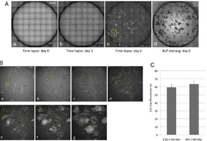
AKT-activated PGCs are efficiently converted into EGCs Our results indicated that about half of E10.5 PGCs could be reprogrammed into EGCs if the AKT signal was strongly activated. However, it is likely that an original single PGC does not exactly correspond to a single EGC colony; multiple PGCs could aggregate to form a single EGC colony and single PGCs could generate multiple EGC colonies. Therefore, we performed time-lapse
analysis of the process of EGC formation from PGCs to more precisely estimate ratios of PGCs that were converted into EGCs (Fig. 4A,B; supplementary material Movies 1, 2). We plated purified PGCs into wells of 96-well plates, and multipoint time-lapse equipment was used to capture fluorescent images of whole wells once every hour for 6 days. At the end of culture, the cells were stained for ALP activity to determine whether each GFP-positive colony was also ALP GFP-positive (Fig. 4A).
As expected, PGCs actively moved around on the feeder layer, and multiple daughter cells that had originated from a single PGC started to form EGC colonies after 3 days in culture (Fig. 4B; supplementary material Movies 1, 2). EGC colonies resulting from a single PGC, those resulting from daughter cells of the original PGCs, or some combination thereof often fused during colony expansion (Fig. 4B). Meanwhile, some PGCs or their daughter cells were fragmented and were lost during culture. Our previous study showed that AKT activation resulted in a marked decrease in the number of apoptotic PGCs in culture (Kimura et al., 2008). Consistent with the result, we found that the expression of BAX was downregulated by AKT activation in cultured PGCs (Fig. 1B).
[image:5.612.100.517.372.656.2]We carefully observed the time-lapse movies, and estimated the proportions of PGCs that contributed to EGC colonies for each whole well culture monitored. About 60% of the original PGCs were incorporated into EGC colonies. We used two different GFP reporter embryos, mil1-GFP (3.0G) and Oct4-deltaPE-GFP (dPE), to detect PGCs; we obtained identical results with both embryo types (Fig. 4C).
Fig. 4. Time-lapse analysis of EGC formation from PGCs.PGCs purified from AKT-MER×mil1-GFP (3.0G) (C) or AKT-MER×Oct4-deltaPE-GFP (dPE) (A-C) embryos at E10.5 were cultured with medium containing KSR, LIF, bFGF and 4OHT. (A) Reconstituted fluorescent images of a culture well after 0 (a), 3 (b) and 6 (c) days in culture. GFP-expressing PGCs and their descendants are seen as white signals. (d) ALP staining of the culture shown in c. (B) Higher magnification images of the area indicated by the yellow rectangle in A,c after 0 (a), 1 (b), 2 (c), 3 (d), 4 (e), 5 (f ) days or 5 days 18 h (g) in culture. Groups of cells or colonies enclosed by colored dotted lines originated from cells indicated by the arrows with the corresponding numbering in B,a. Scale bars: 1 mm (A), 10μm (B). (C) The ratios of initial PGCs that were reprogrammed and were incorporated into EGC colonies were manually determined from the reconstituted movies. We used two different reporter transgenic mice [mil1-GFP (3.0G) and Oct4-deltaPE-GFP (dPE)] to identify PGCs. Data represent four independent experiments using AKT-MER×3.0G or AKT-MER×dPE transgenic embryos. Data show the mean±s.e.m.
DEVEL
O
We next examined whether AKT was indeed activated in PGCs by addition of 4OHT. We cultured PGCs purified from E10.5 embryos for 1 day with or without 4OHT, and then used an antibody against phosphorylated AKT (Ser473-pAKT) to stain them. Each GFP-expressing PGC with dot-like signals of activated pAKT near the cell membrane was counted as a pAKT-positive PGC, and ratios of pAKT-positive PGCs to all PGCs were estimated. In the presence of 4OHT,∼55% of PGCs were pAKT positive (Fig. 5Ad-f,C); in the absence of 4OHT,∼8% of PGCs were pAKT positive (Fig. 5Ag-i,C). We also examined AKT activation in E12.5 PGCs cultured with 4OHT; the ratio of pAKT-positive E12.5 PGCs was similar to the ratio of pAKT-positive E10.5 PGCs (Fig. 5C).
We also stained cultured PGCs of E10.5 AKT-MER transgenic embryos with antibody against AKT protein to examine whether all PGCs overexpressed AKT;∼85% of all PGCs were positive for AKT (Fig. 5Aa-c,B). This finding was consistent with our previous findings (Kimura et al., 2008). The results indicated that, for unknown reasons, 4OHT did not fully activate AKT-MER in some PGCs even though the 4OHT concentration was at saturation levels (Fig. 1C).
Pluripotency of EGCs derived from AKT-activated PGCs To examine pluripotency of EGCs derived from AKT-activated PGCs, single EGC colonies derived from E10.5 PGCs of AKT-MER transgenic embryos were randomly selected and were
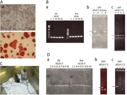
cultured in the LIF+2i medium without 4OHT, to establish EGC lines. We selected 38 colonies; each of which gave rise to an EGC line (AE10-1–AE10-38) with morphology characteristic of pluripotent stem cells, and each line had ALP-positive cells (Fig. 6A).
[image:6.612.52.563.347.653.2]For each randomly selected cell line (AE10-1, 7, 25, 30, 35), we injected EGCs into blastocysts of C57Bl/6 mice to test their ability to contribute to the formation of chimeric mice. We found that injected cells could contribute to chimera formation (Table 1; Fig. 6Bb); notably, the AE10-7, 25 and 35 lines each gave rise to male and female chimeras (Table 1). Germline transmission of AE10-7 or AE10-35 cells through female or male chimeras, respectively, has been confirmed thus far (Table 1; Fig. 6Bc). We used X and Y chromosome-specific primers and PCR to check the sex chromosomes of AE10-1, 7, 25, 30 and 35; each cell line had both X and Y chromosomes (Fig. 6Ba). Therefore, it is likely that at least line AE10-7, which gave rise to female chimera, was a mixture of XX and XY cells and, therefore, this cell line was not clonal. This conclusion was consistent with the time-lapse observation that multiple PGCs could contribute to a single EGC colony. We also injected AE10-7 cells into blastocysts from MCH mice to confirm chimera formation and germline transmission by coat color (Fig. 6C). We further subcloned XY 7-1) and XX (AE10-7-2) cell lines from the AE10-7 line. The sex chromosomes of lines AE10-7-1 and AE10-7-2 were confirmed by PCR (Fig. 6Da), and
Fig. 5. The expression of AKT and phosphorylated AKT in cultured PGCs.PGCs purified from AKT-MER×Oct4-deltaPE-GFP embryos at E10.5 (A-C) and AKT-MER×mil1-GFP (3.0G) embryos at E12.5 (C) were cultured for 1 day with medium containing KSR, LIF and bFGF either with or without 4OHT. (A) Cultured PGCs (green in a,c,d,f,g,i) at E10.5 were stained with antibody against AKT (red in b,c) or pAKT (red in e,f,h,i) and DAPI (blue in c,f,i).
GFP-expressing PGCs are indicated by white arrows or yellow arrowheads. AKT and pAKT expression was detected as dot-like signals near the cell membrane (b,c,e,f ). Without 4OHT, the dot-like signals were rarely observed in GFP-positive PGCs (g-i). Merged images are shown in c, f and i. Yellow arrowheads indicate PGCs with weak AKT signal. Asterisks indicate false GFP signals. Scale bars: 50μm for a-i. (B,C) Ratios of AKT-positive (B) or pAKT-positive (C) PGCs were estimated for individual whole culture wells. Data represent two (B), three (C, E10.5) or four (C, E12.5) independent experiments. Data show the mean±s.d.
(B) or mean±s.e.m. (C); *P<0.0005.
DEVEL
O
cells from each line contributed to male or female, respectively, chimera formation (Fig. 6Db,c).
DISCUSSION
AKT activation greatly enhances the acquisition of pluripotency by PGCs
PGCs are monopotent cells that are destined to become gametes, but they are easily reprogrammed into pluripotent EGCs in culture. About 20% of PGCs at E7.5 convert into EGCs when exposed to 2i,
LIF and bFGF, and the LIF-STAT3 pathway is critical to this conversion (Leitch et al., 2013). Our data demonstrate that AKT activation greatly increases the potential of PGCs to undergo reprogramming, and ∼60% of PGCs purified from AKT-MER transgenic E10.5 embryos were converted into EGCs following 4OHT-mediated activation of AKT (Fig. 4C). This result indicates that the majority of E10.5 PGCs have the potential to acquire pluripotency when AKT signaling is strongly activated. Notably, the potential of these PGCs to acquire pluripotency is similar to that of ESCs derived from early epiblast cells, which is 54% (Nichols et al., 2009).
Additionally, the EGC colonies that emerged in primary cultures of AKT-MER PGCs efficiently established EGC lines (Fig. 6; Table 1). Normal ESC culture conditions, which lack 4OHT, were used to maintain these PGC-derived EGC lines, and these EGCs could contribute to chimera formation when injected into blastocysts; taken together, these observations indicate that the PGC-derived EGCs are indeed pluripotent. The results also indicate that the AKT-MER transgene did not affect development of chimeric mice in the absence of 4OHT. The results together strongly support the idea that early PGCs are closely associated with pluripotency.
[image:7.612.104.514.58.363.2]The percentage of PGCs with hyperactive AKT among all 4OHT-treated PGCs correlated with that of EGC-forming PGCs Fig. 6. Establishment of germline-competent EGC lines from AKT-MER transgenic PGCs.The appearance of AE10-7 EGC colonies derived from PGCs of AKT-MER×mil1-GFP (3.0G) embryos at E10.5. Phase contrast images (a) and ALP staining (b) are shown. Scale bar: 100μm. (B) Identification of the presence of sex chromosomes in AE10-1, 7, 25, 30 and 35 EGCs based on the detection of Y-chromosome-specificZfyor X-chromosome-specificXistby PCR (a). Identification of chimeric mouse (b) and of germline transmission (c) of AE10-7 EGCs based on detection of GFP transgene by PCR. Two female chimeric mice (5 and 8 in b) and four pups of chimeric mouse 8 (3-6 in c) had the GFP transgene. Arrows indicate PCR products for the GFP transgene. Asterisk indicates primer dimer. (C) Chimeric mice derived from AE10-7 EGCs injected in MCH blastocysts showed coat color chimerism, and a pup from a chimeric father showed germline transmission (agouti coat color) (D) Identification of the presence of sex chromosomes in sub lines of AE10-7 EGCs based on PCR amplification of Y-chromosome-specificZfyor X-chromosome-specificXist. AE10-7-2 and AE10-7-3 did not haveZfy, and therefore were female (a). Identification of chimeric mouse of AE10-7-1 and AE10-7-2 EGCs based on PCR amplification of the GFP transgene. One male from AE10-7-1 (1 in b) and one female from AE-10-7-2 (1 in c), each chimeric, had the GFP transgene.
Table 1. Contribution of EGCs derived from AKT-MER PGCs to chimeric mice
EGC line Blastocyst
Chimeras/ pups
Sex of chimeras
Germline transmission
AE10-1 B6 2/4 ♂#1, #2 –
AE10-7 B6 5/12 ♂#1,♀#1, #3, #5, #8
♀#1, #5, #8
AE10-7 MCH 7/17 ♂#1, #2, #3, #4
ND
♀#5, #6, #7 ND
AE10-25 B6 2/11 ♂#1,♀#4 –
AE10-30 B6 1/11 ♀#7 –
AE10-35 B6 3/5 ♂#7, #8,♀#6 ♂#7
ND, not determined.
DEVEL
O
[image:7.612.48.300.615.729.2](Fig. 4C; Fig. 5C). This correlation suggests that the efficiency of AKT activation in PGCs is a limiting factor with regard to the reprogramming of PGCs into EGCs. We used saturating concentrations of 4OHT (Fig. 1C); therefore, for reasons we do not currently understand, 4OHT probably could not fully activate the AKT-MER protein in some AKT-MER-overexpressing PGCs. It is also possible that some PGCs could not convert into EGCs even with activated AKT. We previously showed that the PGC population was heterogeneous and consisted of cells in different differentiation stages (Morita-Fujimura et al., 2009), and that the differentiated PGC subpopulation was resistant to reprogramming (Matsui and Tokitake, 2009). In this study, we show that cell cycle status in PGCs is changed according to PGC development, and the ratio of PGCs in S phase is relatively high at E10.5 compared with that at E12.5 (Fig. 2). The result suggests that highly proliferative PGCs or PGCs in S phase are susceptible to reprogramming, We observed the behavior of AKT-MER PGCs that underwent apoptosis in the time-lapse images, and found that 71% of them died without cell division, whereas the rest of them divided more than once before dying (supplementary material Movie 3). The result is consistent with the notion that proliferative PGCs are susceptible to reprogramming, but some AKT-MER PGCs could not escape apoptosis even though they transited through the cell cycle.
We showed that the expression of BAX was downregulated in PGCs in the presence of 4OHT (Fig. 1B), suggesting that AKT activation prevents apoptosis of PGCs by repression of apoptosis regulators such as BAX and, consequently, more PGCs can initiate the conversion into EGCs. Meanwhile, the expression of BLIMP1, c-MYC and KLF4 in PGCs was not affected by AKT activation (Fig. 1B). Therefore, the reprogramming process might not be accelerated in PGCs by activation of AKT signaling.
The later PGCs lose the potential to form EGCs even with AKT activation
Notably, AKT activation did not enhance EGC formation from E14.5 or E15.5 PGCs; these later stage PGCs rarely converted to EGCs (Fig. 1A) (Kimura et al., 2008). After E14.5, male and female PGCs exhibit growth arrest and meiotic initiation, respectively, and once PGCs exit the mitotic cell cycle, AKT activation might not be able to potentiate the formation of EGCs from PGCs. Additionally, the efficiency of EGC formation from AKT-activated E12.5 PGCs was significantly lower than that from AKT-activated E10.5 PGCs (Fig. 1A), though E12.5 PGCs were still proliferating. However, we found that the cell cycle of PGCs was changed between E10.5 and E12.5, and the ratio of PGCs in S phase was high at E10.5 (Fig. 2). As discussed above, the results suggest that less-proliferative PGCs at E12.5 are resistant to reprogramming.
PGCs might also lose crucial factors necessary for reprogramming during the course of embryonic development, and that this loss could not be reversed by AKT activation. The epigenetic status of PGCs, including DNA methylation as well as histone methylation and acetylation, changes substantially by E12.5, and is different from that in pluripotent cells (Seisenberger et al., 2012; Ng et al., 2013), which suggests that particular epigenetic changes might be crucial for PGCs to be converted into EGCs. Consistent with this notion, previous findings indicate that an inhibitor of histone deacetylase, TSA, enhances EGC formation (Durcova-Hills et al., 2006). Another recent report also demonstrated that, in PGCs, decreased expression of MBD3, a key component of the NuRD complex that has histone deacetylation activity, resulted in highly enhanced EGC formation from E8.5 PGCs compared with PGCs with normal levels of MBD3 (Rais et al., 2013). These results suggest that later-stage PGCs could
potentially be reprogrammed through intense epigenetic manipulation such as forced changes in DNA and histone methylation status.
Relationship between the AKT pathway and LIF or bFGF signaling
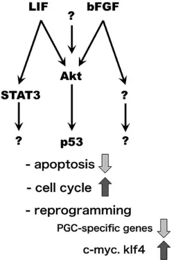
Our current findings demonstrate that AKT activation results in synergistic enhancement of EGC formation with bFGF and LIF (Fig. 1D). In some cases, LIF (Magni et al., 2007) and bFGF (Wang et al., 2009) transmit signals through the AKT pathway, and it is likely that forced activation of AKT might have strengthened LIF and bFGF signals (Fig. 7). Alternatively or in addition, AKT activation might have facilitated reprogramming through molecular pathways that are independent of LIF and bFGF. Previously, we suggested that AKT activation enhances EGC formation through inhibition of p53 (Kimura et al., 2008), which likely suppresses apoptosis and stimulates the cell cycle in PGCs (Fig. 7).
[image:8.612.376.503.478.665.2]However, AKT activation alone was not sufficient to drive EGC formation (Fig. 1D). We previously observed EGC derivation with AKT activation but without bFGF (Kimura et al., 2008). In that case, we estimated the number of EGC-forming culture wells with different combination of cytokines after subculture, a method that might result in over-estimation of the efficiency of EGC formation. In this study, we quantitatively estimated EGC formation in primary culture, and the results indicate that the efficiency of EGC formation without either bFGF or LIF was much lower than that with both bFGF and LIF, even after AKT activation, although EGCs were formed (Fig. 1D). The results indicate that signaling pathways other than the AKT pathway, specifically pathways that are activated by LIF, such as the STAT3 pathway (Leitch et al., 2013), and by bFGF, might have been necessary for the events that occurred during PGC reprogramming; such events include downregulation of PGC specific genes and upregulation of c-MYC and KLF4 (Durcova-Hills et al., 2008) (Fig. 7). Downstream signaling pathways of receptor tyrosine kinases, including FGF receptors, encompass the MAPK pathway and the phospholipase C (PLC) γ pathway. The AKT pathway might synergize with those signaling pathways to enhance EGC formation.
Fig. 7. Signaling pathways involved inthereprogramming of PGCs into EGCs.AKT might be one of multiple downstream targets of bFGF and LIF, but pathways independent of AKT should also be activated by LIF and bFGF. Previous studies demonstrated that p53 and STAT3 are downstream targets of AKT and LIF, respectively, in PGCs. Those signaling pathways suppress apoptosis and stimulate the proliferation of PGCs and might also control the expression of crucial genes for reprogramming.
DEVEL
O
Those pathways might also be involved in Steel factor-cKit signaling for reprogramming PGCs into EGCs.
Because previous reports indicate that LIF+2i can substitute for bFGF and result in higher efficiency of EGC formation than LIF+bFGF (Leitch et al., 2010, 2013), we compared the rate of EGC formation caused by LIF+2i with that caused by the conventional LIF+bFGF combination; notably, under our culture conditions, LIF+2i did not result in a higher rate than LIF+bFGF with or without AKT activation (Fig. 3). Moreover, EGC formation by PGCs was not as greatly potentiated by LIF+2i as by LIF+bFGF+AKT activation. Inconsistencies between our results and previous results might have been due to differences in culture conditions. In the previous study, mixtures of E8.5 PGCs and somatic cells were used (Leitch et al., 2010), whereas we cultured FACS-purified E10.5 PGCs. The responsiveness of PGCs to 2i might be dependent on their developmental stages, and/or contaminating embryonic somatic cells might affect the function of 2i in EGC formation. Another report has also demonstrated that addition of 2i to medium containing KSR enhances EGC formation from E11.5 PGCs on the Sl/Sl4-m220 feeder layer (Nagamatsu et al., 2012). In the aforementioned study, the cells were subcultured onto a STO feeder layer after 3 days in culture, but we continuously cultured on a Sl/Sl4-m220 feeder layer without subculture. Because 2i was somewhat toxic for Sl/Sl4-m220 feeder cells, subculture on STO might enhance the efficiency of EGC formation, although estimation of the efficiency might be less quantitative.
Signaling by bFGF is important for the initial 24-48 h of culture for EGC formation, whereas LIF signals and inhibition of MAPK signaling is required after 48 h in culture (Durcova-Hills et al., 2006; Leitch et al., 2013). Our results show that initial activation of the AKT signal is important (Fig. 1C), and this observation is consistent with the idea that the AKT signal strengthened the bFGF signal. Meanwhile, MAPK signaling could be downstream of bFGF, and inhibition of MEK by PD0325901 for the initial period of culture might inhibit reprogramming of PGCs. Consistent with this notion, the efficiency of EGC formation decreased when PGCs were cultured in the 2i-LIF medium from the beginning of culture (Leitch et al., 2010). In conclusion, our current findings demonstrate that the majority of early PGCs had the potential to acquire naïve pluripotency, and that AKT activation, in combination with the LIF and bFGF signaling pathways, greatly potentiated the ability of PGCs to be reprogrammed.
MATERIALS AND METHODS
Collection of embryos
MCH and C57BL/6 mice were purchased from Japan SLC. The mil-1-GFP (3.0G) (Tanaka et al., 2004) and Oct4-deltaPE-GFP (Yoshimizu et al., 1999) transgenic mice were maintained in a C57BL/6J genetic background. AKT-MER transgenic mice (Kimura et al., 2008) were maintained in a B6D2F1 genetic background. The mice were kept and bred in an environmentally controlled and specific pathogen-free facility, the Animal Unit of the Institute of Development, Aging and Cancer (Tohoku University), according to the guidelines for experimental animals defined by the facility. Animal protocols were reviewed and approved by the Tohoku University Animal Studies Committee. Noon on the day of the plug was defined as E0.5. Embryos of the indicated stages were obtained from female AKT-MER transgenic mice or female MCH mice mated with male mil-1-GFP (3.0G) or Oct4-deltaPE-mil-1-GFP transgenic mice. Embryos were collected and dissected in Dulbecco’s modified Eagle medium (DMEM) containing 10% fetal bovine serum (FBS). Weak expression of GFP in whole embryo bodies was used to identify AKT-MER transgenic embryos (Kimura et al., 2008). Appropriate regions containing PGCs, i.e. the dorsal mesentery of
E10.5 embryos and the genital ridges of E11.5-E15.5 embryos, were dissected from individual embryos.
Flow cytometry
Tissue samples containing PGCs, prepared as described above, were incubated with 1.2 mg/ml collagenase in PBS containing 10% FBS for 1 h at 37°C. To prepare single-cell suspensions for flow cytometry, cells within the samples were dissociated by pipetting, and samples were filtered through a 40μm pore nylon mesh (BD Falcon). A Beckman Coulter ALTRA cell sorter was used to sort and collect PGCs with intense GFP expression.
Culture of PGCs
The sorted PGCs were cultured on a feeder layer of Sl/Sl4/m220 cells (Matsui et al., 1991) pre-treated with mytomycin C in 4-well or 24-well tissue culture dishes with EG medium, which was based on the previously reported GSC culture medium (Kanatsu-Shinohara et al., 2004) with modifications [StemPro34 SFM containing StemPro34 Nutrient, 100μg/ml transferrin, 2 mM L-glutamine, 25μg/ml insulin, 50μM 2-mercaptoethanol, 20 ng/ml EGF, 25 ng/ml human bFGF, 1×10³ U/ml LIF (ESGRO), 100 U/ml
penicillin-streptomycin and 10% FBS or 10% KSR]. In some experiments, 10μM Y27632 or 250 ng/ml human BMP4 was added. In some cases, culture medium was changed to N2B27 medium containing 2i (1μM PD0325901 and 3μM CHIR99021) and 1×10³U/ml LIF after the third day
in culture. After 8-9 days in culture, staining for alkaline phosphatase activity was used to identify EGC colonies, as described previously (Matsui et al., 1991). The efficiency of EGC formation was determined as a ratio of the number of EGC colonies to every 100 sorted PGCs that were seeded in a culture well. For the time-lapse analysis, we seeded GFP-positive sorted PGCs, as described in the flow cytometry section above, into 96 well tissue culture dishes with an Sl/Sl4-m220 feeder layer, and cultured the cells in an incubation chamber equipped with a time-lapse microscope (Leica MDW). We acquired tiled images covering the entire surface of a well of the culture plate every 1 h for 6 days, and constructed a movie from the series of images in which conversion of PGCs into EGCs was recorded. We used the Metamorph software for constructing movies. The fate of each PGC recorded in the movies was manually examined, and ratios of initial PGCs that form EGC colonies were estimated.
Establishment of EGC lines and generation of chimeric mice
EGC colonies in a primary culture were picked and subcultured under the ESC culture conditions; mouse embryonic fibroblast (MEF) cells were used as a feeder layer with N2B27 medium containing 2i and LIF. After subcloning, the cells were maintained without feeder cells, but in culture wells coated with Synthemax II substrate and in the same culture medium. Approximately ten EGCs were injected into each blastocyst obtained from C57BL/6 or MCH mice, and then transferred into the uterus of pseudopregnant female MCH mice.
Genotyping of EGCs and chimeric mice
Chimeric mice and their progeny were identified by PCR by using tail DNA and GFP-specific primers (forward primer, 5′ -TGAACCGCATCGAGC-TGAAGGG-3′; reverse primer, 5′ -TCCAGCAGGACCATGTGATCGC-3′). Coat color was not reliable for identifying mice that carried the EGC-derived genome because we established EGC lines from embryos of AKT-MER transgenic mice mated with 3.0G transgenic mice, the genetic background of which was a mixture of C57BL/6 and B6D2F1, and injected those EGCs into blastocysts derived from C57BL/6 mice. The sex of EGCs was determined by PCR–Zfy primers (forward primer, 5′ -GACTAGA-CATGTCTTAACATCTGTCC-3′; reverse primer, 5′ -CCTATTGCATGGA-CAGCAGCTTATG-3′) andXistprimers (forward primer, 5′ -AGGATAA-TCCTTCATTATCGCGC-3′; reverse primer, 5′ -AAACGAGCAAACATG-GCTGGAG-3′) were used.
Cell cycle analysis
Cell suspension containing GFP-positive PGCs obtained from E10.5 and E12.5 embryos of MCH females mated with Oct4-deltaPE-GFP transgenic males was prepared as described above. We used the reagents from the
DEVEL
O
APC-BrdU Flow Kit (BD Biosciences), but cells were stained only with 7-amino-actinomycin D (7AAD) for estimating DNA amount in cells, according to the manufacturer’s instructions. Flow cytometric analysis was performed using an FC500 unit (Beckman Coulter) and FlowJo software. The ratios of GFP-positive PGCs in the G1/G0, S and G2/M phases were estimated using the Dean-Jett-Fox model.
Quantitative RT-PCR
Cell suspension containing GFP-positive PGCs obtained from E12.5 embryos of AKT-MER transgenic females mated with Oct4-deltaPE-GFP transgenic males was prepared as described above, and was cultured for 2 days with or without 4OHT. After culture, GFP-positive PGCs were purified by cell sorting. Total RNA was purified from the sorted PGCs by using the RNeasy micro kit (Qiagen) and was used to synthesize cDNA. PCR was performed using Power SYBR Green master mix with the primers shown in supplementary material Table S1, according to the manufacturer’s instruction. PCR signals were detected by using CFX Connect (Bio-Rad).Arbp(Rplp0–Mouse Genome Informatics) gene expression was used as an internal control.
Immunofluorescent staining of cultured PGCs
PGCs in 96 well plates cultured for 1 day were washed with PBST (PBS containing 0.1% Triton X-100), and fixed in 4% paraformaldehyde for 1 h at room temperature. After washing with PBST, the cells were treated with blocking solution (PBST containing 10% FBS and 1% BSA) for 1 h at room temperature, and then were incubated with anti-pAKT (Cell Signaling; 1:200) or anti-AKT (Cell Signaling; 1:400) antibody diluted with the blocking solution overnight at 4°C. Cells were then washed with PBST and subsequently incubated with goat anti-rabbit IgG conjugated to Alexa Fluor 568 in the blocking solution containing 1μg/ml DAPI for 2 h at 4°C. The cells were again washed with PBST and observed under a fluorescent microscope (Leica AF6000). Numbers of AKT- or pAKT-positive and -negative PGCs in whole individual wells were counted, and the ratios of positive PGCs were estimated.
Statistical analysis
Statistical differences were calculated using Student’st-test.P<0.05 was considered statistically significant.
Acknowledgements
We thank Mr Nobuhide Tsurumaki for programming Metamorph, Dr Natsuko Chiba, Dr Kozo Tanaka and all the members of Cell Resource Center for Biomedical Research for helpful discussions.
Competing interests
The authors declare no competing financial interests.
Author contributions
Y.M. performed PGC culture, time-lapse analysis and immunostaining. A.T. performed cell cycle analysis and qPCR. Y.T. and M.I. performed the fluorescence activated cell sorting. Y.T. generated and cared for chimeric mice, Y.O. was involved in the initial part of this study, Y.M.-F. supported cell cycle analysis. T.K. and T.N. established and provided the AKT-MER transgenic mice. Y.M. designed the experiments, analyzed the data and wrote the manuscript.
Funding
Y.M. and T.N. were partly supported by Grants-in-Aid for Scientific Research from the Ministry of Education, Culture, Sports, Science and Technology (MEXT) of Japan; and by CREST from Japan Science and Technology Agency (JST). T.K. was supported by Grants-in-Aid for Scientific Research from MEXT of Japan.
Supplementary material
Supplementary material available online at
http://dev.biologists.org/lookup/suppl/doi:10.1242/dev.113779/-/DC1
References
Durcova-Hills, G., Adams, I. R., Barton, S. C., Surani, M. A. and McLaren, A. (2006). The role of exogenous fibroblast growth factor-2 on the reprogramming of primordial germ cells into pluripotent stem cells.Stem Cells24, 1441-1449. Durcova-Hills, G., Tang, F., Doody, G., Tooze, R. and Surani, M. A.(2008).
Reprogramming primordial germ cells into pluripotent stem cells.PLoS ONE3, e3531.
Kanatsu-Shinohara, M., Inoue, K., Lee, J. Yoshimoto, M., Ogonuki, N., Miki, H., Baba, S., Kato, T., Kazuki, Y., Toyokuni, S. et al.(2004). Generation of pluripotent stem cells from neonatal mouse testis.Cell119, 1001-1012. Kimura, T., Suzuki, A., Fujita, Y. Yomogida, K., Lomeli, H., Asada, N., Ikeuchi,
M., Nagy, A., Mak, T. W. and Nakano, T.(2003). Conditional loss of PTEN leads to testicular teratoma and enhances embryonic germ cell production. Development130, 1691-1700.
Kimura, T., Tomooka, M., Yamano, N. Murayama, K., Matoba, S., Umehara, H., Kanai, Y. and Nakano, T. (2008). AKT signaling promotes derivation of embryonic germ cells from primordial germ cells.Development135, 869-879. Koshimizu, U., Taga, T., Watanabe, M., Saito, M., Shirayoshi, Y., Kishimoto, T.
and Nakatsuji, N.(1996). Functional requirement of gp130-mediated signaling for growth and survival of mouse primordial germ cellsin vitroand derivation of embryonic germ (EG) cells.Development122, 1235-1242.
Lawson, K. A., Dunn, N. R., Roelen, B. A. J., Zeinstra, L. M., Davis, A. M., Wright, C. V. E., Korving, J. P. W. F. M. and Hogan, B. L. M.(1999). Bmp4 is required for the generation of primordial germ cells in the mouse embryo.Genes Dev.13, 424-436.
Leitch, H. G. and Smith, A.(2013). The mammalian germline as a pluripotency cycle.Development140, 2495-2501.
Leitch, H. G., Blair, K., Mansfield, W., Ayetey, H., Humphreys, P., Nichols, J., Surani, M. A. and Smith, A.(2010). Embryonic germ cells from mice and rats exhibit properties consistent with a generic pluripotent ground state.Development
137, 2279-2287.
Leitch, H. G., Nichols, J., Humphreys, P., Mulas, C., Martello, G., Lee, C., Jones, K., Surani, M. A. and Smith, A.(2013). Rebuilding pluripotency from primordial germ cells.Stem Cell Rep.1, 66-78.
Leitch, H. G., Okamura, D., Durcova-Hills, G., Stewart, C. L., Gardner, R. L., Matsui, Y. and Papaioannou, V. E.(2014). On the fate of primordial germ cells injected into early mouse embryos.Dev. Biol.385, 155-159.
Magni, P., Dozio, E., Ruscica, M., Watanobe, H., Cariboni, A., Zaninetti, R., Motta, M. and Maggi, R. (2007). Leukemia inhibitory factor induces the chemomigration of immortalized gonadotropin-releasing hormone neurons through the independent activation of the Janus kinase/signal transducer and activator of transcription 3, mitogen-activated protein kinase/extracellularly regulated kinase 1/2, and phosphatidylinositol 3-kinase/Akt signaling pathways. Mol. Endocrinol.21, 1163-1174.
Matsui, Y. and Tokitake, Y.(2009). Primordial germ cells contain subpopulations that have greater ability to develop into pluripotential stem cells.Dev. Growth Differ.51, 657-667.
Matsui, Y., Toksoz, D., Nishikawa, S., Nishikawa, S.-I., Williams, D., Zsebo, K. and Hogan, B. L. M.(1991). Effect of Steel factor and leukaemia inhibitory factor on murine primordial germ cells in culture.Nature353, 750-752.
Matsui, Y., Zsebo, K. and Hogan, B. L. M.(1992). Derivation of pluripotential embryonic stem cells from murine primordial germ cells in culture.Cell 70, 841-847.
Morita-Fujimura, Y., Tokitake, Y. and Matsui, Y.(2009). Heterogeneity of mouse primordial germ cells reflecting the distinct status of their differentiation, proliferation and apoptosis can be classified by the expression of cell surface proteins integrin a6 and c-Kit.Dev. Growth Differ.51, 567-583.
Nagamatsu, G., Kosaka, T., Saito, S., Takubo, K., Akiyama, H., Sudo, T., Horimoto, K., Oya, M. and Suda, T.(2012). Tracing the conversion process from primordial germ cells to pluripotent stem cells in mice.Biol. Reprod.86, 182. Ng, J.-H., Kumar, V., Muratani, M., Kraus, P., Yeo, J.-C., Yaw, L.-P., Xue, K.,
Lufkin, T., Prabhakar, S. and Ng, H.-H.(2013).In vivoepigenomic profiling of germ cells reveals germ cell molecular signatures.Dev. Cell24, 324-333. Nichols, J. and Smith, A.(2009). Naïve and primied pluripotent states. Supression
of Erk signaling promotes ground state pluripotency in the mouse embryo. Development136, 3215-3222.
Nichols, J., Sliva, J., Roode, M. and Smith, A.(2009). Supression of Erk signaling promotes ground state pluripotency in the mouse embryo.Cell Stem Cell4, 487-492.
Ohinata, Y., Payer, B., O’Carroll, D., Ancelin, K., Ono, Y., Sano, M., Barton, S. C., Obukhanych, T., Nussenzweig, M., Tarakhovsky, A. et al.(2005). Blimp1 is a critical determinant of the germ cell lineage in mice.Nature436, 207-213. Okamura, D., Tokitake, Y., Niwa, H. and Matsui, Y.(2008). Requirement of Oct3/4
function for germ cell specification.Dev. Biol.317, 576-584.
Rais, Y., Zviran, A., Geula, S., Gafni, O., Chomsky, E., Viukov, S., Mansour, A. A., Caspi, I., Krupalnik, V., Zerbib, M. et al.(2013). Deterministic direct reprogramming of somatic cells to pluripotency.Nature502, 65-70.
Resnick, J. L., Bixler, L. S., Cheng, L. and Donovan, P. J.(1992). Long-term proliferation of mouse primordial germ cells in culture.Nature359, 550-551. Sasaki, H. and Matsui, Y.(2008). Epigenetic events in mammalian germ-cell
development: reprogramming and beyond.Nature Rev. Genet.9, 129-140. Seisenberger, S., Andrews, S., Krueger, F., Arand, J., Walter, J., Santos, F.,
Popp, C., Thienpont, B., Dean, W. and Reik, W.(2012). The dynamics of genome-wide DNA methylation reprogramming in mouse primordial germ cells. Mol. Cell48, 849-862.
DEVEL
O
Tanaka, S. S., Nagamatsu, G., Tokitake, Y., Kasa, M., Tam, P. P. L. and Matsui, Y. (2004). Regulation of expression of mouse interferon-induced transmembrane protein like gene-3, Ifitm3 (mil-1, fragilis), in germ cells.Dev. Dyn.230, 651-659. Wang, X., Lin, G., Martins-Taylor, K., Zeng, H. and Xu, R.-H.(2009). Inhibition of caspase-mediated anoikis is critical for basic fibroblast growth factor-sustained culture of human pluripotent stem cells.J. Biol. Chem.284, 34054-34064. Watanabe, K., Ueno, M., Kamiya, D., Nishiyama, A., Matsumura, M., Wataya, T.,
Takahashi, J. B., Nishikawa, S., Nishikawa, S.-i., Muguruma, K. et al.(2007). A ROCK inhibitor permits survival of dissociated human embryonic stem cells.Nat. Biotech.25, 681-686.
Weber, S., Eckert, D., Nettersheim, D., Gillis, A. J. M., Schafer, S., Kuckenberg, P., Ehlermann, J., Werling, U., Biermann, K., Looijenga, L. H. J. et al.(2010). Critical function of AP-2gamma/TCFAP2C in mouse embryonic germ cell maintenance.Biol. Reprod.82, 214-223.
Yamaji, M., Seki, Y., Kurimoto, K. Yabuta, Y., Yuasa, M., Shigeta, M., Yamanaka, K., Ohinata, Y. and Saitou, M.(2008). Critical function of Prdm14 for the establishment of the germ cell lineage in mice.Nat. Genet.40, 1016-1022. Ying, Q.-L., Nichols, J., Chambers, I. and Smith, A.(2003). BMP induction of Id
proteins suppresses differentiation and sustains embryonic stem cell self-renewal in collaboration with STAT3.Cell115, 281-292.
Yoshimizu, T., Sugiyama, N., De Felice, M., Yeom, Y. I., Ohbo, K., Masuko, K., Obinata, M., Abe, K., Schoer, H. R. and Matsui, Y.(1999). Germline-specific expression of the Oct-4/green fluorescent protein (GFP) transgene in mice.Dev. Growth Differ.41, 675-684.
Youngren, K. K., Coveney, D., Peng, X. Bhattacharya, C., Schmidt, L. S., Nickerson, M. L., Lamb, B. T., Deng, J. M., Behringer, R. R., Capel, B. et al. (2005). TheTermutation in the dead end gene causes germ cell loss and testicular germ cell tumours.Nature435, 360-364.