with Hand, Foot, and Mouth Disease by a GeXP Analyzer-Based
Multiplex Reverse Transcription-PCR Assay
Xiumei Hu,a,bYong Zhang,aXiaomian Zhou,bBanglao Xu,bMengjie Yang,aMiao Wang,aChen Zhang,aJin Li,aRuyin Bai,a Wenbo Xu,aand Xuejun Maa
State Key Laboratory for Genetic Engineering and Molecular Virology, National Institute for Viral Disease Control and Prevention, Chinese Center for Disease Control and Prevention, Beijing, People’s Republic of China,aand Clinical Laboratory of Guangzhou First Municipal People’s Hospital, Affiliated Hospital of Guangzhou Medical College, Guangzhou, People’s Republic of Chinab
Hand, foot, and mouth disease (HFMD) is a contagious enteroviral disease occurring primarily in young children and caused by
enterovirus 71 (EV71), coxsackievirus A16 (CVA16), and other serotypes of coxsackievirus and echovirus. In this study, a GeXP
analyzer-based multiplex reverse transcription (RT)-PCR assay (GeXP assay) consisting of chimeric primer-based PCR
amplifi-cation with fluorescent labeling and capillary electrophoresis separation was developed to simultaneously identify nine
sero-types of enteroviruses associated with HFMD in China, including EV71, CVA16, CVA4, -5, -9, and -10, and CVB1, -3, and -5. The
RNAs extracted from cell cultures of viral isolates and synthetic RNAs via
in vitro
transcription were used to analyze the
specific-ity and sensitivspecific-ity of the assay. The GeXP assay detected as little as 0.03 tissue culture infective dose (TCID
50) of EV71 and
CVA16, 10 copies of panenterovirus, EV71, CVA16, CVB1, and CVB5, and 100 copies of 10 (including panenterovirus) premixed
RNA templates. A total of 180 stool specimens collected from HFMD patients and persons suspected of having HFMD were used
to evaluate the clinical performance of this assay. In comparison with the results of conventional methods, the sensitivities of the
GeXP assay for detection of panenterovirus, EV71, and CVA16 were 98.79% (163/165), 91.67% (44/48), and 91.67% (33/36),
re-spectively, and the specificities were 80.00% (12/15), 98.48% (130/132), and 100% (144/144), respectively. The concordance of
typing seven other serotypes of enteroviruses with the results of conventional methods was 92.59% (25/27). In conclusion, the
GeXP assay is a rapid, cost-effective, and high-throughput method for typing nine serotypes of HFMD-associated enteroviruses.
H
and, foot, and mouth disease (HFMD) is a common acute
enteroviral infectious disease which usually affects infants
and young children below 10 years old, characterized by a brief
febrile illness with a vesicular rash and cutaneous vesicles on the
hands, feet, mouth, and buttocks. Some complications such as
myocarditis, aseptic meningitis, encephalitis, pulmonary edema,
circulatory disturbance, and even death have occurred in a few
patients (3, 8, 22). HFMD is caused by enteroviruses, which are
members of the picornavirus family (single-stranded RNA,
non-enveloped), and is most commonly associated with coxsackievirus
A16 (CVA16) and human enterovirus 71 (EV71) (12). Other
members of this group, including CVA2, CVA4 to CVA6, CVA9,
CVA10, CVB1 to CVB3, CVB5 (1, 3, 5–6, 10, 14, 23), and partial
echovirus (ECHO) (2, 9, 22, 32), have also been associated with
outbreaks or sporadic cases of HFMD.
The identification and serotyping of enteroviruses have been
based on the time-consuming and labor-intensive procedures of
viral isolation in cell culture and neutralization with mixed
hyper-immune equine serum pools and specific monovalent polyclonal
antisera for confirmation (4, 13, 21). Recently, a series of
molec-ular typing methods were developed for rapid identification of
enteroviral serotypes, including reverse transcription (RT)-PCR
combined amplicon sequencing (17–18), real-time RT-PCR (26–
27), nested and seminested PCR (16), PCR-RFLP (restriction
fragment length polymorphism) assay for typing of enteroviruses
causing aseptic meningitis in Korea (11), microwell
oligonucleo-tide arrays (24), and an RT-PCR-based reverse line blot (RLB)
hybridization assay (31). These molecular methods could
sensi-tively identify different serotypes of enteroviruses; however, the
real-time RT-PCR and nested and seminested PCR were used for
detecting only a limited number of serotypes. The PCR-RFLP,
microwell oligonucleotide arrays, and RLB hybridization assay
could simultaneously detect several viral serotypes; however,
costly, time-consuming, and labor-intensive manual procedures
were needed. Therefore, a rapid, cost-effective, and
high-throughput method for typing the HFMD associated
enterovi-ruses was needed.
The GeXP analyzer is a multiplex gene expression profiling
analysis platform developed by Beckman Coulter Company (Brea,
CA) which was previously used in the rapid identification of gene
expression prostate cancer biomarker signatures in biological
samples, rapid and sensitive detection of 68 unique
varicella-zoster virus gene transcripts (15), and detection of pandemic
in-fluenza A H1N1 virus (19). The principle of the GeXP multiplex
amplification assay is based on the amplification of two sets of
primers, the universal primers and the gene-specific chimeric
primers (gene-specific primers linked to the universal primer
se-Received18 September 2011Returned for modification4 October 2011
Accepted13 November 2011
Published ahead of print23 November 2011
Address correspondence to Xuejun Ma, [email protected], or Wenbo Xu, [email protected].
X.H., Y.Z., and X.Z. contributed equally to the study.
Copyright © 2012, American Society for Microbiology. All Rights Reserved.
doi:10.1128/JCM.05828-11
on May 16, 2020 by guest
http://jcm.asm.org/
quences at the 3
=
end). During the first few cycles of PCR,
ampli-fication is carried out by chimeric forward and reverse primers. In
the later stages of PCR, amplification is predominantly carried out
by universal forward and reverse primers. All gene targets in the
multiplex panel are amplified by universal primers. The forward
universal primer is labeled with a fluorescent dye, enabling
subse-quent fluorescence detection of amplicons by capillary
electro-phoresis.
In this study, a GeXP analyzer-based multiplex RT-PCR assay
(GeXP assay) was developed to simultaneously detect nine
com-mon serotypes of enteroviruses associated with HFMD in China,
including EV71, CVA16, CVA4, CVA5, CVA9, CVA10, CVB1,
CVB3, and CVB5. This assay can be implemented effectively in
routine testing environments by allowing users to process more
samples in less time than existing assays and platforms.
MATERIALS AND METHODS
Viral isolates and RNA extraction. Twenty-eight serotypes of cell-cultured enterovirus isolates used for evaluating the sensitivity and spec-ificity of the GeXP assay were obtained from National Laboratory for Poliomyelitis, National Institute for Viral Disease Control and Preven-tion, Chinese Center for Disease Control and Prevention. These isolates were used as control viruses and were identified previously by sequencing or neutralization tests. These control viruses included EV71, CVA16, CV2, CVA4, CVA5, CVA6, CVA9, CVA10, CVA12, CVA14, CVA24v, CVB1 to CVB6, ECHO1 to ECHO7, ECHO11, ECHO13, ECHO19, and ECHO30. Among them, the EV71 isolate (strain FY17.08/AN/CHN/ 2008; GenBank accession no.EU703812) and CVA16 isolate (strain FY18/AN/CHN/2008; GenBank accession no.EU812514) were used as reference viruses in this study; both reference viruses had an infectivity
titer of 106.550% tissue culture infective doses (TCID
50)/ml on human rhabdomyosarcoma (RD) cells. The viral RNAs were extracted from 140l of cell culture of various reference virus stocks using a QIAamp viral RNA minikit (Qiagen) according to the manual and eluted in 50
l of nuclease-free water. The eluted RNA was aliquoted and stored at
⫺80°C until needed (7).
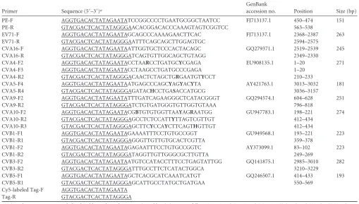
Primers.The GeXP multiplex assay consisted of 11 pairs of chimeric primers (including one pair of panenterovirus primers and 10 pairs of human enterovirus serotype-specific primers) and one pair of universal primers (Tag-F/Tag-R) (Table 1.). Each of these chimeric primers con-sisted of a gene-specific sequence for each virus fused at the 5=end to the universal sequence. Both the forward and reverse universal sequences were quasi-T7 sequences and selected by default using the GeXP eXpress Profiler software. Tag-F/Tag-R was the same as the forward or reverse universal sequence. This strategy was first developed by Beckman Coulter Company and was reported in our previous study (19). The panenterovi-rus primers (PE-F and PE-R) were designed in a highly conserved region of the 5=untranslated region (UTR) of the enterovirus genomes as re-ported by Yang et al. (28). Ten pairs of human enterovirus serotype-specific primers for the nine serotypes of enteroviruses were designed in relatively conserved VP1 regions of each serotype. Serotype-specific prim-ers sequences were evaluated using the NCBI Primer-Blast, Primer Pre-mier 5.0, and Oligo 6.0 software. Degenerate bases were introduced to cover different strains. The 5=end of the forward universal primer (Tag-F) was labeled with the fluorescent dye Cy5 and purified with high-pressure liquid chromatography. All chimeric primers and the reverse universal primer (Tag-R) were synthesized and purified by polyacrylamide gel elec-trophoresis (Invitrogen, Shanghai, China).
[image:2.585.38.547.79.368.2]Evaluation of the specificity of the GeXP assay.Firstly the mono RT-PCR assay was developed individually with extracted RNA from 28 cell-cultured serotypes of enterovirus strains to evaluate the specificity of each TABLE 1Oligonucleotide primers for the GeXP assay
Primer Sequence (5=–3=)a
GenBank
accession no. Position Size (bp)
PE-F AGGTGACACTATAGAATATCCGGCCCCTGAATGCGGCTAATCC FJ713137.1 450–474 151 PE-R GTACGACTCACTATAGGGAACACGGACACCCAAAGTAGTCGGTCC 563–538
EV71-F AGGTGACACTATAGAATAGCAGCCCAAAAGAACTTCAC FJ713137.1 2368–2387 263 EV71-R GTACGACTCACTATAGGGAATTTCAGCAGCTTGGAGTGC 2594–2575
CVA16-F AGGTGACACTATAGAATAATTGGTGCTCCCACTACAGC GQ279371.1 2519–2539 245 CVA16-R GTACGACTCACTATAGGGATCAGTGTTGGCAGCTGTAGG 2349–2330
CVA4-F2 AGGTGACACTATAGAATACCTAARCCTGATGCYCGAGA EU908135.1 1–20 271 CVA4-F3 AGGTGACACTATAGAATACCTAAGCCTGATGCCCGAGA 1–20
CVA4-R2 GTACGACTCACTATAGGGACAACTCTAGCTGRGAATGTYCCT 210–233
CVA5-F4 AGGTGACACTATAGAATAATGAGCCCAGCYAGYACYTA AY421763.1 3013–3032 181 CVA5-R4 GTACGACTCACTATAGGGAGATACHCCTGASACCATGCG 3036–3157
CVA9-F2 AGGTGACACTATAGAATATTTGATCAGAAGGGCTCATACGGGT GQ294574.1 604–628 251 CVA9-R2 GTACGACTCACTATAGGGATCTGTGATGGGTGTTGGTGTAAA 796–818
CVA10-F2 AGGTGACACTATAGAATACGBTGTGTGGTTAAYAGRAATGG GU947783.1 198–221 274 CVA10-R2 GTACGACTCACTATAGGGAGCCTCTCCATTYTTAGTCGTTGT 412–434
CVA10-R3 GTACGACTCACTATAGGGAGCTTCYCCAYCTTCAGTHGTTGT 412–434
CVB1-F1 AGGTGACACTATAGAATAGAAAATTTCCTGTGCCGGT GU949568.1 193–221 223 CVB1-R1 GTACGACTCACTATAGGGAGGGTTGTTGTGCACTCGTTA 359–378
CVB1-F2 AGGTGACACTATAGAATAGAGAATTTCCTGTGCCGGTC AY373099.1 83–102 223 CVB1-R2 GTACGACTCACTATAGGGATAGGTTGTTGGGCGCTTGTTA 249–269
CVB3-F2 AGGTGACACTATAGAATAATGTCCATACCTTTCCTGAGTATTGG GQ141875.1 2985–3010 282 CVB3-R2 GTACGACTCACTATAGGGATTTGCCTTCTCATACTGGCA 3210–3229
CVB5-F1 AGGTGACACTATAGAATAGCTCACGCATCAAATCATGT GQ246507.1 414–433 193 CVB5-R1 GTACGACTCACTATAGGGAGCATTGCCTATGCTGATGAA 550–569
Cy5-labeled Tag-F AGGTGACACTATAGAATA Tag-R GTACGACTCACTATAGGGA
aUniversal tag sequences are underlined. Bold type shows degenerate sites. Abbreviations are as follows: M⫽A or C; R⫽A or G; W⫽A or T; S⫽G or C; Y⫽C or T; K⫽G or
T; V⫽A, G, or C; H⫽A, C, or T; D⫽A, G, or T; B⫽G, C, or T.
on May 16, 2020 by guest
http://jcm.asm.org/
pair of gene-specific primers (including PE-F and PE-R) and ascertain the actual amplicon size of each target region. Both the mono-RT-PCR assay and the multiplex PCR were performed with a Qiagen one-step RT-PCR kit in a 25-l volume containing 5l of 5⫻Qiagen one-step RT-PCR buffer, 1l of RT-PCR enzyme mix, 1l of deoxynucleotide triphos-phate (10 mM each) mix, 5 units of RNase inhibitor, and 1l of extracted viral RNA from each serotype. The mono-RT-PCR assay contained a 50 nM concentration of each pair of gene-specific chimeric primers individ-ually, while the GeXP assay contained 50 nM concentrations of 11 pairs of gene-specific chimeric primers and a 500 nM concentration of the uni-versal tag primers (final concentrations); nuclease-free water was added to a volume of 25l. The RT-PCR was performed under the following con-ditions: 50°C for 30 min (the RT reaction) and 95°C for 15 min, followed by three steps of amplification according to the temperature switch PCR (TSP) strategy (25): step 1, 10 cycles of 95°C for 30 s, 55°C for 30 s, and 72°C for 30 s; step 2, 10 cycles of 95°C for 30 s, 65°C for 30 s, and 72°C for 30 s; step 3, 20 cycles of 95°C for 30 s, 48°C for 30 s, and 72°C for 30 s.
Separation by capillary electrophoresis and fragment analysis.PCR product separation and detection were performed on a GenomeLab GeXP genetic analysis system (Beckman Coulter) by capillary electrophoresis, following the protocols described previously (20). After amplified frag-ments were separated, the peaks were initially analyzed using the fragment analysis module of the GeXP system software and matched to the appro-priate genes. The peak height for each gene was reported in the electro-pherogram.
Evaluation of the sensitivity of the GeXP assay.Sensitivity was tested by following the method described previously (16), using titrated refer-ence viruses (EV71 and CVA16) obtained from infected human RD cells. Serial 10-fold dilutions of the EV71 and CVA16 stock were made in Hanks’ balanced salt solution, and RNA from 140l of each dilution was extracted with the QIAamp viral RNA minikit (Qiagen). The RNA prep-arations, ranging from 105.5to 10⫺1.5TCID
50(0.03 TCID50) per microli-ter, were tested with the GeXP assay. The assay at each template concen-tration was repeated three times.
Absolute sensitivity was measured by usingin vitrotranscribed syn-thetic RNAs derived from 10 recombinant plasmids containing the VP1 sequences of the nine serotypes of enteroviruses or the 5=UTR. Thein vitro-synthesized RNA products were purified with an RNeasy MinElute cleanup kit (Qiagen) and quantitated by spectrophotometry (NanoDrop
ND-1000). The concentrated product for each serotype was diluted to final concentrations ranging from 105copies to 1 copy per microliter and then individually subjected to the GeXP assay. The concentrations of spe-cific primers were optimized according to the amplification efficiency of the GeXP assay using a single template. The sensitivity of the optimized GeXP assay was re-evaluated by using 10 premixed RNA templates rang-ing from 105copies to 10 copies for each serotype per microliter for three times on three different days.
Application to clinical specimens.RNAs were extracted from 180 original clinical stool specimens obtained from HFMD patients and per-sons suspected of having HFMD to illustrate the application of the opti-mized GeXP assay. All the clinical stool specimens were assayed in parallel by conventional methods, including isolation of the viruses followed by neutralization testing and conventional RT-PCR followed by amplicon sequencing (21, 29–30).
RESULTS
Evaluation of the specificity of the GeXP assay.
The RNAs of 28
cell-cultured serotypes of enterovirus isolates were individually
used as templates to evaluate the specificity of each pair of
gene-specific primers. In mono-RT-PCR assays, the panenterovirus
universal primers could amplify the target genes of all 28 serotypes
of enteroviruses, but each pair of serotype-specific primers
gener-ated corresponding VP1 genes only of the targeted serotype,
without cross-amplification. The amplicon sizes for the
sero-types were as follows: panenterovirus, 150 to 153 bp; EV71, 264
to 267 bp; CVA16, 246 to 248 bp; CVA4, 270 to 272 bp; CVA5,
180 to 181 bp; CVA9, 251 to 254 bp; CVA10, 274 to 276 bp;
CVB1, 224 to 225 bp; CVB3, 281 to 284 bp; and CVB5, 193 to
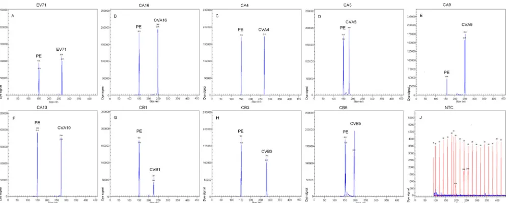
196 bp (electropherograms not shown). The nine serotypes of
enteroviruses associated with HFMD were detected via the
GeXP assay. Two specific amplification peaks were observed,
representing the panenterovirus target amplicon and the
serotype-specific target amplicon (Fig. 1).
Sensitivity of the GeXP assay.
The sensitivity of the GeXP
as-say was measured using titrated reference viruses (EV71 and
CVA16 stocks) or
in vitro
-transcribed RNAs of nine serotypes of
FIG 1Specificity of the multiplex RT-PCR assay. Cy5-labeled PCR products were separated via GeXP capillary electrophoresis and detected by fluorescence spectrophotometry, given as dye signals in arbitrary units on theyaxis. Each peak was identified by comparing the expected to the actual PCR product size on the
xaxis. EV71, CVA16, CVA4, CVA5, CVA9, CVA10, CVB1, CVB3, and CVB5 were assayed by using enterovirus RNAs extracted from various cell-cultured strains. Nuclease-free water was used as the negative control (NTC) (J).
on May 16, 2020 by guest
http://jcm.asm.org/
[image:3.585.44.543.65.265.2]enteroviruses and panenterovirus. The GeXP assay detected as
little as 0.03 TCID
50of EV71 and CVA16, 10 copies of
panentero-virus, EV71, CVA16, CVB1, CVB5, and more than 100 copies of
CVA4, CVA5, CVA9, CVA10, and CVB3 with a single RNA
tem-plate.
Based on all the sensitivity results of GeXP assay with a single
RNA template, the concentration of each chimeric primer in the
new GeXP assay was adjusted as follows (F, forward; R, reverse):
PE (F/R), 20 nM; EV71 (F/R), 50 nM; CVA16 (F/R), 40 nM; CVA4
(F2, F3/R2), 80 nM; CVA5 (F4/R4), 50 nM; CVA9 (F2/R2), 70
nM; CVA10 (F2/R2, R3), 100 nM; CVB1 (F1/R1; F2/R2), 30 nM;
CVB3 (F2/R2), 100 nM; CVB5 (F1/R1), 40 nM. The optimized
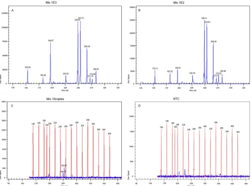
GeXP assay achieved a sensitivity of 100 copies with 10 premixed
RNA templates in three independent experiments on three
differ-ent days (Fig. 2), and the coefficidiffer-ent of variation was less than
10.15% (not shown).
Application to clinical specimens.
A total of 180 specimens
collected from HFMD patients and suspects were assayed
simul-taneously by both the GeXP assay and the conventional methods,
including viral isolation in cell culture, neutralization, and
con-ventional RT-PCR followed by amplicon sequencing (21, 29–30).
The numbers of panenterovirus and each serotype of enterovirus
detected by different methods are shown in Table 2 and 3. In
comparison with the results of conventional methods, the
sensi-tivities of the GeXP assay for detection of panenterovirus, EV71,
and CVA16 were 98.79% (163/165), 91.67% (44/48), and 91.67%
(33/36), respectively, and the specificities were 80.00% (12/15),
98.48% (130/132), and 100% (144/144), respectively. The
concor-dance between the detection results of GeXP assay and the results
of conventional methods for typing seven other serotypes of
en-teroviruses was 92.59% (25/27).
DISCUSSION
In our study, 10 pairs of serotype-specific primers and one pair of
panenterovirus universal primers were designed to develop a
GeXP assay for simultaneous identification of nine serotypes of
enteroviruses, including EV71, CVA16, CVA4, CVA5, CVA9,
CVA10, CVB1, CVB3, and CVB5. The selection of enteroviruses
associated with HFMD was based on the research data from the
National Notifiable Disease Reporting System (NNDRSe) in
China and the previous study (10, 23). The serotype-specific
primers were designed to target relatively conserved VP1 regions
of each enterovirus serotype, and the panenterovirus universal
primers were designed from the 5
=
UTR (28). Due to the high
FIG 2Sensitivity of GeXP detection of 10 premixed RNA templates with multiplex primers. All 10 target genes could be detected at 103copies/l (A) and 102 copies/l levels (B); only CVA16 and CVA9 could be detected at 10 copies/l levels (C). Nuclease-free water was used as the negative control (D).
on May 16, 2020 by guest
http://jcm.asm.org/
[image:4.585.44.543.66.436.2]degree of diversity among VP1 sequences, some degenerate bases
were introduced, and more than two primers for some enterovirus
serotypes were designed to cover the majority of the viral
se-quences, such as the primers for CVA4, CVA10, and CVB1.
The original standard workflow of GeXP was performed in two
separate tubes with an RT reaction and a subsequent PCR
ampli-fication, as described in the previous reports (15, 19–20), which is
a costly, time-consuming, tedious process and apt to bring
carry-over contamination. In order to eliminate these problems, a
one-step RT-PCR kit (Qiagen) was adopted and replaced the GeXP
start kit (Beckman Coulter Company). The amplification
proce-dure of the GeXP assay was improved via temperature switch PCR
(TSP) strategy (25), including three steps with different annealing
temperatures (Fig. 3): step 1 was carried out with gene-specific
sequences of chimeric forward and reverse primers, step 2 was
carried out mainly with chimeric forward and reverse primers,
and step 3 was carried out predominantly with universal forward
and reverse tag primers. The GeXP assay, based on the use of
chimeric and fluorescent-dye-labeled universal tag primers and
the TSP strategy, offered highly sensitive and specific
amplifica-tions of different genes in one multiplex RT-PCR assay, avoided
preferred and inferior amplification, and minimized nonspecific
reactions. The resolution of GeXP analyzer-based capillary
elec-trophoresis is superior to that of conventional capillary
electro-phoresis. The forward universal primer was fluorescently labeled
in the GeXP assay. The resulting dye-labeled PCR products were
separated and detected with a Beckman Coulter GenomeLab
GeXP genetic analysis system using capillary electrophoresis.
Af-ter amplified fragments were separated, the data were initially
an-alyzed using the fragment analysis module of the GeXP system
software. Then each amplified fragment would be present as a sole
peak with an accurate size on the electropherogram. One can
clearly differentiate two peaks with a 3-nucleotide or greater
dif-ference in a practical way. In theory, the target PCR productions of
CVA4 and CVA10 are 271 bp and 274 bp, respectively. The
nucle-otide difference between them is 3 bp.
The improved GeXP assay was further optimized by adjusting
the concentration of each chimeric primer in the reaction based
on the individual sensitivity results of the GeXP assay with a single
RNA template to overcome the potential interference due to
pref-erential amplification in mixed infections. In this study, the
rela-tive and absolute sensitivities were analyzed to evaluate the
detec-tion limit of the GeXP assay. The optimal GeXP assay detected as
little as 0.03 TCID
50of EV71 and EVA16, which is comparable to
[image:5.585.299.544.65.212.2]the detection sensitivity of the real-time RT-PCR assay published
recently (26) and slightly lower than the sensitivity reported for
seminested RT-PCR (16). The absolute sensitivity of the optimal
GeXP assay was 100 copies for simultaneously detecting 10 target
genes without cross or nonspecific amplification. In a test of 180
samples, the sensitivities of the GeXP assay for detection of
pan-enterovirus, EV71, and CVA16 were 98.79% (163/165), 91.67%
(44/48), and 91.67% (33/36), respectively, and the specificities
were 80.00% (12/15), 98.48% (130/132), and 100% (144/144),
respectively, compared with conventional methods, revealing a
high sensitivity and specificity in the detection of these viruses.
Twelve negative specimens were identified by both the GeXP assay
and the conventional methods. Two specimens that were negative
by the GeXP assay were enterovirus positive by the conventional
TABLE 2Comparison of GeXP assay and conventional methods for detecting panenterovirus, EV71, and CVA16a
GeXP assay result and virus
No. of samples with result by conventional methods
Total Positive Negative
Positive
Panenterovirus 163 3 166
EV71 44 2 46
CVA16 33 0 33
Negative
Panenterovirus 2 12 14
EV71 4 130 134
CVA16 3 144 147
Total
Panenterovirus 165 15 180
EV71 48 132 180
CVA16 36 144 180
aAll 180 stool specimens had been identified previously by cell culture and classical
PCR followed by sequencing. A neutralization test was performed for samples positive for EV71 (n⫽48) and CVA16 (n⫽36). Twelve negative specimens were detected by both the GeXP assay and the conventional methods. Two specimens that were negative by the GeXP assay were enterovirus positive by the conventional methods. The false negatives of GeXP assay might due to the RNA degradation or occurrence of PCR inhibition with the samples. Three specimens that were negative by the conventional methods were enterovirus positive by the GeXP assay, which were confirmed later by independent RT-PCR and sequencing to be true positives. As a measure of agreement, kappa values for panenterovirus (P⫽0.000), EV71 (P⫽0.000), and CVA16 (P⫽
[image:5.585.40.286.86.278.2]0.000) were 0.813, 0.914, and 0.946, respectively (using SPSS13.0).
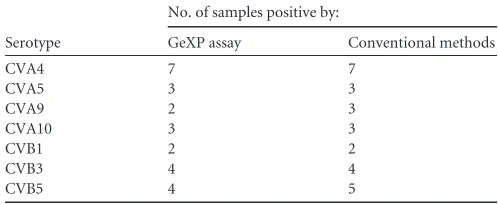
TABLE 3Comparison of the GeXP assay and conventional methods for detecting seven serotypes of enteroviruses
Serotype
No. of samples positive by:
GeXP assay Conventional methods
CVA4 7 7
CVA5 3 3
CVA9 2 3
CVA10 3 3
CVB1 2 2
CVB3 4 4
CVB5 4 5
FIG 3Diagram of the GeXP amplification workflow using a temperature switch PCR strategy.
on May 16, 2020 by guest
http://jcm.asm.org/
[image:5.585.38.287.621.723.2]methods. The false-negative results in the GeXP assay might be
due to RNA degradation or to the occurrence of PCR inhibition
with the samples. Three specimens that were negative by the
con-ventional methods were enterovirus positive by the GeXP assay,
and these were confirmed later by independent RT-PCR and
se-quencing to be true positives. Because of the limited number of
positive samples for the other seven serotypes of enteroviruses
(CVA4, CVA5, CVA9, CVA10, CVB1, CVB3, and CVB5), the
GeXP assay needs to be validated with a larger number of clinical
samples for these viruses.
Two distinct advantages of the GeXP assay are the time savings
and cost effectiveness. The cost of the GeXP assay for
simultane-ous detection of 9 serotypes of enteroviruses is approximately $8
per test, versus $8 per test for each virus using a commercial
real-time RT-PCR kit. The whole reaction was completed in one tube
in a one-step multiplex RT-PCR within 2.5 h, followed by
capil-lary electrophoresis separation. In addition, two 96-well plates can
be placed in parallel in a GeXP machine at the same time to further
increase the throughput of the samples.
Conclusion.
This study has demonstrated that the GeXP assay
is a rapid, cost-effective, high-throughput method with high
sen-sitivity and specificity for typing most HFMD-associated
entero-viruses, which may be adopted for general use in the Department
of Viral Disease Control and Prevention for molecular
epidemio-logic survey of enteroviruses.
ACKNOWLEDGMENTS
This work was supported by the China Mega-Project for Infectious Dis-ease (2011ZX10004-001, 2009ZX10004-101, 202, 2008ZX10004-002, 001, 004).
A Chinese patent (application number 201110089404.9) has been filed for the combination of all the listed primers specific to 10 HFMD associ-ated enteroviruses and experimental parameters. Xuejun Ma and Wenbo Xu are inventors on the patent application. The technology is available for research-only purposes.
REFERENCES
1.Abubakar S, Chee HY, Shafee N, Chua KB, Lam SK.1999. Molecular detection of enteroviruses from an outbreak of hand, foot and mouth disease in Malaysia in 1997. Scand. J. Infect. Dis.31:331–335.
2.Apisarnthanarak A, Kitphati R, Pongsuwann Y, Tacharoenmueng R, Mundy LM.2005. Echovirus type 11: outbreak of hand-foot-and-mouth disease in a Thai hospital nursery. Clin. Infect. Dis.41:1361–1362. 3.Barriere H, Berger M, Billaudel S.1976. Hand, foot and mouth disease.
Sem. Hop.52:2215–2220. [In French.]
4.Bell EJ, McCartney RA, Basquill D, Chaudhuri AK.1986. Mu-antibody capture ELISA for the rapid diagnosis of enterovirus infections in patients with aseptic meningitis. J. Med. Virol.19:213–217.
5.Blomqvist S, Klemola P, Kaijalainen S, Paananen A, Simonen ML, Vuorinen T, and Roivainen M.2010. Co-circulation of coxsackieviruses A6 and A10 in hand, foot and mouth disease outbreak in Finland. J. Clin. Virol.48:49 –54.
6.Chen SP, et al.2010. Comparison of clinical features between coxsacki-evirus A2 and enterovirus 71 during the enterovirus outbreak in Taiwan, 2008: a children’s hospital experience. J. Microbiol. Immunol. Infect.43: 99 –104.
7.Chen TC, et al.2006. Combining multiplex reverse transcription-PCR and a diagnostic microarray to detect and differentiate enterovirus 71 and coxsackievirus A16. J. Clin. Microbiol.44:2212–2219.
8.Frydenberg A, Starr M.2003. Hand, foot and mouth disease. Aust. Fam. Physician32:594 –595.
9.Han JF, et al.2011. Echovirus 30 in EV71-associated hand, foot and mouth disease outbreak, Guangxi, China. J. Clin. Virol.50:348 –349. 10. Hu YF, et al.2011. Complete genome analysis of coxsackievirus A2, A4,
A5, and A10 strains isolated from hand, foot, and mouth disease patients
in China revealing frequent recombination of human enterovirus A. J. Clin. Microbiol.49:2426 –2434.
11. Lee YS, et al.2002. PCR-RFLP based molecular typing of enteroviruses isolated from patients with aseptic meningitis in Korea. Arch. Virol.147: 1711–1720.
12. Li L, et al.2005. Genetic characteristics of human enterovirus 71 and coxsackievirus A16 circulating from 1999 to 2004 in Shenzhen, People’s Republic of China. J. Clin. Microbiol.43:3835–3839.
13. Lin TL, et al.2008. Rapid and highly sensitive coxsackievirus A indirect immunofluorescence assay typing kit for enterovirus serotyping. J. Clin. Microbiol.46:785–788.
14. Lindenbaum JE, Van Dyck PC, Allen RG.1975. Hand, foot and mouth disease associated with coxsackievirus group B. Scand. J. Infect. Dis. 7:161–163.
15. Nagel MA, Gilden D, Shade T, Gao B, Cohrs RJ. 2009. Rapid and sensitive detection of 68 unique varicella zoster virus gene transcripts in five multiplex reverse transcription-polymerase chain reactions. J. Virol. Methods157:62– 68.
16. Nix WA, Oberste MS, Pallansch MA.2006. Sensitive, seminested PCR amplification of VP1 sequences for direct identification of all enterovirus serotypes from original clinical specimens. J. Clin. Microbiol.44:2698 – 2704.
17. Oberste MS, Maher K, Kilpatrick DR, Pallansch MA.1999. Molecular evolution of the human enteroviruses: correlation of serotype with VP1 sequence and application to picornavirus classification. J. Virol.73:1941– 1948.
18. Oberste MS, Nix WA, Maher K, Pallansch MA.2003. Improved molec-ular identification of enteroviruses by RT-PCR and amplicon sequencing. J. Clin. Virol.26:375–377.
19. Qin M, et al.2010. Detection of pandemic influenza A H1N1 virus by multiplex reverse transcription-PCR with a GeXP analyzer. J. Virol. Meth-ods168:255–258.
20. Rai AJ, Kamath RM, Gerald W, Fleisher M.2009. Analytical validation of the GeXP analyzer and design of a workflow for cancer-biomarker discovery using multiplexed gene-expression profiling. Anal. Bioanal. Chem.393:1505–1511.
21. Rigonan AS, Mann L, Chonmaitree T.1998. Use of monoclonal anti-bodies to identify serotypes of enterovirus isolates. J. Clin. Microbiol.36: 1877–1881.
22. Russo DH, Luchs A, Machado BC, Carmona RC, Timenetsky MC. 2006. Echovirus 4 associated to hand, foot and mouth disease. Rev. Inst. Med. Trop. Sao Paulo48:197–199.
23. Shi HJ, et al.2010. Biological characters of a coxsackievirus B3 variant strain isolated from a hand-foot-mouth disease patient with severe clinical symptoms. Zhonghua Yi Xue Za Zhi90:1141–1144.
24. Susi P, et al.2009. Typing of enteroviruses by use of microwell oligonu-cleotide arrays. J. Clin. Microbiol.47:1863–1870.
25. Tabone T, Mather DE, Hayden MJ. 2009. Temperature switch PCR (TSP): robust assay design for reliable amplification and genotyping of SNPs. BMC Genomics10:580.
26. Xiao XL, et al.2009. Simultaneous detection of human enterovirus 71 and coxsackievirus A16 in clinical specimens by multiplex real-time PCR with an internal amplification control. Arch. Virol.154:121–125. 27. Xiao XL, et al.2009. Simultaneous detection of enterovirus 70 and
cox-sackievirus A24 variant by multiplex real-time RT-PCR using an internal control. J. Virol. Methods159:23–28.
28. Yang CF, et al.1992. Genotype-specific in vitro amplification of se-quences of the wild type 3 polioviruses from Mexico and Guatemala. Virus Res.24:277–296.
29. Zhang Y, et al.2009. An outbreak of hand, foot, and mouth disease associated with subgenotype C4 of human enterovirus 71 in Shandong, China. J. Clin. Virol.44:262–267.
30. Zhang Y, et al. 2010. Molecular evidence of persistent epidemic and evolution of subgenotype B1 coxsackievirus A16-associated hand, foot, and mouth disease in China. J. Clin. Microbiol.48:619 – 622.
31. Zhou F, et al.2009. Identification of 20 common human enterovirus serotypes by use of a reverse transcription-PCR-based reverse line blot hybridization assay. J. Clin. Microbiol.47:2737–2743.
32. Zhu Z, et al.2007. Molecular epidemiological analysis of echovirus 19 isolated from an outbreak associated with hand, foot, and mouth disease (HFMD) in Shandong Province of China. Biomed. Environ. Sci.20:321– 328.