Transcriptase PCR Assay for Pan-Dengue Virus Detection and
Comparison of Four Molecular Dengue Virus Detection Assays
Jesse J. Waggoner,aJanaki Abeynayake,bMalaya K. Sahoo,bLionel Gresh,cYolanda Tellez,dKarla Gonzalez,dGabriela Ballesteros,d Angel Balmaseda,dKumudu Karunaratne,eEva Harris,fBenjamin A. Pinskya,b
Department of Medicine, Division of Infectious Diseases and Geographic Medicine, Stanford University School of Medicine, Stanford, California, USAa; Department of
Pathology, Stanford University School of Medicine, Stanford, California, USAb; Sustainable Sciences Institute, Managua, Nicaraguac; National Virology Laboratory, Centro
Nacional de Diagnóstico y Referencia, Ministry of Health, Managua, Nicaraguad; Department of Medical Microbiology, Lady Ridgeway Hospital, Colombo, Sri Lankae;
Division of Infectious Diseases and Vaccinology, School of Public Health, University of California, Berkeley, California, USAf
A number of diagnostic tests are available for dengue virus (DENV) detection, including a variety of nucleic acid amplification tests (NAATs). However, reports describing a direct comparison of different NAATs have been limited. In this study, we report the design of an internally controlled real-time reverse transcriptase PCR (rRT-PCR) that detects all four DENV serotypes but does not distinguish between them (the pan-DENV assay). Two hundred clinical samples were then tested using four different DENV RT-PCR assays: the pan-DENV assay, a commercially produced, internally controlled DENV rRT-PCR (the Altona assay), a widely used heminested RT-PCR, and a serotype-specific multiplex rRT-PCR assay. The pan-DENV assay had a linear range extending from 1.0 to 7.0 log10cDNA equivalents/l and a lower limit of 95% detection ranging from 1.7 to 7.6 cDNA equiva-lents/l, depending on the serotype. When measured against a composite reference standard, the pan-DENV assay proved to be more clinically sensitive than either the Altona or heminested assays, with a sensitivity of 98.0% compared to 72.3% and 78.8%, respectively (P<0.0001 for both comparisons). The pan-DENV assay detected DENV in significantly more samples collected on or after day 5 of illness and in a subgroup of patients with detectable anti-DENV IgM at presentation. No significant difference in sensitivity was observed between the pan-DENV assay and the multiplex rRT-PCR, despite the presence of an internal control in the former. The detection of DENV RNA late in the course of clinical illness should serve to lengthen the period during which a confirmed molecular diagnosis of DENV infection can be provided.
D
engue virus (DENV) is the most common vector-bornehu-man viral pathogen worldwide (1). Infection with one or
more of the four closely related virus serotypes (designated DENV-1 to -4) results in a range of clinical manifestations span-ning asymptomatic infection, dengue fever (DF), and severe gue, a category that includes entities previously classified as den-gue hemorrhagic fever (DHF) and denden-gue shock syndrome (DSS)
(1,2). Infection with one serotype (primary infection) results in
immunity to that serotype, but infection can still occur with any of the remaining serotypes (secondary infection). Secondary DENV infection has been shown to be a significant risk factor for the
development of DHF or DSS (3,4). DENV transmission largely
occurs in the tropical and subtropical regions of the world, though the number of countries where DENV is endemic has been
in-creasing (1,5). Recent reports estimate that 230 million DENV
infections occur annually worldwide, including 2 million cases of
severe disease and 21,000 deaths (6). Over 3.6 billion people live in
regions where this pathogen is endemic and are at risk for
infec-tion (6). DF is also one of the most common causes of a systemic
febrile illness in travelers returning from countries where the virus is endemic and remains a major concern for military personnel
stationed in these areas (7–9).
A wide array of diagnostic tests for DENV have been developed and remain in use throughout the world. These include laborato-ry-based testing for anti-DENV IgM, anti-DENV IgG, and the DENV nonstructural protein 1 (NS1). Point-of-care tests for these analytes are also available. In addition, molecular methods are performed, including heminested RT-PCR and real-time
re-verse transcriptase RT-PCR (rRT-PCR) assays (1,10). Currently,
the WHO supports the use of a number of these tests depending on individual laboratory capabilities and the goals of testing, as no
gold standard for DENV diagnosis has been established (1).
Consistent with the state of DENV diagnostics as a whole, a variety of nucleic acid amplification tests (NAATs) have been re-ported in the literature. A heminested RT-PCR for the detection and serotyping of DENV, a version of which remains in wide use,
was first reported in 1992 (11,12). Though numerous DENV
spe-cies-specific and serotype-specific assays have since been devel-oped, direct comparisons between these assays are notably rare
(11,13–17). Rather, studies evaluating new DENV molecular
di-agnostic tests typically use samples collected within the first 5 days of fever from patients who had positive testing by viral isolation,
seroconversion, or both (10, 18–25). This practice ensures that
only the highest viral loads are evaluated and it likely does not reflect clinical reality, where patients may present at any time dur-ing their illness, includdur-ing five or more days after fever onset.
The majority of DENV NAATs have been designed with the
Received13 March 2013Returned for modification15 April 2013
Accepted23 April 2013
Published ahead of print1 May 2013
Address correspondence to Benjamin A. Pinsky, [email protected]. Copyright © 2013, American Society for Microbiology. All Rights Reserved.
doi:10.1128/JCM.00548-13
on May 16, 2020 by guest
http://jcm.asm.org/
dual goals of sensitive DENV detection and the ability to provide a
serotype-specific diagnosis (11,13,14,23,24). While serotyping
capability is useful for epidemiologic surveillance, the relative contribution of serotype-specific information to the care of
indi-vidual patients remains unclear (1,10,26). For laboratories
out-side regions where the disease is endemic that primarily test re-turning travelers, serotype-specific DENV diagnosis may not be
required (10,20,26). Pan-DENV assays may still be able to
pro-vide information that has been associated with the development of severe disease, including the detection of DENV RNA at
deferves-cence (27–30).
Despite the large number of reported DENV NAATs, few of these assays have been designed to include an internal control (IC), either as an extrinsic molecule spiked into each sample be-fore or after extraction or as a heterologous intrinsic target that is
coextracted with DENV RNA (14,17, 21,25, 31). It has been
advocated that ICs be used in settings where PCR inhibitors pres-ent a significant source of false-negative results, which may be particularly important in the performance of NAATs in countries
where dengue is endemic (26,32).
In this study, we report the development of a species-specific internally controlled rRT-PCR utilizing hydrolysis probes for DENV detection (here referred to as the pan-DENV assay). We also report an independent evaluation of the Altona RealStar den-gue RT-PCR kit, which is a commercially produced internally controlled rRT-PCR kit for DENV detection. Furthermore, using 200 clinical samples, we directly compared four molecular tests for DENV: the pan-DENV and Altona assays, a version of the
hemin-ested RT-PCR (11), and the serotype-specific DENV multiplex
rRT-PCR (16).
MATERIALS AND METHODS
Pan-DENV assay design.The pan-DENV assay utilizes the same primers as the multiplex rRT-PCR assay, though the primer concentrations differ.
The primer design has been described previously, and these primers are listed inTable 1(16). Separate hydrolysis probes were designed for the pan-DENV assay based on the 95% consensus sequence of the 5= untrans-lated region (UTR) and capsid gene. RealTimeDesign software (Biosearch Technologies, Novato, CA) was used to create BHQplus probes, which utilize a duplex stabilizing technology to increase melting temperature and allow for reduced probe length. Four probes were designed to account for sequence diversity; these probe sequences are listed inTable 2and were given letter designations A to D. The probes were testedin silicousing BLASTn to query the NCBI nucleotide database. Searches excluding the DENV group (identification no. 11052) were also performed to identify the best non-DENV sequence matches in the database, as well as matches in theFlaviviridaefamily.
A previously described assay for the detection of RNase P was modified for use as the IC in this assay (33). The primer and probe sequences were maintained as originally published (seeTables 1and2); however, the RNase P probe was labeled with Cal Fluor Red 610 to facilitate multiplex-ing. The IC primer and probe concentrations were selected based on titra-tion experiments, such that samples that were negative for DENV rou-tinely had crossing threshold (CT) values between 25 and 30.
[image:2.585.40.548.77.195.2]RT-PCR assays.The pan-DENV assay was performed using the Su-perScript III Platinum one-step qRT-PCR kit (Invitrogen, Carlsbad, CA). Reaction mixtures were scaled down from the manufacturer-recom-mended 50-l volume to 25l per reaction mixture, and 5l of RNA template was added to each reaction mixture. The primer and probe con-centrations in the final reaction mixtures are listed inTables 1and2, respectively. During the analytical evaluation, 5l of pooled RNA extracts from domestic patient plasma samples was spiked into the reaction mix-ture to mimic the IC in the clinical samples. RT-PCRs were performed using the Rotor-Gene Q instrument (Qiagen, Valencia, CA). Cycling con-ditions differed from those of the multiplex rRT-PCR and were as follows: 52°C for 15 min (RT step), 94°C for 2 min, 45 cycles of 94°C for 15 s, 55°C for 20 s, and 68°C for 20 s (run time, 110 min). Detection was performed in the green and orange channels at 55°C, and the gain was set at 4.67 for green and at 10 for orange. During analysis, the first five cycles were cropped from the green channel to improve baseline normalization; slope TABLE 1Primer sequences for the pan-DENV assay
Primer name Primer sequence (5=¡3=) Concn (nM)a DENV genomic locationb
DENV-1, -2, -3 forward CAGATCTCTGATGAACAACCAACG 350 86 DENV-2 forward C¡T CAGATCTCTGATGAATAACCAACG 350 87 DENV-3 forward C¡T CAGATTTCTGATGAACAACCAACG 300 85 DENV-4 forward GATCTCTGGAAAAATGAAC 450 81
DENV-1, -3 reverse TTTGAGAATCTCTTCGCCAAC 300 199 (DENV-1), 198 (DENV-3) DENV-2 reverse AGTTGACACGCGGTTTCTCT 350 171
DENV-2 reverse A¡G AGTCGACACGCGGTTTCTCT 350 171 DENV-4 reverse AGAATCTCTTCACCAACC 450 190 RNase P forward AGATTTGGACCTGCGAGCG 100 NA RNase P reverse GAGCGGCTGTCTCCACAAGT 100 NA
aThe concentration of each primer in the final reaction mixture. Predicted amplicon sizes are as follows: DENV-1, 114 bp; DENV-2, 85 bp; DENV-3, 114 bp; DENV-4, 110 bp.
b
Genomic locations are provided for the 5=base complementary to each primer using the following reference virus sequences: DENV-1 US/Hawaii/1944 (GenBank accession no. EU848545.1), DENV-2 New Guinea C strain (GenBank accession no.AF038403.1), DENV-3 strain H87 (GenBank accession no.M93130.1), and DENV-4 strain H241 (GenBank accession no.AY947539.1). NA, not applicable.
TABLE 2DENV and RNase P probe sequences for the pan-DENV assay
Probe 5=Fluor Probe sequence (5=¡3=) 3=Quencher Concn (nM)b
DENV probe A FAMa CTCGCGCGTTTCAGCATAT BHQplus 200
DENV probe B FAM CTCTCGCGTTTCAGCATAT BHQplus 200 DENV probe C FAM CTCTCACGTTTCAGCATATTG BHQplus 200 DENV probe D FAM CTCACGCGTTTCAGCATAT BHQplus 200 RNase P Cal Fluor Red 610 TTCTGACCTGAAGGCTCTGCGCG BHQ-2 50
a
FAM, 6-carboxyfluorescein.
bThe concentration for each probe in the final reaction mixture.
on May 16, 2020 by guest
http://jcm.asm.org/
[image:2.585.42.549.638.707.2]correction was performed for both channels. The threshold was set at 0.025 for the green channel, and any curve crossing this threshold prior to cycle 40 was considered to be positive for DENV. All results after cycle 40 were evaluated individually, and any sample generating an exponential curve that crossed the threshold after cycle 40 was also considered posi-tive. The threshold was set at 0.1 for the orange RNase P channel. In samples that were negative for DENV, RNase P was considered to be detected if the curve crossed this threshold at any cycle. Samples in which DENV and RNase P RNA were not amplified were considered to have failed extraction or to contain an inhibitory substance.
The Altona assay was performed using the RealStar dengue PCR kit 1.0 (Altona Diagnostics, Hamburg, Germany). Reaction mixtures were scaled down from the recommended 50-l volume to 25l, with each reaction mixture containing 2.5l master mix A, 10l master mix B, 1.25l of internal control solution, and 6.25l of water. Five microliters of RNA template was added to each reaction mixture, which was decreased from the manufacturer-recommended 25l. An IC, included with the kit, was added to the reaction mixture following the setup of the negative-control sample on each run. RT-PCRs were again performed using the Rotor-Gene Q. Cycling conditions were: 50°C for 10 min (RT step), 95°C for 2 min, 45 cycles of 95°C for 15 s, 55°C for 45 s, and 72°C for 15 s (run time, 120 min). Detection was performed in the green and yellow channels at 55°C, and the gain was set at 10 for both channels. During analysis, slope correction was performed for both channels. The threshold was set at 0.1 for the green channel, and curves were interpreted with the same algo-rithm used for the pan-DENV assay. The threshold was set at 0.05 for the yellow IC channel. In samples that were negative for DENV, the Altona IC was required to be detected within 45 cycles; samples in which the Altona IC was not amplified were considered inhibited.
The heminested RT-PCR and the serotype-specific multiplex rRT-PCR were performed as previously described (16). However, for the heminested assay, 5l of RNA template was added in the RT-PCR step, and 5l of RT-PCR product was added to the heminested reaction mix-ture. For all clinical samples, the pan-DENV, multiplex rRT-PCR, and heminested assays were performed concurrently. Samples underwent one further freeze-thaw cycle prior to use in the Altona assay.
Reference virus RNA.Genomic RNA from reference strains of the four DENV serotypes, DENV-1 Hawaii 1944, DENV-2 New Guinea C strain, DENV-3 strain H87, and DENV-4 strain H241 were obtained from Vircell (Granada, Spain). Genomic RNA of three strains of West Nile virus (WNV) (NY 1999, a clinical isolate previously reported as NAL strain, and B956) and a single strain each of Japanese encephalitis virus (JEV) and tick-borne encephalitis virus (TBEV) was obtained from the St. Orsola-Malpighi Hospital, Regional Reference Centre for Microbiological Emer-gencies, in Bologna, Italy (34).
Plasmid generation, quantitation, and sequencing.The generation and sequencing of plasmids has been described previously (16). Plasmid sequences were identical to the expected sequences for each DENV refer-ence strain. The concentration of plasmid DNA was quantified using the Quant-iT PicoGreen double-stranded DNA (dsDNA) reagent and kit (In-vitrogen, Carlsbad, CA). Twenty-five- and 50-fold dilutions were tested in triplicate. A standard curve was generated, and the concentration of plas-mid in the initial eluate was calculated.
Linearity, lower limit of detection, and precision.The analytical eval-uation of the pan-DENV assay was performed according to previously published recommendations (32). For each serotype, linearity studies were performed on serial 10-fold dilutions of both quantified plasmid DNA and reference virus RNA. For the plasmid DNA, dilutions from 7.0 log10copies/l to 1 copy/l were tested in triplicate on a single run. Ref-erence virus RNA concentrations were originally quantified by the man-ufacturer in ng/l of total RNA. Ten-fold dilutions from 1 ng/l to 0.01 pg/l RNA were tested in triplicate on a single run. Using the standard curve generated with dilutions of plasmid DNA, the concentration in DENV cDNA equivalents/l was calculated for the highest concentration of RNA (1 ng/l) for each serotype. The linear range was established by
fitting a best-fit line to the data by regression analysis and included the range where theR2value for this line wasⱖ0.99.
To establish the lower limit of 95% detection (95% LLOD), the lowest concentrations of RNA at which all replicates were detectable during the linear range study were used as the starting point. Ten replicates of four 2-fold dilutions from this concentration were tested on a single run. The 95% LLOD was then calculated using probit analysis.
The precision of the pan-DENV assay was determined using three dilutions of RNA controls (high positive, low positive, and limit of quan-titation). These were performed five times on three separate days. Fresh dilutions were made on the day of each run from aliquots of high-concen-tration stocks (1 ng/l, high positive). Intra- and interrun variability were calculated from the log10concentration of the samples.
To establish the linear range and estimate the intrarun precision for the Altona assay, serial 10-fold dilutions from 1 ng/l to 0.01 pg/l of reference virus RNA for each DENV serotype were tested in triplicate on a single run. Testing to establish the 95% LLOD was performed as described for the pan-DENV assay.
Specificity.Specificity was evaluated by testing genomic RNA from the WNV, JEV, and TBEV isolates, as well as from the yellow fever 17D (YF-17D) vaccine strain. The JEV, TBEV, and WNV strains are described above. For YF-17D, genomic RNA (Vircell, Granada, Spain) was tested at concentrations ofⱖ12,500 copies/l and 250 copies/l, as quantitated by the manufacturer. An additional 60 domestic samples with detectable levels of hepatitis C virus (HCV) were extracted and tested. All specificity experiments were performed for the pan-DENV and Altona assays.
Clinical samples.A total of 201 clinical samples from patients pre-senting with a systemic febrile illness were tested, including 161 archived deidentified samples from Nicaragua and 40 deidentified prospectively collected samples from Sri Lanka. The Nicaraguan samples were collected between 23 September 2008 and 23 December 2011 as part of the Nicara-guan Pediatric Dengue Cohort study, as well as a hospital-based study to assess the risk factors for severe dengue in inpatients of the infectious diseases ward of the Hospital Infantil Manuel de Jesús Rivera (Managua, Nicaragua). The study design and methods for both of these studies have been described previously (35–37). Of these patients, 121 were suspected dengue cases and had been tested with an earlier version of the heminested RT-PCR assay on site (12). For 40 patients, another etiology was felt to be more likely than DENV (referred to as C cases). C cases have been de-scribed previously; these samples had not been tested for DENV (36). Results from the C cases were included in the overall sensitivity analysis for each assay but were separated for analysis based on the day of illness on which the sample was obtained. For all samples, RNA was extracted from aliquots of plasma that had been stored at⫺80°C.
Samples were also collected from 40 children presenting to the Lady Ridgeway Hospital (Colombo, Sri Lanka) with an acute febrile illness that was clinically suspected to be dengue. These samples have been described elsewhere (16). Briefly, samples were collected from 18 March to 28 May 2012, and patients were tested with the Hexagon GmbH dengue assay (HUMAN Diagnostics, Wiesbaden, Germany), which is a rapid assay de-tecting anti-DENV IgM and IgG antibodies.
For the clinical samples, a composite reference was utilized as the reference standard. Samples that tested positive by two or more RT-PCR assays were considered to be positive by the composite reference. Those that tested positive by only one RT-PCR assay or that tested negative by all assays were considered negative by the composite reference. The clinical sensitivity and specificity of each assay were then calculated using this composite reference standard. Throughout the paper, sensitivity refers to clinical sensitivity. The phrase analytical sensitivity is used when this char-acteristic is being explicitly discussed.
Nucleic acid extraction from clinical specimens.Nucleic acid extrac-tion was performed using the QIAamp viral RNA mini kit (Qiagen, Va-lencia, CA). All extractions were carried out according to the manufactur-er’s recommendations. Extractions were performed using 140 l of patient plasma and eluted into 60l of buffer AVE.
on May 16, 2020 by guest
http://jcm.asm.org/
A second centrifugation step that is recommended following the ad-dition of buffer AW2 to the columns was performed for all clinical sam-ples. In order to assess the causes of IC failures in the Altona assay, a subset of HCV-positive plasma samples were reextracted, and this centrifugation step was lengthened to 5 min. HCV-positive plasma samples that dis-played evidence of inhibition were also heat treated at 65°C for 10 min, and two 10-fold dilutions were performed to eliminate potential inhibi-tors.
Ethics.The protocols for the Nicaraguan Pediatric Dengue Cohort study and the Pediatric Hospital-Based Dengue study were reviewed and approved by the institutional review boards (IRB) of the University of California—Berkeley and the Nicaraguan Ministry of Health. The parents or legal guardians of all subjects provided written informed consent, and subjects agedⱖ6 years provided assent. The IRB at Stanford University waived the review of this study, as all samples were precollected and de-identified.
Statistics.Basic statistical analysis, including the calculation of means and standard deviations was performed using Excel software (Microsoft, Bellevue, WA). Two-tailed Fisher’s exact tests and pairedttests were per-formed using GraphPad software (GraphPad, La Jolla, CA). Probit anal-ysis was performed using SPSS (IBM, Armonk, NY).
RESULTS
Pan-DENV analytical evaluation.The primers and probes used
in the pan-DENV assay are listed inTables 1and2, respectively.
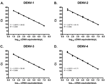
Using serial dilutions of plasmid DNA, the linear range for each
serotype extended from 1.0 to 7.0 log10cDNA equivalents/l (Fig.
1). Based on the standard curves generated using these plasmids,
the concentration of DENV in cDNA equivalents/l for the
high-est-concentration reference virus RNA was calculated. Using se-rial dilutions of reference virus RNA, the linear range of the
pan-DENV assay for each serotype, expressed in cDNA equivalents/l,
was 0.16 to 5.16 log10for DENV-1, 0.83 to 4.83 log10for DENV-2,
0.03 to 5.03 log10for DENV-3, and 1.11 to 4.11 log10for DENV-4
(data not shown).
The 95% LLOD was determined for each serotype in the pan-DENV assay by probit analysis using reference virus RNA dilu-tions. The 95% LLOD was calculated to be 1.7 for DENV-1, 1.7 for
DENV-2, 2.2 for DENV-3, and 7.6 cDNA equivalents/l for
DENV-4. Both intra- and interrun precision measures were cal-culated for each serotype at three concentrations (high positive, low positive, and limit of quantitation) using reference virus RNA.
Geometric mean concentrations in log10cDNA equivalents/l,
as well as intra- and interrun precision measures, are shown in
Table 3.
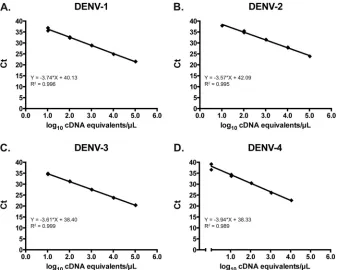
Altona assay analytical evaluation.The linear range of the Altona assay was established using serial dilutions of reference virus RNA for each DENV serotype. The linear range for this
as-say, expressed in cDNA equivalents/l, extended from 1.04 to 5.04
to log10for DENV-1, 1.61 to 4.61 log10for DENV-2, 1.02 to 5.02
log10for DENV-3, and 0.04 to 4.04 log10for DENV-4 (Fig. 2). The
95% LLOD was determined for each serotype in the Altona assay by probit analysis using reference virus RNA dilutions. The 95% LLOD was calculated to be 7.6 for DENV-1, 21.7 for DENV-2, 1.2
for DENV-3, and 0.97 cDNA equivalents/l for DENV-4.
Intrarun precision was determined for the Altona assay using DENV reference virus RNA. The standard deviation (percent co-efficient of variation) at the high and low ends of the linear range, respectively, for each serotype was 0.02 (0.37%) and 0.20 (20.17%) for DENV-1, 0.005 (0.1%) and 0.11 (6.72%) for DENV-2, 0.02 (0.39%) and 0.06 (5.38%) for DENV-3, and 0.01 (0.13%) and 0.10 (9.54%) for DENV-4. These results for the Al-tona assay were comparable to the results obtained with the pan-DENV assay.
FIG 1Linearity of the pan-DENV assay using serial 10-fold dilutions of plasmid containing the amplicons for DENV-1 (A), DENV-2 (B), DENV-3 (C), and DENV-4 (D). Ct, crossing threshold.
on May 16, 2020 by guest
http://jcm.asm.org/
[image:4.585.123.461.63.335.2]Specificity.The pan-DENV primers and probes were evalu-atedin silicoby querying the NCBI nucleotide database for related sequences. As described previously, the primers demonstrated
limited homologies to other members of theFlaviviridae
fam-ily, as well as to non-DENV sequences (16). No organism
rep-resented the best match for both the forward and reverse prim-ers for any of the primer pairs. The best non-DENV matches for
probes A, B, C, and D wereBrassica rapa,Schistosoma mansoni,
Drosophila ananassae, andDesulfatibacillum alkenivoransAK-01, re-spectively (query coverage, 89 to 94%; E value, 0.28 to 4.4). The
best matches within theFlaviviridaefamily were strains of JEV
(query coverage, 71 to 78%; E value, 0.44 to 0.52), though these sequences would not be predicted to amplify using the primers in this assay.
No amplification was observed in the pan-DENV or Altona assays when RNA from other closely related flaviviruses (three strains of WNV, one strain each of JEV and TBEV, and the YF-17D strain tested at two concentrations) was used. Sixty HCV-positive clinical samples were also tested by both assays; these samples had
[image:5.585.41.545.78.268.2]a median HCV viral load of 6.05 log10IU/ml of patient plasma
TABLE 3Precision of the pan-DENV assay
DENV serotype Dilution
Interrun precision Intrarun precision
Concn
(log10cDNA equivalents/l) SD % CoVa Concn
(Range, log10cDNA equivalents/l) SD range % CoV rangea
DENV-1 High positive 5.05 0.17 3.4 4.95–5.16 0.06–0.18 1.2–3.7 Low positive 3.25 0.16 4.8 3.05–3.38 0.04–0.07 1.3–2.3 Limit of quantitation 1.20 0.15 12.2 1.03–1.32 0.05–0.10 4.0–9.5
DENV-2 High positive 4.61 0.19 4.1 4.43–4.83 0.02–0.10 0.1–2.0 Low positive 2.62 0.08 3.2 2.57–2.74 0.051–0.054 1.9–2.1 Limit of quantitation 0.57 0.28 48.1 0.39–0.76 0.18–0.29 23.9–56.0
DENV-3 High positive 5.02 0.18 3.7 4.89–5.03 0.07–0.21 1.3–4.2 Low positive 2.35 0.13 5.5 2.18–2.44 2.8–7.8 1.2–3.2 Limit of quantitation 0.32 0.24 7.6 0.20–0.45 0.27–0.17 38.0–127
DENV-4 High positive 4.04 0.14 3.6 3.96–4.16 0.06–0.18 1.4–4.6 Low positive 2.32 0.12 5.1 2.20–2.39 0.07–0.09 3.1–3.8 Limit of quantitation 1.43 0.18 12.6 1.27–1.60 0.08–0.16 4.9–12.8
a
% CoV, percent coefficient of variation.
FIG 2Linearity of the Altona dengue assay using serial 10-fold dilutions of reference virus RNA for DENV-1 (A), DENV-2 (B), DENV-3 (C), and DENV-4 (D). Ct, crossing threshold.
on May 16, 2020 by guest
http://jcm.asm.org/
[image:5.585.124.464.430.700.2](range,⬍1.63 to 7.66). No amplification was observed in either assay for any of these samples.
IC performance.The performance of the ICs for the pan-DENV and Altona assays was evaluated using 201 clinical samples from Nicaragua and Sri Lanka. A single sample resulted in an IC failure on the pan-DENV assay, indicating a failed extraction or the presence of inhibitors. This sample was not included in further analysis for any assay. All samples that were negative for DENV
RNA by the pan-DENV assay (n⫽45) had similar amounts of
RNase P RNA (meanCT, 29.69; standard deviation, 2.76). Samples
withCTvalues of⬍20 in the green channel (DENV) did not have
a detectable RNase P signal, whereas samples with aCTof⬎25 in
the green channel yielded consistently detectable signals for RNase P (data not shown).
The Altona assay experienced a high number of IC failures during testing with the clinical samples. Overall, IC failure oc-curred in 49 samples (24.5%). These samples were not interpreta-ble by the Altona assay and were excluded from further analysis for this test. Of these samples, DENV RNA was detected in 25 (51.0%) samples by the pan-DENV assay, 24 (49.0%) by the multiplex rRT-PCR, and 12 (48.0%) by the heminested assay. The percent-ages of IC failures were similar in both groups of clinical samples (Nicaragua, 39/160 [24.4%], and Sri Lanka, 10/40 [25%]). Three IC failures also occurred in the HCV-positive plasma samples de-scribed above (3/60 [5%]). These failures were repeated and con-firmed on separate runs (data not shown). After 10- and 100-fold dilution of these samples, IC results were similar to the IC-only control. Prolonged centrifugation during the extraction or heat-ing of the eluate did not improve results (data not shown).
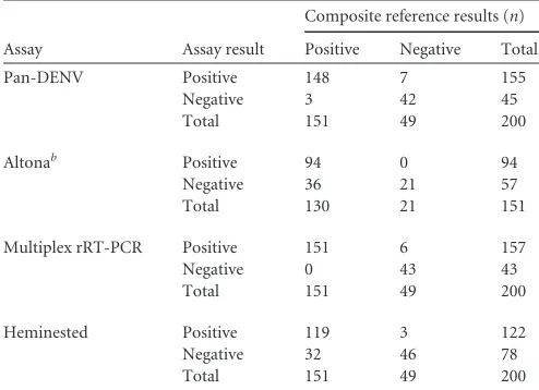
Samples from areas of endemicity.All clinical samples were tested by the pan-DENV, Altona, heminested, and DENV multi-plex rRT-PCR assays. Each assay was compared against a compos-ite reference standard, defined as the detection of DENV RNA in
two or more RT-PCR assays (Table 4). The composite reference
comprised 200 samples for the pan-DENV, multiplex rRT-PCR, and heminested assays. The sensitivity and specificity of each assay were then calculated using this reference. The pan-DENV and multiplex rRT-PCR had similar sensitivities (98.0% and 100%,
respectively;P⫽0.248). Both assays proved to be more sensitive
than the heminested assay (sensitivity, 78.8%;P⬍0.0001 for both
comparisons). The clinical specificities of the pan-DENV (85.7%), multiplex rRT-PCR (87.8%), and heminested assays
(93.9%) were not significantly different from one another (P⬎
0.05 for all comparisons).
The sensitivity and specificity for the Altona assay were calcu-lated using a composite reference comprising 151 samples. Test characteristics for the other assays were also calculated using this subset for comparison. The Altona assay had the lowest sensitivity
of any assay tested (72.3%;P⬍0.0001 for comparison with the
pan-DENV or multiplex rRT-PCR assays,P⫽0.035 for
compar-ison with the heminested assay). All samples detected by the Al-tona assay were also detected using the pan-DENV and multiplex rRT-PCR assays. The clinical specificity of the Altona assay was 100% compared against the composite reference, but this was not significantly different from the specificities of the other three
as-says (P⬎0.05 for all comparisons). A total of 16 samples had
DENV RNA detected by only a single assay (Table 5). The mean
CTvalues for these samples, when applicable, are also displayed in
Table 5.
To understand the clinical performance of these tests in more detail, we specifically analyzed the results for the C cases and IgM-positive samples, and we evaluated test performance based on the day of illness of sample collection.
C cases.Of the 40 samples collected from C cases, which were febrile patients thought to have a nondengue disease, a total of 19 (47.5%) samples had DENV RNA detected by at least one assay. This included 12 (30%) positive samples as detected by either the pan-DENV or multiplex rRT-PCR and 10 (25%) positive samples as detected by the heminested assay (data not shown). Seven sam-ples (all DENV-3) tested positive by the pan-DENV, heminested, and multiplex rRT-PCR assays, which include the five samples that tested positive using the Altona assay. Five samples were de-tected only by the pan-DENV assay, and five different samples were detected only using the multiplex rRT-PCR assay. The
aver-ageCTfor these 10 samples was 40.2 (standard deviation, 1.8)
compared to 20.8 for the seven samples that tested positive by
both assays (standard deviation, 9.3;P⬍0.0001). Two samples
(both DENV-3) were detected only using the heminested assay. The alternative diagnoses for these patients were not available for this study.
[image:6.585.39.286.87.266.2]IgM-positive Sri Lankan samples.Given the high analytical and clinical sensitivity of the pan-DENV assay, we next compared assay performance measures in the subset of specimens that were positive for anti-DENV IgM antibodies. The 40 samples from Sri Lanka were collected relatively late in the clinical course (mean day of illness, 5.9; standard deviation, 1.4), and 36 patients already
TABLE 4Comparison of the pan-DENV, Altona, multiplex rRT-PCR, and heminested assays with a composite reference
Assay Assay result
Composite reference results (n)
Positive Negative Total
Pan-DENV Positive 148 7 155 Negative 3 42 45 Total 151 49 200
Altonab Positive 94 0 94
Negative 36 21 57 Total 130 21 151
Multiplex rRT-PCR Positive 151 6 157 Negative 0 43 43 Total 151 49 200
Heminested Positive 119 3 122 Negative 32 46 78 Total 151 49 200
a
A single sample failed RNase P amplification and was excluded from all comparisons. bForty-nine IC failures were excluded from the Altona analysis.
TABLE 5Results for 16 clinical samples with DENV RNA detected using a single assay
Assay n
MeanCT
(SD)a Serotype (no.)
Pan-DENV 7 38.88 (1.34) NAb
Multiplex rRT-PCR 6 41.19 (1.66) DENV-1 (5), DENV-3 (1) Heminested 3 NAb DENV-3 (3)
a
CT, crossing threshold. bNA, not applicable.
on May 16, 2020 by guest
http://jcm.asm.org/
[image:6.585.296.545.87.145.2]had detectable anti-DENV IgM at presentation. Complete IgM data were not available for the Nicaraguan samples.
In this IgM-positive group, the pan-DENV assay detected DENV RNA in 35 samples (97.2%), whereas the heminested assay
detected DENV RNA in 18 samples (50.0%;P⬍0.0001). The
pan-DENV assay was also more sensitive than the Altona assay when only those samples with interpretable results by the latter
were considered (26/26 [100%] versus 13/26 [50.0%]; P ⬍
0.0001). The multiplex rRT-PCR assay detected DENV RNA in the same samples detected by the pan-DENV assay; these results
have been reported previously (16).
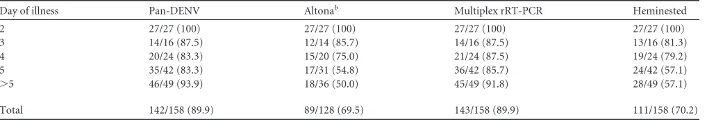
Day-of-illness analysis. Test results for 158 samples with available data were stratified according to the day of illness of sample collection (40 C cases plus 2 additional samples did not have the day of illness recorded). The difference in sensitivity be-tween the pan-DENV or multiplex rRT-PCR assays and either the Altona or the heminested RT-PCR varied based on the day of
illness on which the sample was obtained (Table 6). In samples
collected onⱕ4 days of illness, DENV RNA was detected in 61/67
(91.0%) samples by the pan-DENV assay compared to 59/67 (88.1%) samples by the heminested assay. This difference was not
significant (P⬎0.05). There was also no significant difference
between the assays when only the subset of samples with
interpre-table Altona results was considered (P⬎0.05, data not shown).
When the sample was obtained onⱖ5 days of illness (range, 5 to 9
days), DENV RNA was detected in 81/91 (89.0%) samples by the pan-DENV assay compared to 52/91 (57.1%) samples by the
heminested RT-PCR (P⬍0.0001). For samples with interpretable
Altona results, the pan-DENV was more sensitive than the Altona
assay with samples obtained onⱖ5 days of illness (64/67 versus
35/67, respectively;P⬍0.0001). The difference between the
Al-tona and heminested assays (44/67 detected) did not reach
statis-tical significance (P⫽0.16). There was no significant difference
between the pan-DENV and multiplex rRT-PCR assays based on the day of illness (data not shown).
DISCUSSION
This study presents the development and evaluation of the pan-DENV assay, which is a species-specific internally controlled rRT-PCR for pan-DENV detection. A second commercially produced internally controlled rRT-PCR assay, the Altona assay, was also independently evaluated. Both assays were then included in a large clinical comparison of four molecular assays for the detection of DENV, including a heminested RT-PCR that remains in wide use throughout the world and a serotype-specific multiplex rRT-PCR
previously developed in our laboratory (1,16).
Comparisons of different DENV molecular tests, such as those
performed in the current study, are scarce, despite the large
num-ber of diagnostic assays that have been reported for DENV (11,
13–15,17,38). Two earlier studies, published in 1998 and 2002,
compared conventional RT-PCR assays to the original version of the heminested RT-PCR. In both of these studies, the heminested assay was equivalent to or more sensitive than the newer assays
(17, 38). Four other groups have reported the comparison of
DENV NAATs (three rRT-PCRs and one transcription-mediated
amplification assay [TMA]) to a second molecular test (11,13–
15). Ito et al. (15) described the development of a hydrolysis
probe-based rRT-PCR for DENV and compared its performance
to that of two conventional RT-PCR assays (39). The new assay
proved more sensitive when tested using 35 clinical specimens
from returning travelers to Japan (15). Two groups have reported
the comparison of serotype-specific multiplex rRT-PCR assays to
a version of the heminested assay used in this study (11,13). In
both of these studies, however, the heminested assay remained
more sensitive than either rRT-PCR assay (11,13). A commercial
TMA assay (14) was compared with one of the rRT-PCRs and
proved to be more sensitive, though it was not compared to the heminested RT-PCR.
The current comparison offers two DENV rRT-PCRs that proved to be more sensitive than the heminested and Altona as-says. The pan-DENV and multiplex assays were more sensitive
when testing clinical samples obtained on ⱖ5 days of illness,
whereas all four assays performed comparably when testing sam-ples collected before that time. Results reported in DENV NAAT
studies are often obtained using samples collected onⱕ5 days of
illness, when, based on our findings, it may be difficult to
distin-guish assays with relatively poor sensitivity (14,23,40). In studies
where samples are tested onⱖ5 days of illness, test sensitivity
decreases markedly (41,42). Both the pan-DENV and multiplex
rRT-PCR assays also remained sensitive (97.2%) when samples obtained from acutely ill patients with detectable anti-DENV IgM levels were tested. It has generally been reported that as anti-DENV IgM levels become detectable, anti-DENV viral load declines
below detectable levels (1). As a result, patients with a positive IgM
are infrequently tested in DENV NAAT studies. In cases where such patients are tested, the sensitivity of DENV detection has
generally been poor, ranging from 0 to 69% (20,22,43–45). The
improved sensitivities of the pan-DENV and multiplex rRT-PCR assays may result from the use of a highly conserved target region
in the 5=UTR and capsid gene, compared to previous assays that
utilized targets in other regions of the genome, including the
C-prM, E, NS3, NS5, and 3=UTR (11,13,15,20,24).
[image:7.585.41.550.77.164.2]Despite the improved sensitivities of the pan-DENV and mul-tiplex rRT-PCR assays, the specificities of these two assays did not
TABLE 6Clinical samples with detectable DENV RNA stratified by the day of illness of sample collectiona
Day of illness Pan-DENV Altonab Multiplex rRT-PCR Heminested
2 27/27 (100) 27/27 (100) 27/27 (100) 27/27 (100) 3 14/16 (87.5) 12/14 (85.7) 14/16 (87.5) 13/16 (81.3) 4 20/24 (83.3) 15/20 (75.0) 21/24 (87.5) 19/24 (79.2) 5 35/42 (83.3) 17/31 (54.8) 36/42 (85.7) 24/42 (57.1)
⬎5 46/49 (93.9) 18/36 (50.0) 45/49 (91.8) 28/49 (57.1)
Total 142/158 (89.9) 89/128 (69.5) 143/158 (89.9) 111/158 (70.2)
a
Results are reported as number detected/total number (%). Data were not available for the 40 C cases and two additional samples. bIC failures were removed from the Altona assay analysis.
on May 16, 2020 by guest
http://jcm.asm.org/
differ significantly from those of the heminested or Altona assays. Furthermore, no amplification was observed in the pan-DENV assay using samples from 60 patients with HCV, as well as with reference virus RNA from other closely related flaviviruses. Simi-lar results for the multiplex rRT-PCR have been published
previ-ously (16). The samples that were detected by only one of these
two assays and counted as false positives had very low concentra-tions of DENV RNA, making it difficult to confirm or refute the results obtained by other means, such as sequencing. While the Altona assay had a specificity of 100%, this came at the expense of 36 false-negative results and poor assay sensitivity in the samples
collected onⱖ5 days of illness.
The pan-DENV assay provides species-specific DENV detec-tion but cannot be used for serotype determinadetec-tion in patient samples. For the management of acutely ill patients, however, the provision of a serotype-specific diagnosis may not be necessary
and currently does not affect clinical care (1,10,20). Also, outside
areas where dengue is endemic, the arguments for needing
sero-typing capability are less pertinent (10,26). Given the differences
in quantitation between the different serotypes, quantitative re-sults from the pan-DENV assay would only be accurate if serotype information was also available. If serial samples are tested over time, however, relative quantitation can be performed, as well as sensitive detection on the day of defervescence, a finding that has
been associated with the development of severe disease (30). With
further testing of the pan-DENV assay, thresholds may be estab-lished for these parameters that identify patients who are at in-creased risk for the development of severe dengue. The somewhat-lower cost of the pan-DENV compared to the multiplex rRT-PCR reagents ($2.00 versus $2.60 per reaction mixture) may also allow for its use as a screening test or for serial monitoring, as mentioned above.
A further benefit of the pan-DENV assay is the multiplex de-tection of RNase P, which serves as a heterologous intrinsic IC. Of the method comparisons mentioned previously, only the conven-tional RT-PCR developed in 1998 and the TMA assay contained
an IC (14,17). IC results were also not reported as a part of an
external quality-control evaluation of laboratories that were
per-forming molecular DENV testing (26). The presence of IC data
may have allowed the examination of extraction efficiency and reaction inhibition as potential contributors to the poor test sen-sitivity that is observed in a number of participating laboratories. The utility of an IC is further highlighted in this study by the large number of failures for the Altona assay, which uses an extrinsic molecule spiked into the reaction mixture. The fact that controls diluted in water (rather than extracted RNA eluted in buffer AVE) yielded consistently positive IC results suggests that the Altona reaction mixture may have been inhibited by eluates from the QIAamp viral RNA minikit. Ethanol contamination is one specific concern, though we decreased this risk by including an optional centrifugation step during RNA extraction. However, prolonged centrifugation did not improve the results. Eluate dilution did eliminate the observed inhibition, though this would not be a practical step for use with clinical samples. While the high IC failure rate did not fully explain the decreased sensitivity of the Altona assay, it remains a point of concern regarding the use of the current version of this test.
A number of samples (7/40 [17.5%]) from acutely ill patients who were initially suspected to have an illness other than dengue (C cases) tested positive for DENV by at least three of the
RT-PCRs used in this study. The viral load was high among these
samples, as indicated by theCTvalues, consistent with
true-posi-tive results. Twelve additional samples (30%) were positrue-posi-tive by only one assay, albeit at very low viral loads. Because the samples were obtained during high DENV activity in Nicaragua, we favor the interpretation that these are also true positive. A large number
of inapparent DENV infections occur, estimated at 50% to⬎90%
of infections, with large variation from year to year and between
studies (depending on the case capture procedures used) (46–49);
thus, it is likely that some proportion of C cases were experiencing an inapparent DENV infection.
Despite the inclusion of 200 clinical samples from two areas where DENV is endemic, there were no clinical samples with de-tectable levels of the DENV-4 serotype. While the analytical sen-sitivity for DENV-4 has been documented in our laboratory for all of the assays included in this study, the performance of these tests needs to be confirmed with DENV-4-positive clinical specimens. In addition, we cannot rule out the possibility that very high levels of IC may inhibit DENV detection, though the analytical and clin-ical sensitivities of the pan-DENV assay suggest that IC competi-tion is unlikely to be a significant problem in plasma samples. Nevertheless, the pan-DENV assay may be even more sensitive if performed without IC primers and probes. Further limitations of this study include concerns that are common to all NAATs regard-ing the emergence of divergent viral strains that do not match the primer or probe designs of a given assay. A specific limitation of the pan-DENV assay in this regard is the use of BHQplus moieties to allow for the design of shorter DENV probes with higher melt-ing temperatures. While this improves the specificity of the assay, the binding of BHQplus probes may be more susceptible to single base-pair changes than longer hydrolysis probes without this
modification (50).
In conclusion, we report here the design and evaluation of the pan-DENV assay, which is a species-specific internally controlled rRT-PCR for pan-DENV detection. In a comparison of four mo-lecular diagnostic assays for DENV, the pan-DENV assay was shown to be equally sensitive to a previously described multiplex rRT-PCR, as well as more sensitive than a commercially produced rRT-PCR and a widely used heminested assay. This was despite the addition of an IC that should serve to improve quality assur-ance, particularly when sample preparation and assay setup are performed under suboptimal conditions. The improved sensitiv-ities of the pan-DENV and multiplex rRT-PCR assays resulted
from the ability to detect DENV RNA in samples collected onⱖ5
days of illness and samples with detectable anti-DENV IgM, thereby lengthening the period of time during which a confirmed molecular diagnosis of DENV infection can be obtained.
ACKNOWLEDGMENTS
This research was supported by the National Institutes of Health grant RC4 TW008781-01. The studies in Nicaragua were supported by R01AI099631 (to A.B.), U54AI65359 (to A.B.), and HHSN2722001000026C (to E.H. and A.B.) from the National Institutes of Health and VE-1 (to E.H.) from the Pediatric Dengue Vaccine Initiative. The funders had no role in the study design, data collection and analysis, decision to publish, or preparation of the manuscript.
We thank the Stanford SPARK program for their support over the course of this project. We also thank Vittorio Sambri, Anna Pierro, and Paolo Gaibani for providing genomic flavivirus RNA for the specificity experiments. Finally, we thank Altona Diagnostics for providing the RealStar dengue RT-PCR kits for evaluation.
on May 16, 2020 by guest
http://jcm.asm.org/
REFERENCES
1.World Health Organization. 2009. Dengue: guidelines for diagnosis, treatment, prevention and control. WHO Press, Geneva, Switzerland. 2.World Health Organization.1997. Dengue haemorrhagic fever:
diagno-sis, treatment, prevention and control, 2nd ed. World Health Organiza-tion, Geneva, Switzerland.
3.Halstead SB, Nimmannitya S, Cohen SN.1970. Observations related to pathogenesis of dengue hemorrhagic fever. IV. Relation of disease severity to antibody response and virus recovered. Yale J. Biol. Med.42:311–328. 4.Martina BEE, Koraka P, Osterhaus AD.2009. Dengue virus
pathogen-esis: an integrated view. Clin. Microbiol. Rev.22:564 –581.
5.Gubler DJ.2012. The economic burden of dengue. Am. J. Trop. Med. Hyg.86:743–744.
6.Beatty ME, Letson GW, Margolis HS.2008. Estimating the global burden of dengue.InAbstract book: dengue 2008. Proceedings of the 2nd Inter-national Conference on Dengue and Dengue Haemorrhagic Fever. Phuket, Thailand.
7.Freedman DO, Weld LH, Kozarsky PE, Fisk T, Robins R, von Sonnen-burg F, Keystone JS, Pandey P, Cetron MS, GeoSentinel Surveillance Network.2006. Spectrum of disease and relation to place of exposure among ill returned travelers. N. Engl. J. Med.354:119 –130.
8.Burnette WN, Hoke CH, Jr, Scovill J, Clark K, Abrams J, Kitchen LW, Hanson K, Palys TJ, Vaughn DW.2008. Infectious disease investment decision evaluation algorithm: a quantitative algorithm for prioritization of naturally occurring infectious disease threats to the U.S. military. Mil. Med.173:174 –181.
9.Flores-Figueroa J, Okhuysen PC, von Sonnenburg F, DuPont HL, Libman MD, Keystone JS, Hale DC, Burchard G, Han PV, Wilder-Smith A, Freedman DO, GeoSentinel Surveillance Network.2011. Pat-terns of illness in travelers visiting Mexico and Central America: the Ge-oSentinel experience. Clin. Infect. Dis.53:523–531.
10. Peeling RW, Artsob H, Pelegrino JL, Buchy P, Cardosa MJ, Devi S, Enria DA, Farrar J, Gubler DJ, Guzman MG, Halstead SB, Hunsperger E, Kliks S, Margolis HS, Nathanson CM, Nguyen VC, Rizzo N, Vázquez S, Yoksan S.2010. Evaluation of diagnostic tests: dengue. Nat. Rev. Mi-crobiol.8(12 Suppl):S30 –S38. doi:10.1038/nrmicro2459.
11. Chien LJ, Liao TL, Shu PY, Huang JH, Gubler DJ, Chang GJJ.2006. Development of real-time reverse transcriptase PCR assays to detect and serotype dengue viruses. J. Clin. Microbiol.44:1295–1304.
12. Lanciotti RS, Calisher CH, Gubler DJ, Chang GJ, Vorndam AV.1992. Rapid detection and typing of dengue viruses from clinical samples by using reverse transcriptase-polymerase chain reaction. J. Clin. Microbiol. 30:545–551.
13. Johnson BW, Russell BJ, Lanciotti RS.2005. Serotype-specific detection of dengue viruses in a fourplex real-time reverse transcriptase PCR assay. J. Clin. Microbiol.43:4977– 4983.
14. Muñoz-Jordán J, Collins CS, Vergne E, Santiago GA, Peterson L, Sun W, Linnen JM.2009. Highly sensitive detection of dengue virus nucleic acid in samples from clinically ill patients. J. Clin. Microbiol.47:927–931. 15. Ito M, Takasaki T, Yamada KI, Nerome R, Tajima S, Kurane I.2004. Development and evaluation of fluorogenic TaqMan reverse transcriptase PCR assays for detection of dengue virus types 1 to 4. J. Clin. Microbiol. 42:5935–5937.
16. Waggoner J, Abeynayake J, Sahoo MK, Gresh L, Tellez Y, Gonzalez K, Ballesteros G, Pierro AM, Gaibani P, Guo FP, Sambri V, Balmaseda A, Karunarante K, Harris E, Pinsky BA.2013. Single-reaction, multiplex, real-time RT-PCR for the detection, quantitation, and serotyping of den-gue viruses. PLoS Negl. Trop. Dis. 7:e2116. doi:10.1371/journal.pntd .0002116.
17. Harris E, Roberts TG, Smith L, Selle J, Kramer LD, Valle S, Sandoval E, Balmaseda A.1998. Typing of dengue viruses in clinical specimens and mosquitoes by single-tube multiplex reverse transcriptase PCR. J. Clin. Microbiol.36:2634 –2639.
18. Callahan JD, Wu SJ, Dion-Schultz A, Mangold BE, Peruski LF, Watts DM, Porter KR, Murphy GR, Suharyono W, King CC, Hayes CG, Temenak JJ.2001. Development and evaluation of serotype- and group-specific fluorogenic reverse transcriptase PCR (TaqMan) assays for den-gue virus. J. Clin. Microbiol.39:4119 – 4124.
19. Drosten C, Göttig S, Schilling S, Asper M, Panning M, Schmitz H, Günther S.2002. Rapid detection and quantification of RNA of Ebola and Marburg viruses, Lassa virus, Crimean-Congo hemorrhagic fever virus,
Rift Valley fever virus, dengue virus, and yellow fever virus by real-time reverse transcription-PCR. J. Clin. Microbiol.40:2323–2330.
20. Dumoulin A, Marti H, Panning M, Hatz C, Hirsch HH.2008. Pan-dengue virus detection by PCR for travelers returning from the tropics. J. Clin. Microbiol.46:3104 –3106.
21. Hue KD, Tuan TV, Thi HT, Bich CT, Anh HH, Wills BA, Simmons CP. 2011. Validation of an internally controlled one-step real-time multiplex RT-PCR assay for the detection and quantitation of dengue virus RNA in plasma. J. Virol. Methods177:168 –173.
22. Kong YY, Thay CH, Tin TC, Devi S.2006. Rapid detection, serotyping and quantitation of dengue viruses by TaqMan real-time one-step RT-PCR. J. Virol. Methods138:123.
23. Lai Y, Chung YK, Tan HC, Yap HF, Yap G, Ooi EE, Ng LC.2007. Cost-effective real-time reverse transcriptase PCR (RT-PCR) to screen for Dengue virus followed by rapid single-tube multiplex RT-PCR for sero-typing of the virus. J. Clin. Microbiol.45:935–941.
24. Leparc-Goffart I, Baragatti M, Temmam S, Tuiskunen A, Moureau G, Charrel R, de Lamballerie X.2009. Development and validation of real-time one-step reverse transcription-PCR for the detection and typing of dengue viruses. J. Clin. Virol.45:61– 66.
25. Naze F, Le Roux K, Schuffenecker I, Zeller H, Staikowsky F, Grivard P, Michault A, Laurent P.2009. Simultaneous detection and quantitation of Chikungunya, Dengue and West Nile viruses by multiplex RT-PCR assays and Dengue virus typing using high resolution melting. J. Virol. Methods 162:1–7.
26. Domingo C, Niedrig M, Teichmann A, Kaiser M, Rumer L, Jarman RG, Donoso-Mantke O.2010. 2nd international external quality control as-sessment for the molecular diagnosis of Dengue infections. PLoS Negl. Trop. Dis.4:e833. doi:10.1371/journal.pntd.0000833.
27. Duyen HT, Ngoc TV, Ha do T, Hang VT, Kieu NT, Young PR, Farrar JJ, Simmons CP, Wolbers M, Wills BA.2011. Kinetics of plasma viremia and soluble nonstructural protein 1 concentrations in dengue: differential effects according to serotype and immune status. J. Infect. Dis.203:1292– 1300.
28. Libraty DH, Young PR, Pickering D, Endy TP, Kalayanarooj S, Green S, Vaughn DW, Nisalak A, Ennis FA, Rothman AL.2002. High circu-lating levels of the dengue virus nonstructural protein NS1 early in dengue illness correlate with the development of dengue hemorrhagic fever. J. Infect. Dis.186:1165–1168.
29. Tricou V, Minh NN, Farrar J, Tran HT, Simmons CP.2011. Kinetics of viremia and NS1 antigenemia are shaped by immune status and virus serotype in adults with dengue. PLoS Negl. Trop. Dis.5:e1309. doi:10 .1371/journal.pntd.0001309.
30. Vaughn DW, Green S, Kalayanarooj S, Innis BL, Nimmannitya S, Suntayakorn S, Endy TP, Raengsakulrach B, Rothman AL, Ennis FA, Nisalak A.2000. Dengue viremia titer, antibody response pattern, and virus serotype correlate with disease severity. J. Infect. Dis.181:2–9. 31. Wang WK, Lee CN, Kao CL, Lin YL, King CC. 2000. Quantitative
competitive reverse transcription-PCR for quantification of dengue virus RNA. J. Clin. Microbiol.38:3306 –3310.
32. Burd EM.2010. Validation of laboratory-developed molecular assays for infectious diseases. Clin. Microbiol. Rev.23:550 –576.
33. WHO/CDC.2009. CDC protocol of realtime RTPCR for swine influenza A (H1N1). WHO Collaborating Centre for Influenza at CDC Atlanta, United States of America, Atlanta, GA.
34. Rossini G, Carletti F, Bordi L, Cavrini F, Gaibani P, Landini MP, Pierro A, Capobianchi MR, Di Caro A, Sambri V.2011. Phylogenetic analysis of West Nile virus isolates, Italy, 2008-2009. Emerg. Infect. Dis.17:903– 906.
35. Gutierrez G, Standish K, Narvaez F, Perez MA, Saborio S, Elizondo D, Ortega O, Nuñez A, Kuan G, Balmaseda A, Harris E.2011. Unusual dengue virus 3 epidemic in Nicaragua, 2009. PLoS Negl. Trop. Dis. 5:e1394. doi:10.1371/journal.pntd.0001394.
36. Kuan G, Gordon A, Avilés W, Ortega O, Hammond SN, Elizondo D, Nuñez A, Coloma J, Balmaseda A, Harris E.2009. The Nicaraguan pediatric dengue cohort study: study design, methods, use of information technology, and extension to other infectious diseases. Am. J. Epidemiol. 170:120 –129.
37. Narvaez F, Gutierrez G, Pérez MA, Elizondo D, Nuñez A, Balmaseda A, Harris E.2011. Evaluation of the traditional and revised WHO classifica-tions of Dengue disease severity. PLoS Negl. Trop. Dis.5:e1397. doi:10 .1371/journal.pntd.0001397.
38. Raengsakulrach B, Nisalak A, Maneekarn N, Yenchitsomanus PT,
on May 16, 2020 by guest
http://jcm.asm.org/
somwong C, Jairungsri A, Thirawuth V, Green S, Kalayanarooj S, Suntayakorn S, Sittisombut N, Malasit P, Vaughn D.2002. Comparison of four reverse transcription-polymerase chain reaction procedures for the detection of dengue virus in clinical specimens. J. Virol. Methods 105:219 –232.
39. Yamada K, Takasaki T, Nawa M, Kurane I.2002. Virus isolation as one of the diagnostic methods for dengue virus infection. J. Clin. Virol.24: 203–209.
40. Dos Santos HW, Poloni TR, Souza KP, Muller VD, Tremeschin F, Nali LC, Fantinatti LR, Amarilla AA, Castro HL, Nunes MR, Casseb SM, Vasconcelos PF, Badra SJ, Figueiredo LT, Aquino VH.2008. A simple one-step real-time RT-PCR for diagnosis of dengue virus infection. J. Med. Virol.80:1426 –1433.
41. Chutinimitkul S, Payungporn S, Theamboonlers A, Poovorawan Y. 2005. Dengue typing assay based on real-time PCR using SYBR Green I. J. Virol. Methods129:8 –15.
42. Gurukumar KR, Priyadarshini D, Patil JA, Bhagat A, Singh A, Shah PS, Cecilia D.2009. Development of real time PCR for detection and quan-titation of dengue viruses. Virol. J.6:10. doi:10.1186/1743-422X-6-10. 43. Hertz JT, Munishi OM, Ooi EE, Howe S, Lim WY, Chow A, Morrissey
AB, Bartlett JA, Onyango JJ, Maro VP, Kinabo GD, Saganda W, Gubler DJ, Crump JA.2012. Chikungunya and dengue fever among hospitalized febrile patients in northern Tanzania. Am. J. Trop. Med. Hyg.86:171–177. 44. Laue T, Emmerich P, Schmitz H.1999. Detection of dengue virus RNA in patients after primary or secondary dengue infection by using the Taq-Man automated amplification system. J. Clin. Microbiol.37:2543–2547. 45. Shu PY, Chang SF, Kuo YC, Yueh YY, Chien LJ, Sue CL, Lin TH,
Huang JH.2003. Development of group- and serotype-specific one-step SYBR green I-based real-time reverse transcription-PCR assay for dengue virus. J. Clin. Microbiol.41:2408 –2416.
46. Balmaseda A, Standish K, Mercado JC, Matute JC, Tellez Y, Saborio S, Hammond SN, Nuñez A, Avilés W, Henn MR, Holmes EC, Gordon A, Coloma J, Kuan G, Harris E.2010. Trends in patterns of dengue trans-mission over 4 years in a pediatric cohort study in Nicaragua. J. Infect. Dis. 201:5–14.
47. Endy TP, Anderson KB, Nisalak A, Yoon IK, Green S, Rothman AL, Thomas SJ, Jarman RG, Libraty DH, Gibbons RV.2011. Determinants of inapparent and symptomatic dengue infection in a prospective study of primary school children in Kamphaeng Phet, Thailand. PLoS Negl. Trop. Dis.5:e975. doi:10.1371/journal.pntd.0000975.
48. Endy TP, Chunsuttiwat S, Nisalak A, Libraty DH, Green S, Rothman AL, Vaughn DW, Ennis FA.2002. Epidemiology of inapparent and symptomatic acute dengue virus infection: a prospective study of pri-mary school children in Kamphaeng Phet, Thailand. Am. J. Epidemiol. 156:40 –51.
49. Standish K, Kuan G, Avilés W, Balmaseda A, Harris E.2010. High dengue case capture rate in four years of a cohort study in Nicaragua compared to national surveillance data. PLoS Negl. Trop. Dis.4:e633. doi:
10.1371/journal.pntd.0000633.
50. Kutyavin IV, Afonina IA, Mills A, Gorn VV, Lukhtanov EA, Belousov ES, Singer MJ, Walburger DK, Lokhov SG, Gall AA, Dempcy R, Reed MW, Meyer RB, Hedgpeth J.2000. 3=-minor groove binder-DNA probes increase sequence specificity at PCR extension temperatures. Nucleic Ac-ids Res.28:655– 661.