From
DEPARTMENT OF MEDICINE SOLNA
Karolinska Institutet, Stockholm, Sweden
CHRONIC KIDNEY DISEASE:
A COMPLEX NETWORK OF LEUKOCYTE
DYSFUNCTION AND INFLAMMATION
Ladan Mansouri
یروصنم ندلاAll previously published papers were reproduced with permission from the publisher. Published by Karolinska Institutet.
Printed by E-print AB 2016, Stockholm Sweden. © Ladan Mansouri, 2016
Departm ent of Medicine Solna
Chronic kidney disease: A complex network of
leukocyte dysfunction and inflammation
THESIS FOR DOCTORAL DEGREE (Ph.D.)
The public defense at Karolinska Institutet will be held at Rolf Luft auditorium, CMM Ground floor (L1:00), Karolinska University Hospital Solna
Friday, November 11
th2016, 09:00
By
Ladan Mansouri, MD.
Principal Supervisor:
Professor Joachim Lundahl Karolinska Institutet
Department of Medicine Solna Unit of Immunology and Allergy
Co-supervisor(s):
Docent Anna Nopp Karolinska Institutet
Department of Medicine Solna Unit of Immunology and Allergy
Professor Stefan Jacobson Karolinska Institutet Danderyd Hospital
Department of Clinical Sciences
Opponent:
Professor Mårten Segelmark Linköping University
Department of Medical and Health Sciences
Examination Board:
Docent Guro Gafvelin Karolinska Institutet
Department of Clinical Neuroscience
Professor Börje Haraldsson University of Gothenburg Department of Medicine
Division of Molecular and Clinical Medicine
Docent Ann-Charlotte Wikström Karolinska Institutet
Department of Medicine Solna Unit of Immunology and Allergy
To my loved ones
,
“Excellence is never an accident.
It is always the result of high intention, sincere effort, and intelligent execution.
It represents the wise choice of many alternatives
- Choice, not chance, determines your destiny”
ABSTRACT
Chronic kidney disease (CKD) is a common medical condition with a prevalence of up to 13% worldwide. The insidious loss of renal function develops over time to reach end stage renal disease (ESRD) when the kidneys are unable to remove toxins and interventions such as dialysis or kidney transplantation are mandatory. These patients are at increased risk of morbidity and mortality, essentially due to cardiovascular diseases and infections, which arise from alterations in both the innate and adaptive immune responses. This thesis focuses on the functional aberrations in leukocytes, inflammation and outcomes of medical interventions in CKD patients. In paper I, we investigated the impact of blood-membrane interaction on circulating basophils and neutrophils in hemodialysis patients (stage 5D) using dialyzers with two different pore sizes; high-flux and low-flux. Passage through the low-flux dialyzer induced a significant up-regulation of the activation marker CD63 on basophils, while this was not the case in high-flux dialysis. No significant differences were observed in the expression of neutrophil activation markers (CD11b, the active epitope of CD11b, and CD88), either comparing the two dialyzers, or compared to healthy controls. In paper II, we analyzed the proliferation of lymphocytes and production of pro-inflammatory molecules following in vitro stimulation of whole blood from pre-dialysis (CKD stage 3-4), hemodialysis patients (stage 5D), and healthy individuals. We found a similar absolute number of lymphoblasts, and CD4+ and CD8+ subpopulation in response to stimulation when comparing the three groups. However, levels of selected cytokines were lower in the staphylococcus enterotoxin A (SEA)-, influenza A vaccine (IAV)-, and pokeweed mitogen (PWM)-stimulated supernatants from hemodialysis patients compared to those in pre-dialysis and healthy controls. In paper III, we aimed to investigate whether the decreased in vitro cytokine production results from dysfunction of T cells per se. We analyzed the T lymphocyte profile in CKD patients (stage 5D) following stimulation of isolated CD4+ cells with an antigen-independent stimulator (SEA). The proportion of CD4+CD25+FOXP3+ (Treg) and CD4+GATA3+ (Th2) cells was significantly lower in patients. Levels of Interleukin (IL)-4 were significantly lower in supernatants from patients, while Interferon (IFN)-γ levels were higher. IL-10 levels did not differ, comparing patients to healthy individuals. In paper IV, we performed a clinical trial for 12 weeks with the vitamin D receptor activator (VDRA)-paricalcitol and placebo in patients with moderate CKD (stage 3-4). We observed that selected pro-inflammatory cytokines decreased following VDRA treatment but not in the placebo group. Micro RNAs: 432-5p, 495-3p, and 576-5p were also significantly down regulated in the active treated group compared to the placebo group.
In conclusion, we demonstrate that chronic renal impairment and hemodialysis leads to a high inflammatory state and alteration in the function of innate and adaptive immune cells. Moreover, we show that treatment with active vitamin D-paricalcitol has a positive impact on dampening inflammatory molecules and miRs involved in atherosclerosis and inflammation. Altogether, these findings provide us with a better understanding of potential factors orchestrating the higher prevalence of cardiovascular diseases and infections, as well as the progression of CKD.
LIST OF SCIENTIFIC PAPERS
I. Aljadi Z*, Mansouri L*, Nopp A, Paulsson JM, Winqvist O, Russom A, Ståhl M, Hylander B, Jacobson SH, Lundahl J.
Activation of basophils is a new and sensitive marker of biocompatibility in Hemodialysis. Artif Organs. 2014 Nov; 38(11): 945-53.
II. Ladan Mansouri, Josefin M. Paulsson, Ali Moshfegh, Stefan H. Jacobson, Joachim Lundahl.
Leukocyte proliferation and immune modulator production in patients with chronic kidney disease. PLoS ONE 2013, 8 (8), e73141.
III. Ladan Mansouri, Anna Nopp, Stefan H. Jacobson, Britta Hylander, Joachim Lundahl.
Hemodialysis patients display a declined proportion of CD4+CD25+ FOXP3+ and CD4+GATA3+ T cells but a high IFN-γ profile in response to superantigen. Manuscript, submitted.
IV. Ladan Mansouri*, Kristina Lundwall*, Ali Moshfegh, Stefan H. Jacobson, Joachim Lundahl, Jonas Spaak.
Vitamin D Receptor Activation Reduces Inflammatory Cytokines and Plasma MicroRNAs in Moderate Chronic Kidney Disease– A Randomized Trial. Manuscript, under revision.
* Shared first authorship
RELATED SCIENTIFIC PAPERS OUTSIDE THE THESIS
Wallquist C, Mansouri L, Norrbäck M, Hylander B, Jacobson SH, and Lundahl J.
Early Changes in Monocyte Adhesion Molecule Expression and Tumor Necrosis Factor-α Levels in Chronic Kidney Disease- A 5-Year Prospective Study. Am J Nephrol. 2016 Sep 8; 44(4):268-275.
Wallquist C, Mansouri L, Norrbäck M, Hylander B, Larsson T, Jacobson SH, , and Lundahl J.
Associations of fibroblast growth factor 23 with markers of inflammation and leukocyte function in chronic kidney disease. Manuscript, submitted.
CONTENTS
1 INTRODUCTION ... 1
1.1 Chronic Kidney Disease (CKD) ... 1
1.1.1 Definition and staging ... 1
1.1.2 Epidemiology... 1
1.1.3 CKD-related complications... 2
1.1.4 Detection ... 2
1.1.5 Management ... 2
1.2 CKD and the immune system ... 3
1.2.1 Immune system ... 3
1.2.2 Immunity in CKD ... 3
1.2.3 Uremia ... 3
1.2.4 Renal replacement therapy ... 4
1.3 CKD and neutrophils ... 5 1.3.1 Neutrophil biology ... 5 1.3.2 Neutrophils in CKD patients ... 7 1.4 CKD and basophils ... 8 1.4.1 Basophil biology ... 8 1.4.2 Basophils in CKD patients ... 10 1.5 CKD and T lymphocytes ... 10 1.5.1 T cell biology ... 10 1.5.2 T cells in CKD patients ... 12
1.6 Gene transcription and epigenetic regulation in CKD ... 12
1.6.1 Transcriptome analysis in CKD ... 13
1.6.2 Role of microRNAs in CKD ... 13
1.7 Impact of medications in CKD ... 14
1.7.1 Angiotensin- converting enzyme inhibitor (ACE-I) and angiotensin receptor blockers (ARBs)... 14
1.7.2 Erythropoiesis-stimulating agents (ESA) ... 14
1.7.3 Vitamin D ... 14
2 AIMS OF THE THESIS ... 17
3 MATERIAL AND METHODS ... 19
3.1 Study population ... 19 3.1.1 Patients... 19 3.1.2 Healthy controls ... 20 3.2 Study methods ... 21 3.2.1 Study I ... 21 3.2.2 Study II ... 21 3.2.3 Study III ... 22 3.2.4 Study IV... 23 3.3 Statistical analysis ... 24
4.1 Study I ... 25 4.2 Study II ... 27 4.3 Study III ... 30 4.4 Study IV ... 31 5 PRELIMINARY RESULTS ... 35 5.1 Part I. ... 35 5.2 Part II... 36 5.3 Future plans ... 36 6 CONCLUSIONS ... 37 7 ACKNOWLEDGEMENTS... 39 8 REFERENCES ... 43
LIST OF ABBREVIATIONS
ACE-I Angiotensin-converting enzyme inhibitor
AhR Aryl hydrocarbon receptor
ARB Angiotensin II receptor blocker
B-FGF Basic-fibroblast growth factor
CAPD Continuous Ambulatory Peritoneal Dialysis
CKD Chronic kidney disease
Con A Concanavalin A
CRP C-reactive protein
CVD Cardiovascular diseases
DCs Dendritic cells
DIP Degranulation inhibiting protein
DM Diabetes Mellitus
ECM Extracellular matrix
EGFR Estimated glomerular filtration rate
ESA Erythropoiesis-stimulating agent
ESRD End stage renal disease FcεRI Fc epsilon receptor type I
FMLP N formyl- Methionyl-Leucyl-Phenylalanine
FOXP3 Forkhead box P3
FPR Formylpeptide receptor
FPRL1 Formyl peptide receptor-like 1
GATA3 GATA Binding Protein 3
G-CSF Granulocyte colony-stimulating factor
GFR Glomerular filtration rate
GM-CSF Granulocyte macrophage colony-stimulating factor
GPx-1 Glutathione peroxidase-1
IAV Influenza A vaccine
IFN Interferon
IL- Interleukin
LDL Low-density lipoprotein
LPS Lipopolysaccharide
LVMI Left ventricular mass index
MCP Monocyte chemoattractant protein
MHC Major histocompatibility complex
MIP Macrophage inflammatory protein
MiR Micro RNA
NET Neutrophil extracellular traps
NF-κB Nuclear factor-kappaB
NKCs Natural killer cells
OxLDL
PAF
Oxidized LDL
Platelet-activating factor
PAMP Pathogen-associated molecular pattern
PBMC Peripheral blood mononuclear cell
PDGF Platelet-derived growth factor
PKD Polycystic kidney disease
PMA Phorbol myristate acetate
PRDX1 Peroxiredoxin-1
PRRs Pattern recognition receptors
PSGL-1 P-selectin glycoprotein ligand 1
PTH Parathyroid hormone
PWM Pokeweed mitogen
RAAS Renin-angiotensin-aldosterone system
RGD Arginine-glycine-aspartic acid
RORγ RAR-related orphan receptor gamma
ROS Reactive oxygen species
SEA Staphylococcus enterotoxin A
SelW Selenoprotein W
SLE Systemic lupus erythematosus
SOD2 Superoxide dismutase 2
TCR T cell receptor
Tfh Follicular helper T cell
TLR Toll-like receptor
TNF Tumor necrosis factor
Treg Regulatory T cell
VDR Vitamin D receptor
VDRA Vitamin D receptor activator
VEGF Vascular endothelial growth factor
1 INTRODUCTION
1.1 CHRONIC KIDNEY DISEASE (CKD)
1.1.1 Definition and staging
The kidneys are vital organs, which remove wastes and extra fluids, keep electrolytes in balance, regulate blood pressure (by renin), manage the production of red blood cells (by erythropoietin), and bone metabolism (by active vitamin D). Chronic kidney disease (CKD) comprises heterogeneous disorders, which affect the performance or structure of the kidney. The presence or absence of kidney damage (albuminuria, kidney biopsy findings, or imaging abnormalities) and declined level of kidney function (glomerular filtration rate, GFR <60 mL/min/1.73 m2) for more than three months, define CKD, irrespective of diagnosis (Levey and Coresh 2012).
A simplified classification of CKD into different stages based on GFR is as follows;
Stage 1: GFR >90 (Kidney damage with normal GFR) Stage 2: GFR 60-89 (Mild)
Stage 3: GFR 30-59 (Moderate) Stage 4: GFR 15-29 (Severe) Stage 5: GFR <15 (Kidney failure)
There is a normal age-related decline of GFR in adults, which follows global glomerular sclerosis and cortical atrophy, but the consequences of this phenomenon have not been thoroughly studied in elderly, age ≥65 years (Denic, Glassock et al. 2016).
1.1.2 Epidemiology
In recent years, guidelines have stated that CKD is a common disorder with various degrees of severity. Currently CKD ranks 19th as mortality cause worldwide, presenting an 82% increase since 1990 (Stanifer, Muiru et al. 2016). The prevalence of CKD stage 1-5 in adults has been reported between 3-17% with a substantial variation across Europe (Bruck, Stel et al. 2016). According to recent estimations in the United States, the disease prevalence in adults will increase significantly in the next decade (Hoerger, Simpson et al. 2015). The prevalence of CKD is not well characterized in low- and middle-income countries, however a higher exposure to occupational and environmental toxins, infections, and medical conditions such as: diabetes mellitus (DM), hypertension, obesity, poly cystic kidney disease, acute kidney injuries, and malignancies ensures an enormous increase of CKD prevalence in these countries (Stanifer, Muiru et al. 2016).
Altogether, these reports indicate that CKD is a growing medical condition, which requires attention, effective prevention, early detection and management. Early stages of the disease are usually asymptomatic and possibly reversible, but when kidney failure is established, only controlling the comorbidities, dialysis or transplantation is applicable.
1.1.3 CKD-related complications
The enormous burden of the disease is not limited to decreased removal of uremic toxins from the body, it also imposes an increased risk of complications associated to the eGFR <60 mL/min/1.73 m2 or albumin/creatinine ratio >30 g/mol.
These complications comprise cardiovascular diseases, hypertension, mineral and bone disorders, anemia, progression to ESRD, infections and low response to vaccinations (Levey, de Jong et al. 2011). Cardiovascular diseases and infections are the most common causes of morbidity and mortality in CKD patients (Thompson, James et al. 2015).
1.1.4 Detection
Since serum creatinine testing is included in the basic metabolic panels, detection of CKD mostly occurs during routine care. There are more accurate assessments than serum creatinine to determine GFR (clearance of inulin, 51Cr-EDTA or iohexol).
1.1.4.1 Estimated Glomerular Filtration Rate
GFR is estimated by equations using serum creatinine and combination of age, sex, ethnicity and body size (estimated GFR; eGFR). The modification of diet in renal disease (MDRD) and the chronic kidney disease epidemiology collaboration (CKD-EPI) equations are accurate at eGFRs <60 and eGFRs >60, respectively. Generally CKD-EPI is less biased compared to the older MDRD equation (Levey, Stevens et al. 2009).
1.1.4.2 Quantification of albuminuria
A spot albumin/creatinine ratio in urine is a sensitive measure of kidney damage, and is crucial to evaluate prognosis (Inker, Astor et al. 2014).
1.1.4.3 Imaging or serologic testing
Imaging is not routinely required, but in the case of urinary tract stones, frequent urinary tract infections, or a family history of polycystic kidney disease (PKD), a kidney and bladder ultrasound should be performed. A systemic or glomerular disease requires serologic workup.
1.1.5 Management
The main goals of CKD management are to delay progressive loss of kidney function and to prevent and manage complications. Currently the most commonly applied strategies to prevent progression of the disease and associated complications include treatment of hypertension and reduction of albuminuria by use of Angiotensin converting enzyme inhibitor (ACE-I), and Angiotensin II receptor blockers (ARBs). Furthermore, diabetes control, anemia management (Erythropoiesis-stimulating agent; ESA), correction of secondary hyperparathyroidism, hypocalcemia, and hyperphosphatemia (supplementation with vitamin D, calcimimetics, and phosphate binders), correction of metabolic acidosis and lipid-lowering therapy (statins) are important measures in CKD patients with risk of
cardiovascular diseases (Uhlig, Berns et al. 2010; National Kidney 2012; Kliger, Foley et al. 2013; Taler, Agarwal et al. 2013; Sarnak, Bloom et al. 2015).
1.2 CKD AND THE IMMUNE SYSTEM
1.2.1 Immune system
The immune system is a complex network of cells and soluble factors, which contribute to protection of host against various microorganisms and diseases. It is not only involved in detection and removal of invading factors, but also contributes to the repair of injured tissues. The immune system comprises two main sectors: the innate and the adaptive immune systems. The innate immunity performs as a ready-to-go defender. Innate immune cells recognize conserved microbial components through their pattern recognition receptors (PRRs), and mediate blockage or elimination of infections at a fast pace. The main components of innate immunity are physical epithelial barriers, leukocytes such as neutrophils, monocytes, macrophages, dendritic cells (DCs), natural killer cells (NKCs), mast cells, eosinophils, basophils, and circulating plasma proteins such as complements. The adaptive immunity is a slower but more specialized and antigen-specific immune defense with a memory to mount a stronger response on re-encounter of the same pathogen. T-lymphocytes and B-T-lymphocytes are players in the adaptive immunity.
Although innate and adaptive immunity are classified separately, they interact very closely, and antigen-presenting cells (APCs) are the major links. APCs process and present the antigenic peptides on major histocompatibility complex (MHC) to the T cells, and activate them to further proliferate, release the inflammatory molecules, boost other immune cells, promote antibody production, or convey apoptosis.
1.2.2 Immunity in CKD
It’s known that loss of renal function is strongly associated with two contrasting features. On the one hand there is a high inflammatory state in CKD patients, which is witnessed by high level of pro-inflammatory molecules in the plasma; such as Interleukin (IL)-1, IL-6 and Tumor necrosis factor (TNF)-α (Honda, Qureshi et al. 2006; Cohen, Phillips et al. 2010), oxidative stress (Locatelli, Canaud et al. 2003), and activation of immune cells such as monocytes. This ongoing inflammation leads to processes such as atherosclerosis and cardiovascular diseases. On the other hand there is an immunodeficiency, which results in a higher susceptibility to infections (Sarnak and Jaber 2000), increased risk of virus-associated cancers (Stewart, Vajdic et al. 2009) and a low response to vaccinations (Robinson 2004).
The immune system aberrations in CKD patients originate mainly from retention of uremic toxins and cytokines or medical interventions such as in dialysis (Betjes 2013).
1.2.3 Uremia
When CKD is progressing to ESRD (eGFR <15 mL/min/1.73 m2), uremia develops. Patients may present characteristic signs and symptoms of uremia such as nausea, significant weight
loss, neurologic dysfunction, bleeding diathesis, and volume overload, which may require renal replacement therapy. However in many cases, advanced CKD may be asymptomatic with very few and uncharacteristic signs and symptoms.
1.2.3.1 Uremic toxins
Since serum urea and creatinine are inadequate markers of uremic toxicity, various classification systems have been developed to further characterize uremia.
One is based on the physicochemical properties of uremic toxins, and their removal by renal replacement therapy, dividing them into small water-soluble molecules (molecular weight ≤500 Da), middle molecules (>500 Da), and protein-bound molecules (Vanholder, De Smet et al. 2003). Another classification divides them, according to their site of origin, into those produced by endogenous metabolism or microbial metabolism and toxins ingested from an exogenous source (e.g. oxalate and advanced glycation) (Evenepoel, Meijers et al. 2009).
The vast majority of uremic toxins are either highly protein bound or middle molecular weight compounds, which are difficult to remove by conventional dialysis.
1.2.3.2 Impact of uremic toxins on the immune system
There is a strong connection between residual renal function and the level of inflammatory markers such as IL-6 (Pecoits-Filho, Heimburger et al. 2003). Loss of renal function leads to retention of cytokines and uremic toxins, which in turn result in activation of immune cells and oxidative stress. Reactive oxygen species (ROS) can interact with the pattern recognition receptors on immune cells, activate them and establish a more sustained inflammatory response (Bierhaus, Schiekofer et al. 2001). Moreover, this inflammatory state is accompanied by an immune system dysfunction, including defective phagocytosis, insufficient antigen presentation, and low T cell-dependent response to vaccination (Eleftheriadis, Antoniadi et al. 2007). There are different underlying mechanisms behind these phenomena, among which are: decreased responsiveness of activated immune cells (tachyphylaxis) (Hendrikx, van Gurp et al. 2009), activation-induced apoptosis, variation in differentiation status, and the new paradigm of premature immunological aging in CKD patients (Meier, Dayer et al. 2002; Majewska, Baj et al. 2003; Betjes and Litjens 2015).
1.2.4 Renal replacement therapy
The ultimate aim of renal replacement therapy is to remove accumulated uremic toxins, minerals and excessive fluid from the blood, and to preserve residual renal function. Replacement therapy is performed by either hemodialysis (HD) (Fig. 1) or peritoneal dialysis (PD), and through diffusion and convection processes. However, blood-dialysis membrane interactions may lead to a variety of adverse reactions in circulating immune cells and plasma proteins as a consequence of bio-incompatibility.
1.2.4.1 Hemodialysis and biocompatibility
Surface material of the applied dialyzer, membrane permeability properties and dialysis fluid (dialyzate) are among the main causes of alterations in adhesion molecule expression on leukocytes, cytokine production, phagocytic efficiency, apoptosis, ROS production, and complement activation (Kazatchkine and Carreno 1988; Rao, Guo et al. 2004; Banche, Allizond et al. 2006). Modified dialysis membranes with improved biocompatibility have been developed, which preserve the functionality of immune cells (Banche, Allizond et al. 2006). Moreover, the membrane permeability affects biocompatibility of the dialyzers. Efficient removal of larger molecules such as β2-microglobulin and toxins has been observed with high-flux membranes (with large pore size and higher ultrafiltration coefficients) rather than with low-flux membranes (with smaller pore size and lower ultrafiltration coefficients). The long-term clinical consequences in terms of morbidity and mortality are still under debate (Cheung, Levin et al. 2003; Locatelli, Martin-Malo et al. 2009).
Figure 1. Schematic view of hemodialysis procedure.
1.3 CKD AND NEUTROPHILS
1.3.1 Neutrophil biology
Neutrophilic granulocytes are the most abundant immune cells, and form a frontier in the immune defense. During maturation, a variety of proteins are synthesized, sorted into different granules, and stored in neutrophils (Borregaard and Cowland 1997; Borregaard 2010). Plasticity and significant phenotypical changes are observed in neutrophils at different
states of maturity. Under normal circumstances, the circulatory neutrophils reside in the resting phase and keep the granules intact. However when primed and activated, they are able to execute a range of functions. This requires the neutrophils to leave the circulation and migrate to the site of inflammation.
Neutrophils engulf and phagocytize the pathogens, following recognition of pathogen-associated molecular patterns (PAMPs) by PRRs or opsonization through FcγR or complement receptors. They neutralize or degrade the microbes by releasing their granule contents: antimicrobial peptides and proteolytic enzymes, as well as generation of ROS. Up on stimulation, they can release chromatin DNA, histones and granular proteins, called neutrophil extracellular traps (NETs), to control the pathogens extracellularly. By producing cytokines and chemokines, they cross talk and activate other immune cells such as monocyte, DCs and NKCs, in addition to the autocrine effect. Moreover, in recent years they have been proposed as MHC-II holders, which can be involved in antigen presentation and activation of T cells (Kambayashi and Laufer 2014).
1.3.1.1 Neutrophil extravasation: Rolling, adhesion and transmigration
Neutrophil extravasation comprises a series of events, and a variety of molecules that facilitate the cascade. Under inflammatory circumstances, endothelial cells become directly or indirectly (by cytokines) activated, upregulate the expression of adhesion molecules such as P-selectin and E-selectin, which interact with ligands such as P-selectin glycoprotein ligand 1 (PSGL1) and L-selectins on neutrophils. This leads to capturing of circulatory neutrophils, and subsequent rolling along the vessel (Yago, Shao et al. 2010). A variety of kinases in neutrophils become activated by this interaction, which results in upregulation of adhesion molecules of β2 integrin family: lymphocyte function-associated antigen 1 (LFA-1; CD11a/CD18) and macrophage-1 antigen (Mac-1; CD11b/CD18). These molecules interact with their endothelial ligand, intercellular adhesion molecule-1 (ICAM-1), and induce a firm adhesion and crawling (Cinamon, Shinder et al. 2004). Finally, upon arrival to an endothelial junction, neutrophils traverse intercellularly, and transmigrate to the site of inflammation under influence of chemoattractants. Neutrophils can also migrate transcellularly, directed differently by projection of pseudopods into the endothelial cells (Cinamon, Shinder et al. 2004). While transmigrating, neutrophils interact with the active domains of extracellular matrix (ECM) proteins such as sequences arginine-glycine-aspartic acid (RGD), through the integrins, which promote integrin-mediated neutrophil adhesion and migration (Gonzalez, Gobin et al. 2004). Having reached the site, neutrophils carry out their actions such as releasing the granule contents, phagocytosis, NETosis and eliminate the invading factors. During these processes, the oxidative burst machinery also becomes activated.
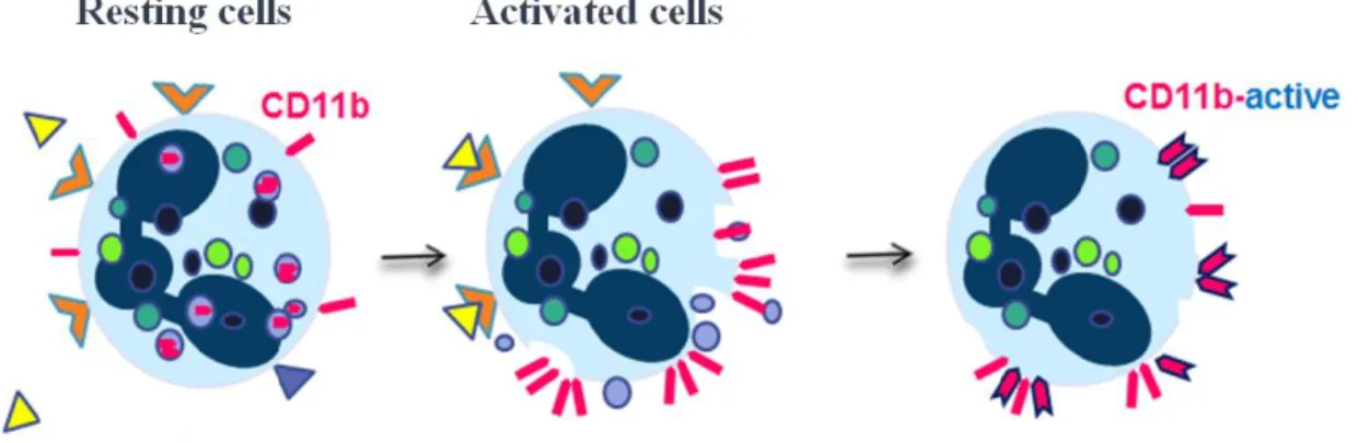
1.3.1.2 CD11b expression on neutrophils
CD11b joins CD18 to form the heterodimeric glycoprotein Mac-1, which is expressed on neutrophils, monocytes, macrophages and NKC. The relative abundance of the molecule on the surface of the cells depends on the cell type and state of activation. There are remarkable
intracellular storage pools within granules of circulating neutrophils. Under resting conditions, the CD11b/CD18 molecules are randomly distributed on the surface of the cells (Fig. 2). When the neutrophil becomes activated, a rapid translocation of granules to the surface results in a tremendous increase in the surface expression and aggregation of CD11b/CD18 (Arnaout 1990). Moreover conformational changes of the CD11b molecule will take place and establish an activation epitope to bind to the ligands such as ICAM and RGD sequences of ECM proteins with a high affinity. CD11b/CD18 interactions lead to adhesion and trans-endothelial migration, chemotaxis, phagocytosis of opsonized particles, and degranulation (Schleiffenbaum, Moser et al. 1989; Le Cabec, Carreno et al. 2002; Feng, Zhang et al. 2011).
Figure 2. Neutrophil activation with stimuli, translocation of CD11b from granules to the surface of the cells, and conformational changes in CD11b (CD11b-active epitope).
1.3.1.3 C5aR expression on neutrophils
The complement system is a part of innate immunity and consists of a variety of serum proteins. Following exposure to PAMPs, a cascade of proteolytic activities occurs, which leads to cleavage of downstream molecules. C5a is one of the complement fragments with different roles, as a conventional anaphylatoxin and a potent neutrophil and macrophage chemoattractant (Snyderman, Pike et al. 1975). A high level of C5a is observed in inflammatory conditions. C5a interacts with its receptor (C5aR or CD88) on the surface of the cells such as neutrophils and induces upregulation of adhesion molecules, release of granule enzymes, and oxidative burst (Mollnes, Brekke et al. 2002; Perianayagam, Balakrishnan et al. 2002; Kourtzelis, Markiewski et al. 2010).
1.3.2 Neutrophils in CKD patients
A wide range of functional aberrations in the circulatory neutrophil has been observed in the CKD patients. Neutrophils present a declined phagocytic capacity with increasing levels of
renal impairment (Mahajan, Kalra et al. 2005; Sardenberg, Suassuna et al. 2006), which improves with hemodialysis (Vanholder, Ringoir et al. 1991). They have a significantly lower antimicrobial killing activity (Anding, Gross et al. 2003). High levels of plasma light chains in advanced CKD can inhibit the chemotaxis of neutrophils (Cohen, Haag-Weber et al. 1995). A high oxidative burst activity is present in neutrophils from CKD patients, indicating a primed state of these cells in the uremic milieu, a systemic oxidative stress, and systemic inflammation (Klein, McLeish et al. 1999; Sela, Shurtz-Swirski et al. 2005).
It has been shown that neutrophils from CKD patients have a preserved capacity in terms of transendothelial migration, but their expression of the adhesion molecule CD11b is significantly reduced at the site of inflammation (Jacobson, Thylen et al. 2002; Dadfar, Lundahl et al. 2004). In the course of hemodialysis, CD11b expression on circulatory neutrophils changes, using a variety of dialyzers with different surface materials (Bonomini, Sirolli et al. 1999), and permeability properties (Moshfegh, Jacobson et al. 2002). Yet, a more biocompatible dialyzer may preserve the transmigration properties (Moshfegh, Jacobson et al. 2002), and may contribute to a conserved CD11b expression on neutrophils (Olsson, Dadfar et al. 2007).
Dialyzer composition has a significant impact on C5a generation (Zhang, Liu et al. 2011). Higher levels of C5a are observed following hemodialysis (Erlenkotter, Endres et al. 2008; Itoh, Takeshita et al. 2008; Inoshita, Ohsawa et al. 2012). Excess stimulation of neutrophils with C5a can lead to downregulation of C5aR due to cleavage of the receptor (van den Berg, Tambourgi et al. 2014), and therefore an impaired complement-dependent phagocytosis and impaired neutrophil function.
Yet, it is noteworthy that there are many contrasting data in terms of neutrophil behavior and expression of different molecules and receptors. This could originate in differences between studied individuals, the stage of renal disease, present comorbidities, treatment modalities, diverse methods and different conditions of the experiments (Hirabayashi, Kobayashi et al. 1988; Iida, Umezawa et al. 1997; Klein, McLeish et al. 1999; Gastaldello, Husson et al. 2000; Pereira, Costa et al. 2010).
1.4 CKD AND BASOPHILS
1.4.1 Basophil biology
Basophils belong to the granulocyte family and constitute less than 1% of circulating leukocytes. They are traditionally known for the storage and release of histamine and leukotrienes C4 (LTC4), involved in allergic reactions and anaphylaxis by induction of
vascular reaction, exudation, and leukocyte accumulation, as well as eradicating helminthes. The mediators are released following cross-linking of immunoglobulin (Ig) E receptors (FcεRI) on basophils, by interaction of receptor-bound IgE with corresponding antigen. However, in recent years, we have learned that the role of basophils is not limited to IgE-mediated reactions. They express a wide range of receptors such as toll-like receptors (TLR)-
2 and 4, IL-3R, IL-33R, C5aR, and chemokine receptors. They are also involved in the regulation of other immune cells (Rivellese, Suurmond et al. 2014) and release cytokines such as IL-4 and IL-13. Furthermore, they have the capacity to induce Th2 immune responses and induce the Ig class switching in B lymphocytes (Denzel, Maus et al. 2008; Schneider, Thieblemont et al. 2010; Sokol and Medzhitov 2010; Karasuyama, Mukai et al. 2011). They are also introduced as antigen-presenting cells for T cells (Sokol, Chu et al. 2009).
There are low affinity receptors for IgG on basophils, FcγRIIA (activating) and FcγRIIB (inhibiting) with which the cell activation deviates from IgE-dependent path (Cassard, Jonsson et al. 2012), and promotes vascular permeability, anaphylaxis and inflammation through release of platelet-activating factor (PAF).
1.4.1.1 FcεRI-mediated activation in basophils
FcεRI is a high-affinity IgE receptor, which plays an important role in activation of basophils. Binding of a multivalent antigen (allergen) to several FcεRI-bound IgE molecules results in aggregation of receptors (cross-linking), initiation of activation and degranulation. Expression of FcεRI is dependent on the level of IgE in the serum. Higher levels of IgE positively correlate to a higher density of the receptor on the basophil surface (MacGlashan, Bochner et al. 1997).
1.4.1.2 N Formyl- Methionyl-Leucyl-Phenylalanine (fMLP) response in basophils
FMLP is a peptide, with both endogenous (mitochondrial proteins from ruptured cells) and exogenous source (bacterial proteins). The interaction of fMLP with its G protein-coupled receptor Formylpeptide receptor (FPR), and variant FPR-like 1 and 2 (FPRL 1, 2) generates signals to induce chemotaxis and release of histamine and leukotrienes from basophils in an IgE-independent manner (MacGlashan, Bochner et al. 1997; Miura and MacGlashan 2000; de Paulis, Montuori et al. 2004).
1.4.1.3 Identification and activation marker
Different surface markers are used for the identification of basophils. CD203c is a glycosylated type II transmembrane molecule, which is expressed exclusively on basophils, mast cells and their precursor cells. Moreover, expression intensity of the molecules is low on resting basophils, but becomes upregulated on activation. Therefore, it can be counted as both an identification and activation marker (Buhring, Streble et al. 2004).
Another marker to identify basophil activation is CD63 (Knol, Mul et al. 1991). CD63 is the most important and applicable activation marker in basophils, which co-exists in the same granule as histamine. Therefore, presence of CD63 on the surface of basophils correlates to basophil degranulation and release of histamine and leukotrienes (MacGlashan 2010).
1.4.2 Basophils in CKD patients
There is no clear picture as to whether basophils are affected by the course of CKD or contribute to the development of complications. Hemodialysis patients, with symptoms such as pruritus, show high levels of IgE antibodies as well as a higher level of histamine release in vitro, regardless of the dialyzer type (Cavaillon, Poignet et al. 1990). Hemodialysis can induce inflammatory mediators such as IL-1β, TNFα and anaphylatoxins that may affect basophil function in terms of histamine release (Subramanian and Bray 1987).
During the dialysis procedure, production of bradykinin may be triggered (Verresen, Fink et al. 1994), which in turn can activate the basophils in an IgE-independent manner and induce anaphylactoid reactions during the session (Ebo, Bosmans et al. 2006).
Interestingly, a high level of autoreactive IgE has been observed in patients with lupus nephritis together with an accumulation of basophils in the spleen and lymph nodes. These IgE antibodies may activate the basophils and induce their migration to lymphoid organs where they promote Th2 differentiation, and subsequently B-cell potentiation and further production of autoantibodies that convey enforcement of the disease (Charles, Hardwick et al. 2010).
1.5 CKD AND T LYMPHOCYTES
1.5.1 T cell biology
T lymphocytes are the mainstay of cell-mediated adaptive immunity. They carry a unique T-cell receptor (TCR) with diverse recognition repertoire. When the APCs; dendritic T-cells, macrophages, or B cells, present the antigenic peptides to T cells, an antigen-specific activation of naive cells occurs, which leads to the development of effector and memory T-cells. Effector cells can convey their function by direct destruction of the target (CD8+ T cells, or cytotoxic T cells) or by supporting recruitment, activation and maturation of other immune cells through secretion of cytokines (CD4+ T cells, or T helper cells). Moreover, T cells are involved in the development of tolerance and anti-inflammatory responses.
1.5.1.1 CD4+ T helper cells
Being activated, helper T cells differentiate into a variety of subsets, depending on the stimuli and the cytokines present in the milieu. These subsets were initially classified based on production of distinct signature cytokines. Later, the master regulators (transcription factors) were introduced, which not only promote the production of signature cytokines by the cell but also dampen the development of opposing lineage in some subsets. When polarized, T cells produce cytokines, which further reinforce the polarization toward that subset.
Traditionally Th1 and Th2 were recognized as the main subsets, but over time other phenotypes were introduced such as Th9, Th17, and Th22, and follicular helper T (Tfh) cells (Chang, Sehra et al. 2010; Basu, O'Quinn et al. 2012; Liu, Nurieva et al. 2013). Additionally, regulatory T cells (Treg) are known as the anti-inflammatory subset (Fig. 3).
Th1 cells are responsible for the activation of macrophages and B-cells through production of IFN-γ and contribute to the immune defense against intracellular microorganisms. The master regulator of Th1 cells, T-box transcription factor (T-bet), promotes the IFN-γ production as well as the suppression of Th2 and Th17 differentiation (Afkarian, Sedy et al. 2002; Djuretic, Levanon et al. 2007). Th2 cells, recognized by their cytokines IL-4, IL-5, IL-13, and transcription factor GATA-Binding Protein 3 (GATA3), are involved in immune reactions against extracellular pathogens, humoral immunity and allergic diseases (Zheng and Flavell 1997). Regulatory T cells are divided into natural thymus-derived subset (nTreg), and peripheral-induced Treg cells (iTreg). They play an important role in maintaining the immunologic tolerance by production of cytokines such as IL-10, which inhibits the pro-inflammatory responses, and suppresses allergic reactions together with TGF-β. The master regulator of Tregs is forkhead box P3 (FOXP3) (Sakaguchi, Ono et al. 2006; Ouyang, Rutz et al. 2011).
Interestingly, it has been shown that the antigen-experienced T cells are not restricted to one terminally differentiated subset, but rather retain some plasticity to acquire new characteristics and functions upon antigenic re-encounter. The factors and environmental characteristics promoting the plasticity or stability of human T cell subsets still remain to be understood (Geginat, Paroni et al. 2014).
1.5.2 T-cells in CKD patients
The disturbances in the cell-mediated immunity start in the early stages of renal failure and CKD patients with mild disease present a different immune pattern, compared to hemodialysis patients. There are two different overviews on what gives rise to these aberrations. There is a T cell dysregulation conception, indicating that abnormality in T cell function results from disturbances in antigen presentation and co-stimulation by APCs, and pro-inflammatory molecules in the milieu (Dinarello 1992; Girndt, Kohler et al. 1993; Kelly 1994). However, another conception claims that these disturbances originate in T cell dysfunction per se. Patients with renal failure exhibit changes in the proliferation and phenotype of T cells, and premature aging of the adaptive immune response (Raska, Raskova et al. 1983; Daichou, Kurashige et al. 1999; Nitta, Akiba et al. 2002; Alvarez-Lara, Carracedo et al. 2004; Costa, Lima et al. 2008; Betjes and Litjens 2015). Yet, there is no common view on these aberrations. In terms of proliferation capacity of T cells in response to different stimuli, there are mixed findings of being either normal or decreased. In some studies Th1 is introduced as the main effector T cell, while others pinpoint alternative phenotypes such as Th2 or Th17 (Sester, Sester et al. 2000; Libetta, Rampino et al. 2001; Yokoyama, Nitta et al. 2001; Zhang, Hua et al. 2010; Ma, Zhang et al. 2015). Variation in the proportion of FOXP3+ Tregs in patients has been observed from being normal to declined (Lisowska, Debska-Slizien et al. 2012). These aberrations have also been connected to the occurrence of complications in CKD patients. For instance, it has been shown that generation of oxidized LDL (oxLDL) in hemodialysis patients enhances the CD4+CD25+ Treg sensitivity to Fas-mediated apoptosis and leads to a decreased proportion of these cells. Meanwhile a Th1-mediated immune response occurs in atherosclerosis when oxLDL is uptaken by APCs in the vessels (Meier, Meier et al. 2008). The changes in T cell function can also affect the humoral immunity and B cell response, which causes low response to vaccinations (Dumann, Meuer et al. 1990). Moreover, the exacerbated immune activation and cytokine network constantly provoke the immune cells, and therefore they become too exhausted to respond to an acute challenge.
The current scattered data can again arise from differences in groups of patients, interventions, and analysis approaches.
1.6 GENE TRANSCRIPTION AND EPIGENETIC REGULATION IN CKD
As mentioned earlier, a defective immune system and a chronic inflammatory state are features of chronic renal failure. Retention of uremic toxins and the dialysis procedure are the main contributors to the occurrence of complications. Many efforts have been made to investigate and demonstrate the biological-cellular alterations, and molecular changes in these patients. Analysis of gene transcription and epigenetic regulations can contribute to an increased understanding of the biological mechanisms behind these aberrations.
Genomic techniques allow us to analyze the whole transcriptome of the cell. Following analysis of gene expression data, a number of significant genes are identified and enrolled to
different molecular pathways and functional categories. Comparison of gene expression profiles between patients and healthy individuals can provide us with valuable findings. Yet, it is difficult to choose the right candidates with clinical relevance for further validation (Perco, Pleban et al. 2006).
The genetic basis of many medical conditions has been studied, however gene polymorphisms are not the only explanation for different traits and complications. There are other factors that can regulate the gene expression and the eventual phenotype. These factors are known as epigenetic changes. Epigenetics comprises changes in gene expression without alteration in the primary genes and is mainly divided into DNA methylation, histone modification and RNA interference such as microRNAs (miRs). These modifications or interferences lead to a change in the level of target protein, and an altered phenotype (Egger, Liang et al. 2004).
1.6.1 Transcriptome analysis in CKD
The underlying biological mechanisms involved in dysfunction of the innate and adaptive immune system in CKD are of interest. There are reports on the transcriptomic profile of peripheral mononuclear immune cells in CKD patients and differences between treatment groups (Zaza, Pontrelli et al. 2008; Zaza, Granata et al. 2013). Polymorphism of different cytokine genes such as TGF-β and IL-6 has been connected to progression of kidney disease and complications (Girndt, Kaul et al. 2002; Rao, Guo et al. 2004; Liu, Berthier-Schaad et al. 2006). Genes involved in oxidative metabolism such as Superoxide dismutase-2 (SOD2) are shown to be expressed differently in activated neutrophils from CKD patients (Olsson, Jacobson et al. 2011).
1.6.2 Role of microRNAs in CKD
MicroRNAs are small non-coding RNAs, which interfere with the translation of mRNA by silencing or degrading them (Bartel 2009). They contribute to the regulation of a variety of cell functions such as proliferation, differentiation and apoptosis, and they are shown to be involved in the regulation of immune function (Chen, Li et al. 2004; Backdahl, Bushell et al. 2009).
Several miRs are presented to play a role in normal kidney function (Backdahl, Bushell et al. 2009), as well as in disease process (Badal and Danesh 2015). The dysregulation of selected miRs can lead to proteinuria, renal failure (Zhdanova, Srivastava et al. 2011), ESRD (Szeto, Ching-Ha et al. 2012), and cardiovascular complications (Ji, Cheng et al. 2007). Moreover, miR inhibition has been shown to have therapeutic effects in different groups (Macconi, Tomasoni et al. 2012; Putta, Lanting et al. 2012; Zhong, Chung et al. 2013).
The circulatory miRs are considered as non-invasive biomarkers for certain human diseases. In acute kidney failure, it has been reported that miRs are not removed during the hemodialysis procedure, perhaps due to transportation by larger proteins or microvesicles. Hence, miRs may be good candidates as biomarkers (Martino, Lorenzen et al. 2012).
However other studies have reported a declined concentration of miRs in more severe CKD patients and following hemodialysis, independent of miR removal by the kidneys or dialysis (Neal, Michael et al. 2011; Emilian, Goretti et al. 2012), probably due to enzymatic degradation. MicroRNAs are suggested as a good indicator for progression of peritoneal fibrosis in patients undergoing peritoneal dialysis (Ge, Xiao et al. 2012), and even as biomarker for cardiovascular changes in hemodialysis patients (Wen, Song et al. 2014). Yet, there is a great need for studies in CKD patients to clarify the profile of miRs and their potential as biomarkers in these patients.
1.7 IMPACT OF MEDICATIONS IN CKD
An important factor, which can impact the analysis and interpretation of experimental data, is the patient selection. Most CKD patients take medications to manage complications such as high blood pressure, anemia, low levels of Vitamin D or high levels of PTH, and so forth. Some of these medications have been proposed to have an impact on the immune system, both negatively and positively. The later may be a treatment advantage to not only be applied for correction of the targeted abnormalities, but also to improve the immunity status in CKD patients.
1.7.1 Angiotensin-converting enzyme inhibitor (ACE-I) and angiotensin II receptor blocker (ARB)
These medications are the first choice for treatment of high blood pressure in CKD patients, particularly in patients with proteinuria (Kunz, Friedrich et al. 2008). They have been proposed to better preserve renal function in patients with CKD (Wright, Bakris et al. 2002; Hou, Zhang et al. 2006). Moreover, further studies have revealed a beneficial effect of these medications to diminish inflammation (Fliser, Buchholz et al. 2004), to augment anti-inflammatory T cells, and to modulate anti-inflammatory T cells (Platten, Youssef et al. 2009).
1.7.2 Erythropoiesis-stimulating agent (ESA)
CKD patients suffer from anemia and therefore ESAs are often prescribed in these patients. Studies have shown that ESAs, such as recombinant human erythropoietin, can decrease inflammation (Bryl, Mysliwska et al. 1998), enhance proliferation and antibody production of activated B cells (Kimata, Yoshida et al. 1991), and improve the activation and proliferation capacity of CD4+ T cells (Lisowska, Debska-Slizien et al. 2010).
1.7.3 Vitamin D
CKD patients show a disturbance in bone mineral metabolism, including hypocalcemia, hyperphosphatemia, high PTH, and reduced levels of active vitamin D. Therefore, many patients are treated with phosphate binders and vitamin D analogues. Studies indicate that vitamin D has broader advantages than merely regulation of calcium and phosphate homeostasis. Reduction of albuminuria and cardiovascular events in ESRD patients has been reported following treatment with vitamin D (Alborzi, Patel et al. 2008; Lishmanov,
Dorairajan et al. 2013). Moreover, a positive impact of vitamin D supplementation on endothelial function has been suggested (Dreyer, Tucker et al. 2014; Zoccali, Curatola et al. 2014; Lundwall, Jorneskog et al. 2015).
Currently, we know that the active form of vitamin D is not only generated in the kidneys but also in many other cells including immune cells (Aranow 2011), which in turn interacts with the vitamin D receptor (VDR) and regulates their function. The VDR is a nuclear receptor, which affects the expression of a range of genes following activation (Hossein-nezhad, Spira et al. 2013). It can induce both inflammatory responses such as antimicrobial peptide production and bacterial killing, and anti-inflammatory responses by suppression of pro-inflammatory cytokines, induction of anti-pro-inflammatory cytokines, and Th2 and regulatory T cell differentiation (Vojinovic 2014).
There are reports suggesting an association between vitamin D levels and inflammation and the role of vitamin D treatment in complications such as cardiovascular diseases (Chitalia, Ismail et al. 2014; Dreyer, Tucker et al. 2014; Yin and Agrawal 2014). Yet many questions remain unanswered concerning the role of vitamin D in controlling inflammation in CKD.
2 AIMS OF THE THESIS
The overall aim of this thesis is to increase the understanding of how the immune system operates in CKD patients and to address key issues in terms of immune insufficiency and inflammation in CKD patients.
This thesis comprises four studies and their respective aims are:
Study I. To investigate whether blood-membrane interaction has any impact on the human peripheral basophils and neutrophils in hemodialysis patients.
Study II. To study the lymphocyte proliferation response and immune modulator production, following in vitro stimulation of cells from pre-dialysis and hemodialysis patients.
Study III. To investigate the profile of CD4+ T cell subsets and cytokine production, following in vitro stimulation in hemodialysis patients.
Study IV. To assess the effect of vitamin D receptor activation on the pro-inflammatory profile and up-stream gene expression regulators, micro RNAs, in moderate CKD.
3 MATERIAL AND METHODS
This section comprises a description of the study participants (patients and healthy individuals), and the methodology used in the studies of this thesis. A more detailed description can be obtained within the ‘Material and Methods’ section of each paper (I-IV). 3.1 STUDY POPULATION
3.1.1 Patients
Patients in studies I, II, and III were recruited from the Department of Nephrology at Karolinska University Hospital Solna, Stockholm, Sweden. Written informed consent was obtained from all participants and the local Ethics Committee in Stockholm, Sweden, approved all the studies. Patients in study IV were recruited from the Department of Nephrology at Danderyd University Hospital, Stockholm, Sweden. All patients provided written informed consent. The regional Ethics Committee in Stockholm, Sweden, approved the study protocol and the clinical trial was registered on clinicaltrials.gov (SOLID study; NCT01204528).
None of the patients had been diagnosed with cancer, ongoing infection, chronic inflammatory and rheumatologic diseases, or received treatment with immunosuppressive drugs.
3.1.1.1 Study I
Patients (n=10) with an eGFR <20 mL/min/1.73 m2 (stage 5D) with a residual GFR from 6-11 mL/min/1.73 m2 were recruited. Patients were undergoing hemodialysis with polysulfone high-flux dialyzers (effective surface area of 2.3 m2, K0A urea: 1421 mL/min, ultrafiltration
coefficient: 76 mL/h/mm Hg), three times per week for four to four and a half hours per dialysis before the study. An arteriovenous fistula or a central dialysis catheter was used for dialysis.
3.1.1.2 Study II
Patients (n=14) with an eGFR <20 mL/min/1.73 m2 (predialysis, CKD stage 4 and 5), and patients (n=14) with eGFR <20 mL/min/1.73 m2 undergoing hemodialysis were recruited. Hemodialysis was performed three times per week for four to four and a half hours per dialysis with polysulfone high-flux dialyzers. Residual GFR ranged from 4-12 mL/min/1.73 m2 (median 8.5). Dialysis was done through an arteriovenous fistula, graft or a central dialysis catheter (n=4).
3.1.1.3 Study III
Patients (n=10) with CKD stage 5D were recruited. Patients were undergoing maintenance hemodialysis for four hours/session, three times per week, using polysulfone high-flux
dialyzers. The residual GFR ranged from 4-12 mL/min/1.73 m2 (median 8.5). Dialysis was done via an arteriovenous fistula, graft or a central dialysis catheter.
3.1.1.4 Study IV
This was a sub-study of a clinical trial, the SOLID-trial. Patients (n=36) with eGFR of 15-59 mL/min/1.73 m2 (CKD stage 3 and 4), a calcium level below 2.6 mmol/L, and a plasma PTH level of 3.7-53 pmol/mL were recruited in three groups: placebo (n=12), 1 µg paricalcitol (n=12), or 2 µg paricalcitol (n=12) daily, in a randomized, double-blinded manner. Further patient characteristics are shown in Table 1.
Male n (%) Age (y), range (median) CVD (n) ACE-I/ARB (n) ESA (n)
Study I (n=10) 5 (50) 37–76 (62.5) Heart disease (4)
Hypertension (9) 5 9 Study II Predialysis (n=14) Hemodialysis (n=14) 9 (64) 7 (50) 33-65 (51) 36-66 (53.5) Heart disease (1) Hypertension (14) Heart disease (3) Hypertension (7) 13 4 6 12
Study III (n=10) 5 (50) 62-83 (71) Heart disease (5)
Hypertension (5) 3 9 Study IV Placebo (n=12) 1 μg paricalcitol (n=12) 2 μg paricalcitol (n=12) 9(75%) 11(92%) 8 (67%) mean (SD) 70.8 (10.0) 66.1 (7.9) 59.1 (11.6) Heart disease (5) Hypertension (3) Heart disease (3) Hypertension (4) Heart disease (1) Hypertension (4) 11 9 9 ND ND ND
Table 1. Demographic characteristics of participants at baseline in study I-IV, CVD: Cardiovascular disease, ACE-I: angiotensin-converting enzyme inhibitor, ARB: angiotensin II receptor blocker, ESA: erythropoiesis-stimulating agent, SD: standard deviation, ND: not determined.
3.1.2 Healthy controls
In study I, II and III, the healthy controls were recruited among healthy blood donors and were age and sex-matched (±5-7 years) with the patients.
3.2 STUDY METHODS
3.2.1 Study I
To perform the study, the dialyzer was shifted from high-flux to a low-flux polysulfone capillary dialyzer (effective surface area of 1.8 m2, K0A urea: 976 mL/min, ultrafiltration
coefficient: 14 mL/h/mm Hg), in one session of treatment. Blood samples were collected before and after the dialysis procedure in each session (one occasion with a high-flux and one occasion with a low-flux dialyzer).
3.2.1.1 Basophil activation
Detection of surface expression of CD203c and CD63 on basophils was done by flow cytometry (Navios, Beckman Coulter), following treatment of whole blood with stimulators fMLP (Sigma Aldrich), and anti-FcεRI antibody (Bühlmann Laboratories).
3.2.1.2 Counting of basophils
Absolute number of basophils was determined using the ImmunoPrep reagent system (Beckman Coulter), cell-counting beads (Flow-Count Fluorospheres, BeckmanCoulter), and flow cytometry (Navios).
3.2.1.3 Neutrophil activation
Detection of surface expression of CD11b and active CD11b on neutrophils was done by flow cytometry (Navios), following treatment of whole blood with IL-8 (CXCL8) (Research & Diagnostics Systems).
3.2.1.4 Expression of CD88 (C5aR) on neutrophils
Whole blood was incubated with anti-CD88 antibody (Becton Dickinson), and expression of CD88 was detected by flow cytometry (Navios).
3.2.2 Study II
3.2.2.1 Cell culturing with mitogens
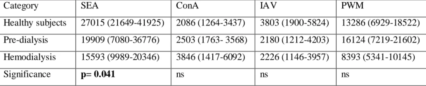
Whole blood was diluted in RPMI 1640 medium (Gibco) containing 2 mM L-glutamine (Gibco), 10 000 IU/mL Penicillin (Gibco) and 10 000 µg/mL Streptomycin (Gibco). 1 µg/mL of Pokeweed mitogen (PWM, Aldrich), 1 µg/mL of Concanavalin A (Con A, Sigma-Aldrich), 100 ng/mL of Staphylococcus enterotoxin A (SEA, Sigma-Sigma-Aldrich), and Influenza A vaccine (IAV, Fluarix, GlaxoSmithKline AB) diluted 1:100, were added to blood-medium suspensions, and incubated at 37°C, 5 % CO2 for six days.
3.2.2.2 Cell counting
Cell count was performed using cell-counting beads (Flow-Count Fluorospheres), and flow cytometry (Navios).
3.2.2.3 Flow cytometric Assay of Specific Cell-mediated Immune response in Activated whole blood (FASCIA)
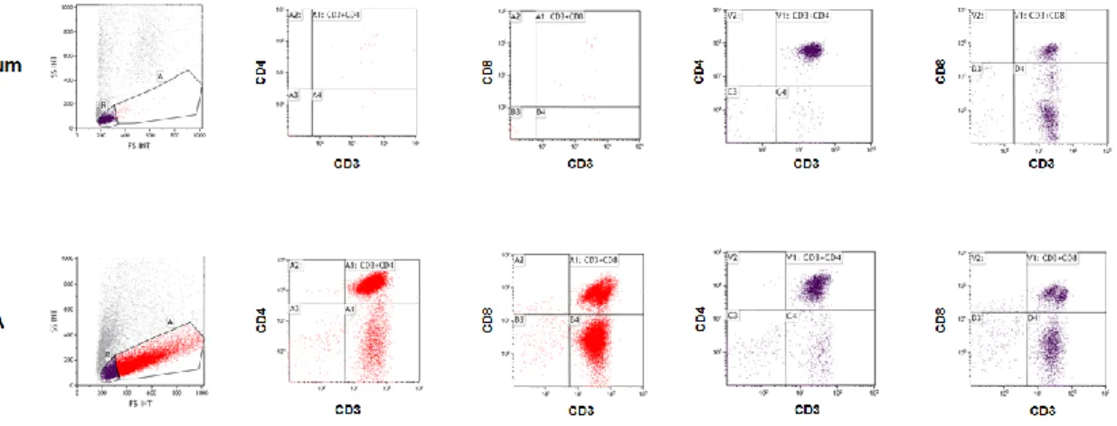
This method was applied for the detection of T lymphocyte proliferation and lymphoblast formation. Following staining with anti-CD3, -CD4, -CD8 and -CD45 (CYTO-STAT, tetra CHROME, Beckman Coulter), cells were analyzed by flow cytometry (Navios) (Fig. 4).
Figure 4. Flow cytometric analysis of cell-mediated immune response in the activated whole blood. The CD45+ resting (blue color) and activated leukocytes (red color) from medium, and staphylococcus enterotoxin A (SEA)-stimulated cultures were gated according to their size and granularity on forward and side scatter plot. The CD3+CD4+ cells and CD3+CD8+ cells were then detected within the resting and lymphoblast population accordingly.
3.2.2.4 Characterization of pro-inflammatory molecules
Milliplex 26-plex (Millipore Corp.) was applied for measuring a broad range of cytokines and chemokines: eotaxin, granulocyte macrophage colony-stimulating factor (GM-CSF), granulocyte colony-stimulating factor (G-CSF), IFN-α2, IFN-γ, 1α, 1β, 2, 3, IL-4, IL-5, IL-6, IL-7, IL-8, IL-10, IL-12 (p40), IL-12 (p70), IL-13, IL-15, IL-17, interferon gamma-induced protein (IP)-10, monocyte chemoattractant protein (MCP)-1, macrophage inflammatory protein (MIP)-1α, MIP-1β, TNF-α and TNF-β (Lymphotoxin α) in plasma and culture supernatants.
3.2.3 Study III
3.2.3.1 Isolation of CD4+ cells
Peripheral blood mononuclear cells (PBMCs) were isolated from whole blood by density-gradient centrifugation (Ficoll-Paque PLUS; GE Healthcare) and CD4+ cells were isolated by positive selection using a magnetic cell-sorting system (MACS, Miltenyi)
3.2.3.2 Cell count and purity check
Cell count was performed using cell-counting beads (Flow-Count Fluorospheres) and flow cytometry (Navios). CD3+CD4+ (Becton Dickinson) staining was applied to check the purity of isolated cells.
3.2.3.3 Cell culturing with superantigen; Staphylococcus enterotoxin A
AIM-V medium (Life technologies), with or without addition of SEA, was utilized to culture the cells in vitro.
3.2.3.4 Profiling the CD4+ T cells
Cells were stained with Live/Dead Fixable Near-IR Dead cell stain kit (ThermoFisher scientific Inc.), anti-CD4 antibody (Becton Dickinson) and anti-CD25 (Becton Dickinson). Intracellular analysis of transcription factors by flow cytometry (Navios) was carried out, following fixation and permeabilization of cells using FOXP3/Transcription Factor staining buffer set kit (eBioecience Inc.), and staining with either T-bet (eBioecience Inc.) anti-GATA3 (Becton Dickinson) or anti-FOXP3 (Becton Dickinson).
3.2.3.5 Characterization of soluble immune modulators in supernatants
ELISA was applied to determine the concentration of cytokines: IFN-γ, IL-4, and IL-10 in supernatants (R&D Systems, USA).
3.2.4 Study IV
The study started with two weeks of placebo, followed by 12 weeks of intervention with placebo, 1 µg paricalcitol, or 2 µg paricalcitol daily. Blood samples were drawn at baseline and after 12 weeks, after 12 hours fasting and 20 min rest.
3.2.4.1 RNA isolation from plasma
The miRCURYTM RNA isolation kit Biofluids, (EXIQON) was applied for purification of total RNA, containing RNAs smaller than 1000 nt (such as miRNAs) from the plasma.
3.2.4.2 Reverse transcription polymerase chain reaction (RT-qPCR) for profiling of microRNA expression
Exiqon miRCURY Ready-to-Use PCR Human panel I + II V1.M (Exiqon miRNA qPCR panel) was applied to identify the potentially affected plasma microRNAs. RNA spike-in for quality control of the RNA isolation and cDNA synthesis was applied. RNA isolation control (UniSp2, UniSp4, UniSp5), cDNA synthesis control (UniSp6), and DNA spike-in (UniSp3) were also included.
3.2.4.3 Reverse transcription polymerase chain reaction (RT-qPCR) for validation of microRNA expression
Universal cDNA synthesis kit II (miRCURY LNATM Universal RT microRNA PCR, EXIQON), was applied for reverse transcription to cDNA. ExiLENT SYBR Green master mix kit (miRCURY LNATM Universal RT microRNA PCR, EXIQON) was used for RT-qPCR. PCR reaction was performed with MicroAmp™ optical 384-well reaction plates using an ABI 7900 (Life Technology).
3.2.4.4 Characterization of soluble immune modulators in plasma
A broad range of cytokines was measured by Milliplex 26-plex (Millipore Corp.) in plasma at the baseline, and following 12 weeks of treatment.
3.3 STATISTICAL ANALYSIS
Statistical analysis in studies I-IV was done in GraphPad Prism 5 (GraphPad Software), STATISTICA version 10 (Stat Soft, Inc.), and SPSS version 22 (IMB, 2011). One-way ANOVA and repeated measures ANOVA was applied for analysis of normally distributed data (study IV). Wilcoxon matched-pairs signed-rank test for two dependent measurements (study I, IV), and Mann-Whitney U test for two independent sample groups (study III, IV) were applied to analyze non-normally distributed values. Nonparametric Kruskal-Wallis test was carried out for comparison of three independent measurements (study I, II, IV).
4 RESULTS AND DISCUSSION
4.1 ALTERATION IN BASOPHIL ACTIVATION: A POTENTIAL MARKER FOR ASSESSMENT OF BIOCOMPATIBILITY IN HEMODIALYSIS (STUDY I) Hemodialysis is required to remove excess electrolytes, fluid and uremic toxins in ESRD. However, interaction between peripheral blood and the surface of the dialyzer membrane may lead to alterations in the cells and plasma proteins (Rao, Guo et al. 2004; Banche, Allizond et al. 2006). This so-called bioincompatibility is dependent on the surface material and the permeability properties of the membrane. In this study, we aimed to investigate the impact of this interaction on the activation of human peripheral basophils and neutrophils in patients undergoing dialysis with high- and low-flux polysulfone dialyzers.
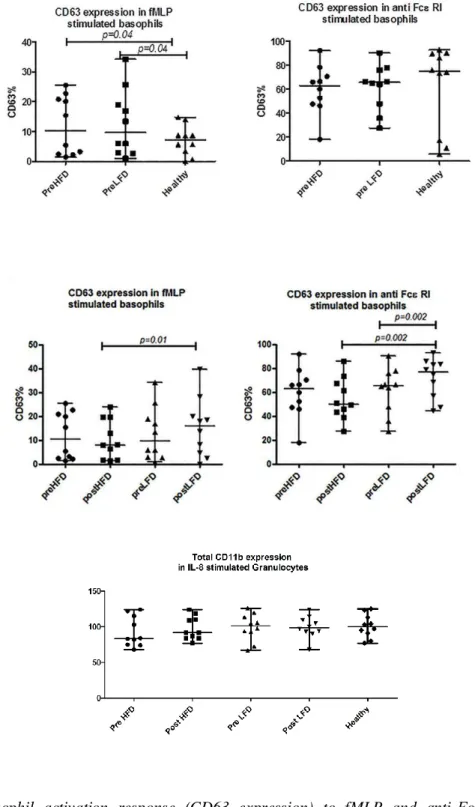
After stimulation with fMLP and anti-FcεRI antibody, basophil CD63 expression was analyzed since its expression represents degranulation of basophils and subsequent histamine release. Samples were obtained before each session of dialysis with high-flux or low-flux dialyzers, and compared to those from healthy controls. The fMLP-activated basophils from patients expressed significantly higher levels of CD63 compared with those in healthy controls (p= 0.04) (Fig. 5, A). However, this was not the case following stimulation with anti-FcεRI antibody.
We observed a higher response of basophils to fMLP in hemodialysis patients compared to healthy individuals. It is not clear whether this increased responsiveness is a consequence of altered fMLP receptor (FPR or FPRL1) expression, or whether the intracellular pathways (MEK–ERK pathway) involved in degranulation are primed in basophils from CKD patients (Miura, Schroeder et al. 1999; de Paulis, Montuori et al. 2004). Therefore, it is noteworthy to further analyze the mechanistic alterations involved in fMLP activation of basophils.
The surface expression of CD63 on basophils was significantly increased after dialysis with low-flux dialyzers compared with high-flux dialyzers in both fMLP (p = 0.01) and anti-FcεRI antibody-activated cells (p= 0.002) (Fig. 5, B). Comparing predialysis and postdialysis samples, significantly higher CD63 expression was observed on anti-FcεRI antibody-activated basophils, following hemodialysis with low-flux membranes (p= 0.002) (Fig. 5, B).
Moreover, we found a similar absolute number of basophils, following hemodialysis with high-flux and low-flux dialyzers compared to that in healthy controls. This indicates that the differential basophil response was due to functional changes, and not the entrapment of low-responding cells in the dialyzer.
Figure 5. Basophil activation response (CD63 expression) to fMLP and anti-FcεRI antibody comparing patients (before dialysis) with healthy controls (A), CD63 expression in activated basophils following dialysis with low-flux (LFD) and high-flux dialyzers (HFD)(B), and total CD11b expression on neutrophils after stimulation with IL-8 (C).
The differences in FcεRI-mediated activation of basophils following passage through the low-flux dialyzer is of interest since anaphylactic reactions have been reported in patients undergoing hemodialysis. This strong response may arise from the cross-linking of Fcε receptors on the basophils and release of mediators such as histamine and Heparin (Ebo, Bosmans et al. 2006). It has also been shown that different membrane surfaces can lead to activation of Hageman factor and bradykinin which in turn may activate basophils in an
IgE-A.
B.
C.