Quis cusîodiet ipsos Custodes?
Who is to guard the guards themselves?
Tolerance Status of Transgenic Mice Expressing a
Major Histocompatibility Complex Class I Molecule
under the Control of Human CD2 Regulatory
Elements
Ruth Margaret Schulz, BSc.
Molecular Immunology
National Institute fo r Medical Research
London
ProQuest Number: 10017249
All rights reserved
INFORMATION TO ALL USERS
The quality of this reproduction is dependent upon the quality of the copy submitted.
In the unlikely event that the author did not send a complete manuscript and there are missing pages, these will be noted. Also, if material had to be removed,
a note will indicate the deletion.
uest.
ProQuest 10017249
Published by ProQuest LLC(2016). Copyright of the Dissertation is held by the Author.
All rights reserved.
This work is protected against unauthorized copying under Title 17, United States Code. Microform Edition © ProQuest LLC.
ProQuest LLC
789 East Eisenhower Parkway P.O. Box 1346
In memory of
Thomas W. Sanderson
Acknowledgements
There are a large number of people who have enabled me to complete my PhD. by assisting me in a number of ways. Firstly, thank you to erstwhile members of Lab 358: Anne-Marit Sponaas for limitless help and advice, and for showing me the mysteries of a CTL assay; Pete Tomlinson and Janey Antoniou for patiently taking CD2K^ tails and for being good company. Many thanks to Andy Mellor for supervision and ideas, and to Tolja Horuszko for reading grafts for me - good luck in the States!
I am also indebted to colleagues from the MRC Clinical Science Centre - Prof. Liz Simpson for advice and ideas, Maggie Millrain for supplying antibodies, and Kevin Corley for being willing to give up an afternoon to stain dead cells. Many thanks to Steve Cobbold at William Dunn School of Pathology in Oxford for antibodies and advice, and to Diana Zelenika for help with TCR primers.
I would like to thank Chris Atkins for his help with FACSplot and sterile sorts, and the NIMR peptide synthesis facility for producing K^[80-92] extra quickly. Rose Zamoyska, Gitta Stockinger and Ann Ager generously donated antibodies, reagents and advice, which I really appreciate. Many thanks also to the MI technical staff in general, without whom life as a PhD. student in MI would be infinitely more arduous, and congratulations and thanks to everyone who stayed awake in my Tuesday meetings.
Contents
Contents: Figures and Tables 11
Abstract 14
Chapter 1 Immunological Tolerance
1.1 Introduction 15
1.1.1 Definitions of immunological tolerance 15
1.1.3 Tolerance and Autoimmunity 16
1.2 Lymphocyte Development 17
1.2.1 Lymphocytes must interact with cells in the bone
marrow or thymus to mature 17
1.2.2 Lymphocyte receptors are generated by somatic gene
rearrangement 17
1.2.3 Somatic hypermutation 19
1.2.4 Isotype switching selects antibody effector function 19 1.2.5 Co-receptor selection affects T cell effector function 20
1.2.6 Thymocyte development 21
1.3 B Cell Tolerance 21
1.4.2 Central tolerance - the mechanism of clonal deletion 27
1.4.3 Theories of clonal deletion 28
1.4.4 The differential affinity model of clonal selection 29 1.4.5 Further mechanisms of central tolerance 30
1.4.6 Peripheral tolerance 30
1.4.7 Clonal deletion in the periphery 31
1.4.8 Anergy in the peripheiy 32
1.4.9 Unresponsiveness towards self antigen 33
1.4.10 Ignorance of self antigen 34
1.4.11 Tolerizing signals may be cumulative 36
Positive Selection 38
1.5.1 Does positive selection occur ? 38 1.5.2 Mechanisms of positive selection 40 1.5.3 The nature of the positively selecting cell 41 1.5.4 The role of co-receptor molecules in positive
selection 42
1.5.5 Summary 43
1.6 The Thesis 45
C h ap ter 2 M aterials and M ethods
2.1 Introduction 47
2.2 Disposable Plastics 47
2.3 Media and Solutions 47
2.3.1 IMDM 47
2.3.2 Air-buffered wash medium 48
2.3.3 Lysis solution 48
2.3.7 Lysis buffer 49
2.3.8 lOXTfl^ buffer 49
2.3.9 lOOX Denhardt's solution 49
2.3.10 Hybridization mix 49
2.3.11 20X Saline sodium citrate 50
2.3.12 lOXTBE 50
2.3.13 Alkaline blotting buffer 50
2.3.14 Neutral blotting buffer 50
2.3.15 lOX Nick mix 50
2.3.16 Tris.EDTA 50
2.3.17 Balanced saline solution 51
2.3.18 ELISA wash buffer 51
2.3.19 Substrate buffer 51
2.3.20 Citrate Buffer 51
2.3.21 Trypsin versene 51
2.4 Experimental Animals 53
2.4.1 Supply and maintenance 53
2.5 DNA Preparation 55
2.6 Genotype Analysis 55
2.6.1 Southern blots 55
2.6.2 Slot blots 56
2.6.3 Production of radioactive probes 57
2.6.4 Polymerase Chain Reactions 57
2.7 Phenotype Analysis - Flow Cytometry 60
2.7.1 Cell samples 60
2.8.2 Biotinylation of monoclonal antibodies 62 2.9 Cell Culture Techniques and Assays 62
2.9.1 Mixed lymphocyte culture 62
2.9.2 Con A blasts 63
2.9.3 Cytotoxic T cell assay 63
2.9.4 Differentiation of bone marrow in vitro 64 2.9.5 Production of GM-CSF supernatant 65
2.10 Skin Grafting 65
Chapter 3 Inheritance and Expression of the CD2K*^ Transgene
3.1 Introduction 68
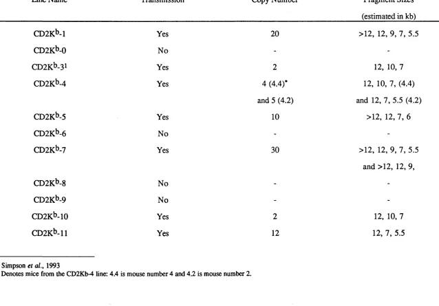
3.2 Establishment of CD2K^ Transgenic Mouse lines 69 3.2.1 Identification of transgenic founders 70 3.2.2 Southern blot analysis of CD2K^ transgenic mice 70 3.2.3 Slot blot analysis of CD2K^ transgenic mice 71 3.3 Phenotype Analysis of CD2K^ Transgenic Mice 78 3.4 Analysis of Copy-number Dependency of H-2Kt> Expression 79 3.5 Selection of CD2K^ Lines for Further Breeding 80
3.6 Discussion 81
Chapter 4 H-2K*^ Expression in CD2K^ Transgenic Mice
4.1 Introduction 87
4.2 Specialized Materials and Methods 89
4.4 Expression of H-2K^ on Bone Marrow-derived Dendritic
cells and Macrophages 96
4.4.1 Staining dendritic cells and macrophages for H-2K^ 96 4.4.2 IFNy release assays for H-2K^ expression 97 4.5 Expression of H-2K^ on Thymic Dendritic Cells 103 4.6 Expression of H-2K^ on Migratory Langerhans' Cells 104 4.7 Expression of H-2K^ on Skin Cells 105
4.8 Discussion 111
Chapter 5 Thymocyte Development in CD2K^ Mice
5.1 Introduction 116
5.2 Negative Selection of the BM3.6 TCR in CD2K^ Mice 117 5.2.1 CD2K^ X BM3.6 FI mice have reduced thymus size 118 5.2.2 Thymocytes bearing self-reactive TCRs are deleted in
CD2Kb X BM3.6 FI mice 118
5.3 Negative Selection of the DES TCR in CD2K^ Mice 122 5.3.1 CD2K*) X DES FI mice have reduced thymus size 122 5.3.2 Thymocytes bearing self-reactive TCRs are deleted in
C D 2K bxD E S F l mice 123
5.4 Are Thymocytes Capable of Positive Selection? 132 5.4.1 The 2C TCR is inefficiently selected in CD2K^-7 x
2C FI mice 132
5.4.2 The 2C TCR is not selected in CD2K^-3 x 2C or
CD2kM x2CF1 mice 133
Chapter 6 t Cell Responses to H-2K*^ in vitro by CD2K** Mice
6.1 Introduction 143
6.2 Assays for Cytotoxicity Mediated by Splenocytes from
CD2KbMice 143
6.3 Transgene Copy Number Affects the Survival of Self-reactive
T Cells 145
6.4 Discussion 152
Chapter 7 Response to H-2K^ in vivo by CD2K^ Transgenic Mice
7.1 Introduction 155
7.2 Specialized Materials or Methods 156
7.2.1 In vivo priming 156
7.2.2 Generation of LPS blasts 156
7.3 CD2K^ Mice Reject CBK Skin Grafts 157 7.4 CD2K^> Mice Accept KAL Skin Grafts 158 7.5 Primed Mice do not Respond to H-2K^ on MHC 11+ Cells in
vitro 158
7.6 Discussion 163
Chapter 8 CD2K*^ CD4+8- T Cell Responses to an H-2K^ Peptide on MHC II
8.1 Introduction 166
8.2 Specialized Materials and Methods 169 8.2.1 Kt>[80-92] peptide synthesis 169
8.2.2 Mouse serum 169
8.4 T Cells from Primed CD2K^-4 Mice Respond to H-2K^ in
vitro 171
8.5 Proliferative Responses to H-2K^ by Primed CD2K^-4 Mice
Involve CD4+8- T Cells 171
8.6 Discussion 179
C h a p te r 9 G eneration of a Cytotoxic T Cell H ybridom a Specific for H-2K*>
9.1 Introduction 181
9.2 Specialized Materials and Methods 182
9.2.1 Hybridoma production 182
9.2.2 Sterile fluorescence activated cell sorting (FACS) 183 9.2.3 Transfection of hybridomas with DNA 183
9.2.4 RNA production 184
9.2.5 cDNA synthesis by RT-PCR 184
9.2.6 Analysis of V region specificities 185 9.2.7 CTLL as say for IL-2 production 185 9.3 Flow Cytometric Analysis of T Cell Hybridomas 185 9.4 T Cell Hybridoma 1.A3 Responds Specifically to Antigen 191 9.5 PCR Analysis of 1.A3 V Region Usage 191
9.6 Discussion 192
C h a p te r
10 Discussion: A utoim m unity in CD 2K ^ T ran sg en ic Mice
10.1 Graft Rejection by CD2K*>-4, CD2Kb-7 and CD2K*>-11 Mice 198
10.2 Tolerance in CD2K'^-3 Mice 201
Contents:
Tables and Figures
Tables
PageTable i Sources of Disposable Plastic Products 52 Table ii Phenotypic Characteristics of Mouse Strains 53
Table iii Transgenic Mice 54
Table iv Probes and Primers for Genotype Analysis of Transgenic Mice 59 Table v Sources and Specificities of Monoclonal Antibodies 67 Table vi Southern Blot Analyses of CD2K^ Transgenic Mice 73 Table vii Phenotype Analyses of CD2K^ Transgenic Mice 79 Table viii Summary of Survival of Skin Grafts from CD2Kb Mice on CBA/Ca
Recipients 110
Table ix Summary o f the Pattern of H-2K^ Expression in CD2K^
Transgenic Mice 115
Table x Summary of CTL Assays carried out on Splenocytes from CD2Kb
Mice 147
Table xi Summary of the survival of skin from CBK mice grafted onto
CD2Kb Mice 159
Table xii Summary of the Survival of Skin Grafts from KAL Mice on CD2Kb
Recipients 160
Figures
PageFigure 1 Southern Blot Analysis of CD2Kb Transgenic Mice Figure 2 Slot Blot Analysis of CD2K^ Mice
Figure 3 Phenotype Analysis of CD2K^ Transgenic Mice
Figure 4 Copy Number Dependence of Transgene Expression in CD2K^
Transgenic Mice 85
Figure 5 Flow Cytometric Analyses of H-2K^ Expression on CD2K^> T
Cells, B Cells and Thymocytes 94
Figure 6 Flow Cytometric Analysis of H-2K^> Expression on Dendritic Cells and Macrophages from CD2K^ Transgenic Mice 99 Figure 7 IFNy Release Assays for H-2K^ Expression on Dendritic Cells and
Macrophages from CD2K^ Transgenic Mice 101 Figure 8 Expression of H-2K^ on Thymic Dendritic Cells 106 Figure 9 Expression of H-2K^ on Migratory Langerhans' Cells from CD2K^
Mice 108
Figure 10 Flow Cytometric Analysis of Thymocytes and Lymph Node Cells from BM3.6 and CD2K^-7 x BM3.6 Transgenic Mice 120 Figure 11 Flow Cytometric Analysis of Thymocytes and Lymph Node Cells
from DES and CD2K^-7 x DES Transgenic Mice 126 Figure 12 Flow Cytometric Analysis of Thymocytes and Lymph Node Cells
from DES and CD2K^-3 x DES Transgenic Mice 128 Figure 13 Flow Cytometric Analysis of Thymocytes and Lymph Node Cells
from DES and CD2K^>-4 x DES Transgenic Mice 130 Figure 14 Flow Cytometric Analysis of Thymocytes and Lymph Node Cells
from 2C and CD2K^-7 x 2C Transgenic Mice 135 Figure 15 Flow Cytometric Analysis of Thymocytes and Lymph Node Cells
from 2C and CD2K^-4 x 2C Transgenic Mice 137 Figure 16 Examples of CTL Assays carried out on Splenocytes from
CD2K^-4, CD2Kb-7 and CD2Kb-l 1 Mice 148
Figure 19 Non-tolerant CD2K^ Mouse Lines Respond to CBA Dendritic Cells
Pulsed with Kb[80-92] 173
Figure 20 Primed CD2K^-4 Mice Respond Strongly to CBK Dendritic Cells in
vitro 175
Figure 21 Proliferative Responses to CBK Dendritic Cells can be Blocked with
Anti-CD4 Monoclonal Antibody 177
Figure 22 Flow Cytometric Analysis o f CD3 Expression on T Cell
Hybridomas 187
Figure 23 Flow Cytometric Analysis of T Cell Hybridomas 189 Figure 24 T Cell Hybridoma 1.A3 Responds Specifically to Antigen 194
Abstract
Chapter One
Immunological Tolerance
1 .1 Introduction
Immunological tolerance is the failure to respond to antigen. Tolerance is desirable when the antigen is self-antigen, but dangerous when the antigen derives from a pathogenic organism. The plasticity of the immune system ensures that it can respond to danger from an immunological challenge and maintain tolerance to self - but it is a delicate balance - a swing towards vigorous response to antigen, or toward an inability to respond to danger causes illness: autoimmunity or pathogenic infection respectively. This chapter will provide a background to successive chapters by reviewing the cellular interactions and mechanisms vital to maintain both self-tolerance, henceforth referred to simply as tolerance, and a functioning immune system.
1.1.1 Definitions o f immunological tolerance
1.1.2 Tolerance and Autoimmunity
1 .2 Lymphocyte Development
Three main steps in lymphocyte development generate the mature lymphocyte repertoire. First, the generation of a large number of unique antigen receptors, which will be covered in this section. Second, the elimination or functional inactivation of lymphocytes bearing self-specific receptors (sections 1.3 and 1.4), and finally, and only in T cells, the selection for maturation of cells bearing receptors restricted to recognising antigen in the appropriate context (1.5).
1.2.1 Lymphocytes must interact with cells in the bone marrow or thymus to mature
B and T cells have a common precursor, a bone marrow-derived stem cell, but as they differentiate pro-T cells move to the thymus - the site of central T cell development, while in mammals pro-B cells remain in the bone marrow, the site of central B cell development. Sites outside the thymus or bone marrow, for example secondary lymphoid organs, are referred to as 'the periphery'. T cell development is dependent on the thymus - nude mice that lack a thymus also lack mature T cells (Pantelouris, 1968).
Thymocyte progenitors can differentiate into one of two distinct T cell lineages expressing different antigen specific receptors: T cell receptor (TCR) a p or yÔ. yÔ T cells are rare, and have a variety of functions - some home to the epidermis, becoming epidermal dendritic cells, other move to the reproductive tract (Itohara et al., 1989) - but yô T cells do not appear to be selected in the thymus like a p T cells. I will not consider yÔ T cells further in this thesis. Thymocytes (immature T cells present in the thymus) that are committed to the a p lineage silence genes encoding the yÔ receptor, and begin to rearrange p genes. In the bone marrow pre-B cells that can interact with stromal cells by expressing cell adhesion molecules mature further by rearranging H genes.
1.2.2 Lymphocyte receptors are generated by somatic gene rearrangement
Vh/Dh and Dh/JH, V o/Ja, Vp/Jp and Jp/Dp joining sites, generates a receptor gene with
one of three characteristics. Firstly, the junction may change the translational reading frame, causing a non-productive rearrangement of the gene. In TCRp genes rearrangement at both Vp/Jp and Jp/Dp joining sites allows frame shifts to occur, some of which cause non-productive rearrangement. This switches on rearrangement of the gene on the second chromosome. Secondly, the junction retains the reading frame but encodes new amino acids at that site, and thirdly, the change at the joining region is conservative, and the amino acid sequence does not change. Junctional diversity in B cells is estimated to have the potential to generate approximately 10^ ^ unique idiotypes, in T cells the total is closer to
10^^ (Janeway and Travers, 1994).
1 2 .3 Somatic hypermutation
DNA rearrangement in B cells is followed by somatic hypermutation (reviewed by Tonegawa, 1983). The latter is unique to B cells, and takes place after contact with foreign antigens and helper T cells in peripheral lymphoid organs (Tonegawa, 1983; Rajewsky et a l, 1987). Point mutations are introduced into the V region genes at a high rate, changing the amino acid sequence only in the complementarity-determining regions (CDRs), which directly contact antigen. Any mutations outside these areas tend to be conservative, not affecting the amino acid sequence. They produce extremely high affinity antibodies that bind foreign antigen from low affinity receptors; this process is referred to as affinity maturation. The extra mutations are necessary because, unlike the TCR, which recognises antigen in the context of another molecule already encountered in the thymus, antibody molecules recognise native antigen, and must bind with high affinity in order to be effective (Goodnow et a/., 1990).
1 2 A Isotype switching selects antibody effector function
five. The first produced is IgM, coded for by the Igp. chain, and is membrane bound. When this molecule contacts antigen, and is not deleted, isotype switching occurs (reviewed by Rajewsky et al., 1987; Shimizu and Honjo, 1984), so that an IgD molecule, with the same variable region, is produced. This can be followed by IgG, IgE or IgA. The constant regions used affect the response to antigen - recognition of antigen with an IgE molecule triggers histamine release from eosinophils and an allergic reaction to antigen ensues. IgG opsonizes pathogens so that they are recognised by phagocytes (reviewed by Kehoe, 1976). IgA lines the mucosa, (reviewed by Lamm, 1978), in defence against pathogens entering the body via nasal passages, for example, while the role of IgD, which can be expressed simultaneously on the cell surface with IgM is unknown.
1.2.5 Co-receptor selection affects T cell effector function
Unlike the BCR and antibodies, the TCR molecule itself does not determine the effector function of the mature T cell, and T cells have only one phenotype - that of helper or cytotoxic cell. (A further effector cell type, suppressor T cells, have been reported but have not been adequately identified, 1.4.1). Although exceptions have been described (Rahemtulla et al., 1994; Schilham et al., 1993), T cells that express CD4 molecules are generally T helper (Ty) cells (Rahementulla et a i, 1991), and those expressing CD8 differentiate into cytotoxic T cells (Tc) (Fung-Leung et al., 1991). CD4-positive (CD4+8-) T cells recognise antigen in the context of MHC II molecules, expressed on antigen presenting cells (APCs), and CD8-positive (CD4-8+) T cells recognise antigen in the context of MHC I molecules, which are ubiquitously expressed (reviewed by Swain,
1983).
upregulation of MHC II expression on neighbouring APCs and attraction of macrophages into the immediate area, for example. Antigen detected by CD4-8+ T cells in the context of MHC I usually derives from the cell itself, and may include viral peptides. If a non-self peptide is recognised by a CD4-8+ T cell the infected cell is rapidly killed by a variety of effector mechanisms mediated by T cells. The decision by the T cell to express either CD4 or CD8 appears to be random, and will be discussed in section 1.5.4.
1.2.6 Thymocyte development
Thymocyte development falls into distinct stages that are characterised by the expression of certain key molecules. The exact expression patterns are extremely complex (Scollay,
1991), and for the purpose of this thesis a summary of the salient points will suffice.
Thymocyte stem cells entering the thymus lack the characteristic T cell marker molecules, and TCR receptor genes are not rearranged. On contact with thymus stroma they begin to express CD2 and Thy-1, which define them as T cell precursors. The TCR p-chain is rearranged first (Kishi et al., 1991), and a few CD8 molecules may also be expressed at this point. Signalling via the TCR p-chain causes CD4 and CD8 upregulation, and the rearrangement of the TCR a-chain (Mombaerts et al., 1992b). The CD3 molecule, which is the signal transducer for the TCR molecule is expressed at this point. These CD4+8+ cells are also known as double positive cells. At this point they are large, cycling cells, and make up approximately 80% of total thymocytes. Cells at the double positive stage undergo positive (1.5) and negative (1.4) selection. Thymocytes which are negatively selected downregulate CD3 expression and shrink, before undergoing apoptosis and phagocytosis by thymic macrophages (Surh and Sprent, 1994). Those that are positively selected downregulate either CD4 or CD8 molecules, (the single positive stage: CD4-8+ or CD4+8-), and emigrate to the periphery as T cells. (Scollay, 1991)
1 .3 B Cell Tolerance
diseases such as systemic lupus erythromatosis and myasthenia gravis arise, caused by inappropriate antibody responses. This section will briefly review the mechanisms of tolerance in B cells that prevent antibody mediated autoimmune diseases occurring frequently, with reference to two elegant transgenic mouse systems.
1.3.1 Elimination o f self-reactive B cells
In order to examine the fate of B cells specific for ubiquitously expressed antigen Nemazee and Biirki (1989) generated transgenic mice encoding a productively rearranged IgM molecule specific for the H-2K^ and H-2D^^ MHC I molecules, on an H-2^ background haplotype. Approximately 50% of peripheral B cells expressed transgenic IgM, and high levels of anti-H-2^ antibodies were detected in the sera of these mice. However, when H- 2^ Ig-transgenic mice were mated to heterozygous H -2 ^ ^ mice, anti-H-2^ antibodies were not detected, and B cells expressing the transgenic IgM were not detected by flow cytometry, suggesting that H-2^-specific B cells were deleted as a result of encountering antigen on cells in bone marrow.
Russell et al. (1991) demonstrated that deletion of self-reactive B cells occurs in the periphery, as well as in bone marrow. They used two transgenic mouse lines, one bearing H-2K^ under the control of the metallothionein promoter (MT-K^; Morahan et al.,
1989b), which directs transgene expression to the liver, and the other bearing H-2K^- specific IgM. Double transgenic mice had no peripheral B cells, while staining with anti idiotype and anti-IgM antibodies revealed H-2K^>-specific B cells in bone marrow. Thus deletion of self-reactive B cells can occur as a result of encounter with antigen in both the bone marrow and in the periphery.
1.3.2 Induction o f anergy in self-reactive B cells
animals that expressed a soluble antigen, hen egg lysozyme (HEL) - ML3 and ML5 mice, and surface IgM and IgD specific for HEL (MD3 mice) showed that there was a threshold level of antigen expression required to tolerize double transgenic mice to HEL (Goodnow et al,, 1989). ML5 x MD3 mice expressed on average 17.4 ng/ml HEL in their sera, and 46% of surface Ig was occupied with HEL molecules. These mice were tolerant - they failed to produce antibodies against HEL, and although B cells could still be detected in these mice they expressed 10 to 20 times less surface IgM than single Ig transgenic mice. Even immature B cells in the bone marrow had downregulated IgM expression. ML3 x MD3 mice, which express on average only 1.4 ng/ml HEL in their sera, had only 4.5% membrane receptors occupied with HEL molecules, including immature B cell receptors. Serum levels of anti-HEL antibody remained high - there was merely a two-fold decrease in surface IgM expression. However, T helper cells were tolerant of HEL, indicating that a lower threshold for tolerance exists in T cells than B cells. This demonstrates that self- reactive cells can be rendered anergic on contact with soluble antigen early in development (Goodnow et al., 1989) - further experiments using the inducible metallothionein promoter were used to determine the effect of increased antigen expression (Goodnow et al., 1989).
ML3 X MD3 mice were given zinc over 4 days, activating the metallothionein promoter and resulting in serum HEL levels reaching 89 - 108 ng/ml. Surface IgM levels had decreased after day 1, and by day 4 they had fallen to the level seen in ML5 x MD3 mice. There was no evidence for bimodal IgM expression, which would have suggested deletion of IgM^i cells and their replacement with IgM^o cells. In contrast to uninduced ML3 X MD3 mice, adoptive transfer of B cells from induced mice, alongside lysozyme primed T helper cells and lysozyme linked to a foreign carrier, into a non-transgenic recipient, did not induce a response to antigen - the cells were fully anergic, even after contact with antigen in adulthood (Goodnow et al., 1989).
1.3.3 Receptor editing avoids deletion
instance B cells from double-transgenic mice expressing both heavy and light chains specific for double stranded DNA were shown to escape deletion by expressing a second light chain (Gay et a l, 1993), and in the other, cells expressing a n t i - H - Z K ^ / k antibody
genes escaped deletion on an H-2k or H-2^ background by switching on RAG genes and rearranging endogenous light chain genes (Tiegs et al., 1993). This implies an ability to override allelic exclusion mechanisms in order to avoid autoreactivity.
1.3.4 Summary
The two steps that form a productively rearranges Ig gene, somatic recombination and hypermutation, take place at two discrete stages in B cell development, and it is likely that separate mechanisms of tolerance induction are used in order to prevent generation of self specific antibodies at each stage. Immature B cells express receptors (surface IgM) with low affinity for antigen and hypermutation increases antigen-specificity, but even in the periphery there is evidence of clonal deletion, in the transgenic system described (Russell et al., 1991). However, the situation in which an MHC molecule is encountered for the first time in the periphery is artificial, since by their nature MHC molecules are encountered by immature lymphocytes, and taken together with the experiments using soluble antigen, supports the hypothesis that the mechanism of tolerance used depends less on the site of antigen expression than on the type of antigen encountered. It is likely that self-reactive immature B cells will undergo apoptosis if they encounter multivalent, surface-bound antigen, such as MHC molecules, for which they have high affinity. This prevents development of B cells that are strongly self-reactive at this stage, and as the experiments described above indicate, mature B cells that encounter multivalent antigen in the periphery, or which hypermutate to become reactive to multivalent self-antigen, are also eliminated.
1 .4 T Cell Tolerance
Many aspects of T cell tolerance are intertwined with the development of the T cell itself, since immature thymocytes are most likely to be tolerized and fully mature, antigen- experienced T cells do not undergo tolerization. This section will dwell on some aspects of T cell development previously glossed over, in an attempt to review the mechanisms of tolerance induction currently known to affect self-reactive T cells.
1.4.1 The need for tolerance toward self
In 1949 Burnet and Fenner described how administration of antigen to neonates prevented response to antigen throughout later life. Billingham et al. (1953) found that cells from one mouse strain injected into foetuses of another prevented rejection of subsequent skin grafts from the donor strain. Niels Jerne (1955) was the first to consider tolerance to self a natural prerequisite of a functional immune system, and not the result of manipulating the immune system. His thinking shaped the work of Burnet, who published 'The Clonal Selection Theory of Acquired Immunity' (1959), which set out the concept now referred to as 'one T cell, one specificity' long before the T cell receptor had been discovered. Lederburg (1959) published a series of postulates that pulled together work on antibody formation and specificity and proposed that antibody-producing cells expressed small amounts of one antibody early in development, and that early encounter with self antigen at that point suppressed further secretion of that antibody.
of giving both signals. Lafferty and Cunningham (1975) also proposed a two-signal system, in which lymphocytes had to receive both 'proliferation' and 'recognition' signals from the presenting cell, one signal was insufficient and led to tolerance.
Gershon and Kondo (1971) proposed that tolerance to self antigen was facilitated by the presence of suppressor T cells that prevented response to self. Green et al. (1983) proposed that a series of complex immunological circuits regulated response to antigen in a positive or negative (suppressive) fashion; at that point suppressor T cells were seen as an essential part of a homeostatic mechanism controlling the size and course of the immune response (Nossal, 1983). 'Veto cells', which prevented CTLs from responding to the antigen which had activated them were also described (Miller, 1986). These theories are controversial, since a suppressor cell has never been isolated, (but tolerant cells have been put forward as suppressor cells, Lombardi et al., 1994 ). Immunology was ready for a simple mechanism of tolerance induction that could be easily verified.
1.4.2 Central tolerance - the mechanism o f clonal deletion
antigens, by generating a transgenic mouse bearing one TCR specificity which recognised the male H-Y antigen in the context of H-2D^. Thymocytes bearing the TCR did not achieve a mature phenotype in male mice - the few present expressed low numbers of CD8
molecules - but were present, with normal CD8 levels, in the periphery of female mice. These experiments strongly suggested that TCRs recognised both MHC and MHC- restricted antigen, and that recognition of antigen during development led to elimination of self-reactive thymocytes.
1 .4 3 Theories o f clonal deletion
epithelial cells expressed 'altered self peptides. This theory has received little support, since thymic epithelial cells have been shown to express antigen capable of negative selection (Bonomo and Matzinger, 1993), and antigen expressed on haematopoietic cells can bring about positive selection (Bix and Raulet, 1992; see 5.4). However, the 'differential affinity' model of selection (below, and reviewed by MacDonald, 1992) has gained most experimental support, and will be discussed in greater detail below.
1 A A The differential avidity model o f clonal selection
Sprent et al. (1988) described a mechanism of tolerization that allowed escape of useful thymocytes, and simultaneous deletion of harmful ones. They proposed that TCR and MHC interact with different affinities depending on the peptide contained in the MHC groove, and that lower TCR-MHC affinity is required for positive selection than for negative selection, thus high affinity interactions signal apoptosis and clonal deletion. TCRs unable to recognise self MHC receive no signal and do not differentiate to a mature T cell. There are some drawbacks with this theory - what is the role of the co-receptor, for example - could a self-reactive T cell escape deletion because it expressed few CD8
avidity, and is supported by recent work by Ashton-Rickardt et al. (1994), who show that the number of TCR molecules available for contact with MHC molecules in the thymus determines whether positive or negative selection occurs as a result of contact with MHC in the thymus.
1.4.5 Further mechanisms o f central tolerance
Clonal deletion is not the only mechanism of tolerance induction to occur in the thymus: frequently anergic cells that have contacted antigen in the thymus have been detected in the periphery. Ramsdell et al., (1989) and others (Roberts et al., 1990; Schonrich et al.,
1992a) showed that expression of antigen on thymic epithelial cells and other radioresistant cell lineages in the thymus brought about anergy (see 1.1.1) in self-reactive thymocytes. Further work with thymus graft chimeras showed that some self-reactive thymocytes were rendered simply unresponsive after contact with antigen on thymic epithelium (Hoffmann et al., 1993). Thus, even in highly manipulated systems, tolerance to self antigen occurs through contact with antigen in the thymus, expressed on both epithelial and haematopoietic cells. Induction of anergy and unresponsiveness in the thymus may be a result of contact with antigen on inappropriate cells, unable to confer signals prompting negative selection (signal one without signal two, Matzinger, 1994), the outcome of permanent co-receptor or TCR downregulation as a means of escaping deletion (Schonrich et al., 1991), or the result of low affinity interactions with antigen (Auphan et al., 1992; Kawai and Ohashi, 1995). Nevertheless, the induction of tolerance in the thymus is 'leaky': some self-reactive T cells escape to the periphery, with the potential for autoimmune responses, and these cells will be the subject of the following sections.
1.4.6 Peripheral tolerance
Boehmer and Kisielow, 1990; Zal et a i, 1994). Peptides from large, insoluble proteins, such as collagen, may not be present in the thymus, and collagen-specific T cells escape to the periphery as a result. It seems likely that mechanisms of tolerance operate in the periphery as well as the thymus to avoid autoimmune disease. The following sections will deal with observations on each mechanism of tolerance induction in turn, and conclude with a theory that links mechanisms of peripheral tolerance by catagorizing each into levels or 'steps' toward deletion of self-reactive thymocytes.
1.4.7 Clonal deletion in the periphery
however, antigen was not present throughout development, and was encountered by an adult immune system.
1.4.8 Anergy in the periphery
Schonrich et al. (1992b), where antigen (H-2K^) was expressed under the control of the mouse albumin regulatory regions on hepatocytes alone (Alb-K^ transgenic mice). Double transgenic offspring of Alb-K^ and DES (H-2Kb-specific TCR-tg mice) parents were tolerant of H-2K^, due to down-regulation of both TCR and CDS molecules that was irreversible in vitro. They did not examine response to antigen by T cells from single transgenic Alb-K^ mice, however, and the effect of antigen on TCR-tg mice does not represent the effect of antigen on an entire repertoire (Sponaas et al., 1994b). This experiment does highlight the advantages of TCR-tg animals, however, since the effect of contact with antigen on antigen-specific TCR+ T cells can be observed.
Thus good examples of anergy induced by contact with extrathymic antigen are rare, perhaps because induction of anergy is unnecessary for tolerance to antigen encountered in the periphery; other mechanisms, such as unresponsiveness, will prevent autoimmune responses. On the other hand, anergy cannot be induced in T cells that do not encounter antigen, and are therefore 'ignorant' of it, and this may be the case in many reported examples of peripherally induced anergy.
1.4.9 Unresponsiveness towards antigen
are artefacts characteristic of the TCR, rather than an effect of antigen expression. While in some cases unresponsiveness could be simply 'ignorance' of antigen, the antigen being sequestered in an immunologically privileged site, such as the pancreas, examples of unresponsiveness in mice expressing antigen on thymocytes and T cells (Simpson et al., 1993), or liver (Bachmann et al., 1994), suggests that this is not always the case. Analysis of double transgenic animals with anti-clonotypic antibodies, reveals that unresponsiveness can be characterized by downregulation of CDS and TCR molecules (Schonrich et al., 1991; Schonrich etal., 1992b), although TCR^^CDS^i T cells could still be detected in the periphery of one double transgenic mouse, suggesting that unresponsiveness could work alongside other tolerance states (Schonrich etal., 1991).
Inducing unresponsiveness in self-reactive T cells makes sense for the immune system. Encounter with antigen on non-APC extrathymic tissues may simply signal transient TCR and CDS downregulation as a mechanism for avoiding autoimmunity, rather than induce apoptosis in an environment where phagocytic monocytes or macrophages are rare and fragmenting T cells would be a problem. In single transgenic systems, in which TCR levels cannot be compared, unresponsive T cells may express a TCR with low affinity for antigen that would not respond to antigen levels present in vivo, but when in contact with large numbers of APCs in vitro, are capable of measurable effector response. Simpson et al. (1993) describe a possible example of this situation (1.6).
1.4.10 Ignorance o f self antigen
After much thought I include the phenomenon of ignorance under the T cell tolerance heading. Ignorance is widely, and perhaps wrongly, used to describe situations, usually in site-specific antigen-transgenic mice, where T cells appear tolerant of antigen - no autoimmunity is detected - until antigen is administered by another route. Instead, 'ignorance' may simply be a lack of immune response to an antigen that cannot be detected
are
Two good examples of ignorance towards transgenic antigen exist. Ohashi et al. (1991) generated a transgenic mouse expressing lymphocytic choriomeningitis viral (LCMV) glycoprotein (GP) in p-islet cells of the pancreas. LCMV-GP transgenic mice were bred to TCR-tg mice expressing a TCR specific for LCMV-GP in the context of H- 2D^. Double transgenic mice did not develop diabetes, nor did they delete or anergize self reactive T cells, nor downregulate TCR or CDS expression. Infection with LCMV led to rapid TCR-tg T cell-mediated diabetes. The other example utilized a TCR specific for myelin basic protein (MBP) to generate TCR-tg mice (Goverman et al., 1993). These mice did not develop experimental allergic encephalomyelitis (EAE - a mouse model of multiple sclerosis), until immunized with MBP, adjuvant and pertussis toxin. However, administration of pertussis toxin alone was capable of causing EAE (Goverman et al., 1993).
Lymphocytes circulate about the body via the lymphatic system and blood stream from lymphoid organ to lymphoid organ . Foreign antigen is presented to T cells in spleen and lymph nodes by APCs, in healthy individuals few lymphocytes extravasate into organs (reviewed by Picker, 1994). Trauma, such as grafting, or administration of pertussis toxin, can cause release of activated self-reactive cells into the circulation (Mu et al., 1994), and thus into contact with tissues and organs expressing antigen.
power of the mechanisms of tolerance described to ensure that autoimmune disease does not occur - provided that antigen is encountered early in development.
1.4.11 Tolerizing signals may be cumulative
The tolerance states described above may not be independent o f each other, indeed peripheral tolerance may be a ’multistep' mechanism (reviewed by Arnold et al., 1993). Some examples of both ignorance and unresponsiveness to antigen used the same promoter to drive expression in the periphery, but with very different results (Ohashi et al., 1991; Lo et al., 1992). This may have been due to the nature of the antigen, although Lo et al. used another viral antigen, influenza haemagglutinin, under RIP control, and showed that naive cells from a non-transgenic grafted thymus were tolerant of haemagglutinin. The level of tolerance exhibited to antigen may depend on the level of antigen expression (Roman et al., 1990). In all, it is unclear why expression of antigen on the same tissue should induce tolerance in one system and not in another. Hammerling et al. (1993) proposed that repeated contact with antigen may drive T cells towards more profound, less reversible, states of tolerance to antigen, citing a transgenic mouse that expressed H-2K^ under the control of part of the keratin-IV promoter (Ferber et al., 1994). H-2Kt> was expressed on a subset of epithelial cells in thymus as well as skin cells, and as a result double transgenic H-2Kt>-specific TCR-tg x 0.8Ker.Kt> FI thymocytes were anergic (Schonrich et al.,
1992b), measured by the lack of response to cross-linking anti-TCR antibodies. However, anergic cells could not be detected in the periphery, and Hammerling et al. concluded that deletion had occurred when anergic cells contacted antigen in the periphery. The problem with this theory is that if a cell is incapable of responding to crosslinked anti-TCR antibodies, how can it respond to antigen with sufficient affinity to induce apoptosis?
splénocytes bearing H-2K^-specific TCRs in vitro. A second encounter with intact H-2K^ molecules induced anergy or deletion in unresponsive cells.
1 .5 Selection of the T Cell R epertoire
This section will take a step back in T cell development to briefly discuss the process by which the T cell repertoire is selected. This process is known as 'positive selection', partly to distinguish it from the process of negative selection, which also shapes the functional repertoire, but also to indicate that the survival of a cell in the thymus is not a default event - thymocytes require interaction with thymic stroma in order to survive. Experiments described in this thesis examine the cell types capable of producing the positively selecting signal, and this aspect of selection will be discussed below.
7.5.7 Does positive selection occur?
cells - the other cells were destined for apoptosis from ontogeny. This seems extremely unlikely, since there would be no selection pressure to conserve the vast majority of TCR genes, nor for most thymocytes to reach the CD4+8+ stage (Sprent et al., 1988).
In 1987 Smith demonstrated that treatment of thymocytes with anti-CD8 mAh eliminated CD4+8- T cells, suggesting a common precursor for both CD4+8- and CD4-8+ cells, and Teh et al. (1988), used TCR-tg mice to demonstrate that contact with MHC in the thymus influenced the expression of the co-receptor. The debate over the exact mechanism by which this occurs continues, and will be discussed below.
In the meantime, further evidence for selection occurring as a result of interaction with MHC in the thymus accumulated. Hiinig and Bevan (1981) used bone marrow-chimeras to suggest that the T cell repertoire is shaped to respond to foreign antigens that are slight variations on self-antigen, and that antigen-MHC interaction may be sufficient to cause perturbations in MHC structure that enable recognition of foreign antigens by TCRs (Hiinig and Bevan, 1982; Zinkernagel and Doherty, 1979). Thus positive selection of the T cell repertoire depends on TCR-MHC interaction in the thymus. Sprent et al. (1988) speculated that positive selection occurred as a result of a protective signal: thymocytes that did not recognise self-MHC in the cortex of the thymus, and were not positively selected as a result, died due to their sensitivity to cortico-steroids. This theory is supported by the observations that early, cortical thymocytes are sensitive to cortico-steroids, while medullary thymocytes are relatively resistant (Scollay etal., 1984).
Evidence for positive selection was obtained by two groups, one using monoclonal antibodies specific for TCR V-regions (Benoist and Mathis, 1989), and the other using TCR-tg animals (Berg et al., 1989). Benoist and Mathis (1989) examined positive selection in three transgenic mouse lines that expressed the MHC II molecule H-
2E ^ a on a genetic background in which a large proportion of thymocytes are positively selected on H-2E^. They showed that positive selection of Vp6+ cells occurred when H-
(1988b), who showed that large numbers of TCR+CD8+ thymocytes accumulated in 2C TCR-tg mice (see 5.1). Berg et al. (1989) used mice that expressed a TCR specific for pigeon cytochrome c in the context of H-2E^. They showed that when the TCR was expressed on an H-2^ background the number of cytochrome c reactive T cells increased 10-fold in comparison to the same TCR on an H-2^ background haplotype. They also demonstrated that T cell development was arrested at the CD4+8+ stage in H-2^ TCR-tg mice, suggesting that encounter with MHC and antigen at this stage determines the fate of thymocytes.
1.5.2 Mechanisms o f positive selection
in her signalling scheme. The strength of TCR-MHC interactions may be affected by co receptors binding to the MHC molecule, and the role of co-receptors in positive selection will be discussed below.
1.5.3 The nature o f the positively selecting cell
Several groups have proposed an alternative theory: that cortical epithelium is non- tolerogenic, and the only outcome of interaction with MHC on thymic cortical epithelium is positive selection (Lo and Sprent, 1986; Benoist and Mathis, 1989). Marrack etal. (1988) suggested that MHC molecules expressed on cortical epithelium contained unusual peptides that would not be encountered elsewhere in the thymus, and thus selected cells escape to the periphery, biased towards recognition of foreign peptides in the context of self-MHC. The work of Vukmanovic et al. (1992) showed that thymus epithelium can process self peptide, and thus must present at least some peptides common to both bone marrow- derived and epithelial cells. While others have proposed that cells do indeed express lineage-specific peptides (Bonomo and Matzinger, 1993), deletion of thymocytes bearing TCRs specific for antigen encountered on thymic epithelium has been observed by several groups (Bonomo and Matzinger, 1993; Speiser et al., 1992; Gao et al., 1990) using bone marrow chimeras. Others have distinguished between the properties of cortical and medullary epithelium (Husbands et al., 1992), and propose that only antigen encountered on medullary epithelium is capable of tolerance induction.
This was contradicted by the work of Longo and Schwartz (1980) who claimed that thymocytes were restricted to self-MHC molecules as a result of encounter with thymic APCs, although this work was not supported by that of others (Fairchild and Austyn,
They suggest that cortical thymocytes could be the haematopoietic cells that drive selection (see 5.4). Markowitz et al. (1993) used a similar system to that of Bix and Raulet to show that haematopoietic cells could not positively select MHC Il-specific thymocytes. They suggested that CD4+8- and CD4-8+ thymocytes differentiate in subtly different ways, which affect their requirements for positively selecting ligands. However, CD4+8- thymocytes can be positively selected by TCR ligation in the absence of MHC molecules (Takahama et a l, 1994), while TCR aggregation inhibits positive selection. These experiments also support the differential affinity hypothesis of positive selection.
1.5.4 The role o f co-receptor molecules in positive selection
The selection of a co-receptor molecule determines the function of a T cell, since MHC co receptor interaction is required for MHC recognition in the periphery. As I discussed above, co-receptor dependence for negative selection may differ according to the nature of the TCR (1.4.4). Co-receptor involvement in positive selection is more complex, since both co-receptors are present on the cell surface. This prompted research into the following question: does contact with a particular MHC molecule instruct the T cell to down-regulate the unnecessary co-receptor, or is co-receptor down-regulation completely random ? These two options form the 'instruction' and 'selection' models of thymocyte selection, and evidence has been amassed to support each model.
thymocyte population that mismatched MHC-specificity and co-receptor (Davis et al., 1993), although constitutive expression of CDS leads to increased selection of a MHC I- specific TCR, and does not affect selection of CD4+8- thymocytes (Robey et al., 1991), suggesting an instructive model of thymocyte development. This model has been supported by evidence from transgenic models of positive selection (Ohashi etal., 1990; Sha et al., 1988b) where CD4+8+ thymocytes express TCR high levels, and reinforced by the work of Corbella et al., (1994), which demonstrated that commitment to an effector cell lineage preceded positive selection. The selective model of positive selection, on the other hand, is supported by experiments in which positive selection is not apparent until the single positive thymocyte stage (Benoist and Mathis, 1989). Further supporting evidence for a selective mechanism includes that of Chan et al., (1993), Bendelac et al. (1994) and van Meerwijk and Germain (1993) who used MHC II- and MHC I-deficient mice respectively to show that both CD4+8- and CD4-8+ thymocytes could develop without interaction with appropriate MHC molecules. CD8 is required for positive selection (Aldrich et al., 1991; Fung-Leung et a l, 1993; Schonrich et al., 1993; Nakayama et al., 1994), but van Meerwijk and Germain suggest that it is required simply to achieve full maturation into functional T cells, and that commitment to a T cell lineage is a stochastic event.
1.5.5 Summary
1 .6 T he Thesis
In 1993 Simpson et al. described a classic example of unresponsiveness to self. Transgenic mice expressing the MHC I molecule H-2K^ under the control of the human CD2 regulatory regions, on an H-2^ MHC background, displayed two distinct phenotypes. Both expressed H-2K^ on thymocytes and T cells, but one line, known as CD2K^-2, was tolerant of H-2K^ both in vitro, measured by cytotoxic T lymphocyte assays (CTL assays), and in vivo, assessed by skin grafts from a CBK mouse (Husbands et al., 1992), which expresses H-2K^ under its own promoter, also on the H-2^ background. The other line, CD2K^-3, tolerated CBK skin grafts, but responded to CBK splenocytes in vitro. They were unable to give a satisfactory explanation for the occurrence of unresponsiveness in one line and not the other, but suggested that the level of H-2K^ expression might affect deletion of self-reactive thymocytes. Auphan et al. (1992) had shown that deletion of a self-reactive transgenic TCR depended on antigen density, where antigen was an MHC I molecule expressed on all cells.
The CD2K^ mice were one of a series of transgenics that expressed genomic H- 2K^> under the control of various promoters. K|3 mice express H-2K^ in erythroid and myeloid cells under the control of human p-globin regulatory regions (Yeoman and Mellor, 1992; Sponaas et al., 1994a). No cytotoxic response to H-2K^ could be detected either in vitro or in vivo, but proliferation by Kp lymph node cells in response to antigen was comparable to response by CBA/Ca (H-2^) mice to H-2K^ on splenocytes. This suggested that a subset of self-reactive T cells were present and detectable in vitro, although the phenotype of the cells is still unknown.
numbers of TCR+CD4+8- or TCR+CD4-8+ cells, though CD4+8+ cells were present in normal numbers (Sponaas etal., 1994a)
These two transgenic lines reinforced the theory that contact with antigen on inappropriate cell lineages facilitates tolerance by a non-deletional mechanism, CD2K^-2 mice, which express H-2K^ on thymocytes, did not respond to H-2K^ in a CTL assay, although capable of proliferating in response to antigen (Simpson et al., 1993). CD2K^-3 mice responded to antigen by producing effector cells in vitro, suggesting that low levels of antigen expression on thymocytes were capable of less effective tolerization than low levels on erythroid or epithelial cells (Simpson et a i, 1993).
The experiments described in this thesis were designed to evaluate the effect that increased level of antigen expression on thymocytes and T cells has on tolerance in CD2K^ mice. These studies are two-pronged: the bulk of this thesis will describe the pattern and level of antigen expression in existing CD2K^-3 mice and the new CD2K^> lines, the effect that antigen expression on thymocytes has on positive and negative selection in the thymus, and on tolerance to self antigen. Finally, one chapter will describe investigations into the nature of self-reactive cells and self-specific TCRs from CD2K^-3 mice.
Chapter Two
Materials and Methods
2 .1 In tro d u c tio n
This chapter is intended as a summary of those materials and techniques used in one or more of the following chapters, or that are routinely employed in order to carry out the experiments described. It is not an exhaustive collection of all techniques used throughout this thesis - descriptions of specialized techniques can be found in the Materials and Methods section at the beginning of each chapter.
2 .2 D isposable P lastics
Disposable plastic products were obtained from a number of sources, as outlined in Table i.
2 .3 M edia an d Solutions
2.3.1 IMDM
necessary to restore L-glutamine concentrations to 2mM. Unless otherwise stated, all cell lines and cultures were grown in this medium.
2.3.2 Air bujfered wash medium (AB wash medium)
For use in preparation of cells for culture, stock solutions were also produced by the Media Division, as above. This solution resembles IMDM, but does not include NaHCOg, PCS or antibiotics.
2.3.3 Lysis solution
Used for lysis of erythrocytes, 0.83% NH4CI in d H 20 was sterilized by means of 0.2pm filters (Sartorius, Cat. SM17597), and warmed to 37°C before use. 5ml of solution was used to resuspend pelleted cells, and incubated at 37®C for 5 minutes. Lysis was stopped by the addition of 15mls AB wash medium.
2.3.4 Trypan blue
Trypan blue (Sigma, Cat. T6146) and used at a stock concentration of 0.2% in PBS. The stock solution was filter sterilized and used to dilute cell suspensions 1:1. The viability of samples was assessed by trypan blue exclusion. Samples were viewed on a haemocytometer (Merck, Cat. 403/0040/03).
2.3.5 Phosphate Buffered Saline (PBS)
2.3.6 Wash solution (WS)
For use in flow cytometry, WS was prepared by the addition of 1% PCS and 0.02% NaNg (BDH, Cat. 30112G) to PBS, to prevent capping and loss of antibody-ligand complexes from the cell surface.
2.3.7 Lysis buffer
For digestion of tissue samples prior to DNA extraction: lOOmM Tris.HCl, 5mM EDTA, 0.2% SDS, 200mM NaCl in dH 20. Samples can also be frozen in lysis buffer and stored at -20°C.
2.3.8 lO XTaq buffer
For use in PCR: 50mM Tris pH 8.3 (at RT), 250mM KCl, lOmM MgCl2, and 0.05% gelatin (Sigma, Cat. G2500). The solution was filtered before adding gelatin.
2.3.9 lOOX Denhardt's solution
This solution is a component of the hybridisation mix given below: 2% BSA (Sigma, Cat. A9418), 2% Ficoll (Pharmacia, Cat. 17-0410-01), and 2% polyvinylpyrallidine (Sigma, Cat. P5288) in dH 20.
2.3.10 Hybridisation mix
This solution is used (with the addition of O.lmg ml“^ denatured calf thymus DNA, Sigma, Cat. D1501), to pre-treat nitrocellulose filters (Anderman, Cat. 401191) before hybridisation with a radioactive probe, in order to prevent non-specific binding of the probe to the filter. It consists of 0.1% sodium pyrophosphate, 3X SSC, lOX Denhardt's solution, 01% SDS, and
2.3.11 20X Saline sodium citrate (SSC)
Produced by the Media Division, as above, this is used at 2X to wet nitrocellulose filters prior to Southern blotting, and as a component of numerous solutions. It comprises 0.15M NaCl and 0.015M sodium citrate or 0.15M citric acid titrated to pH 7.0 with NaOH.
2.3.12 lOX TBE
Used at IX as a running buffer for gel electrophoresis, lOX stocks were made by the Media Division, as above.
2.3.13 Alkaline blotting buffer
This solution is used to prepare agarose gels for Southern blotting: 0.5M NaOH with 1.5M NaCl.
2.3.14 Neutral blotting buffer
This solution neutralizes agarose gels after treatment with alkaline blotting buffer: IM Tris, 1.5M NaCl.
2.3.15 lOX Nick mix
This is a buffer for the nick translation reaction: 500mM Tris pH 7.8, 50mM MgCl2, lOOmM 2ME.
2.3.16 Tris.EDTA (TE)
2.3.17 Balanced saline solution (BSS)
Also known as Hank's BSS this solution was made up by the Media Division, as above: 8g NaCl, 0.4g KCl, 0.14g CaC l2.2H 20, O.lg M g S 0 4 .7 H 2 0 , O.lg M gC l2.6H 20, 0.121g N a2 H P 0 4 .1 2 H 2 0 , 0.06g KH2PO4, Ig glucose, O.lg phenol red and d H 2 0 to 11. lOX solutions were made up with ten times concentrations from the same recipe.
2.3.18 E U S A wash buffer
The ELISA wash buffer was produced by the Media Division, as above. It consists of 0.5% Tween (BDH, Cat. 66369) in PBS (as above). It is used to remove proteins that have not adsorbed to an ELISA plate (Table i), in order to prevent cross-reactions in the ELISA.
2.3.19 Substrate buffer
This buffer was used to dissolve ABTS (Sigma, Cat. A 1888) for use in an ELISA. 0.15M solution A (KH2PO4) was added to 0.15M solution B (Na2HP04) according to the equation x ml A 4-100 - X ml B where x=87.7.
2.3.20 Citrate buffer
For use in elution of antibodies from a Protein A column (Pharmacia, Cat. 170403-01), this solution can be buffered between pH 3.0 and pH 7.0, depending on the requirements of the antibody. For a solution at pH 5.0, O.IM citric acid was added at a ratio of 1:1 with O.IM sodium phosphate, whereas, for a solution with a pH less than 5.0, the citrate was titrated against O.IM sodium phosphate solution.
2.3.21 Trypsin-versene
o .lg EDTA, 1.25g trypsin (Sigma, Cat. T8(X)3), O.Olg phenol red and dH%0 to 11. The solution was brought up to pH 7.8.
Table i Sources of Disposable Plastic Products
Item Source Catalogue No.
96-well U-bottom tissue culture plate Falcon 4-3077-0 96-well flat bottom tissue culture plate Costar 3596 24-well flat bottom tissue culture plate Costar 3524
90mm tissue culture dish Slaughter PD100 40
50ml centrifuge tubes ELKAY 000-2090-STR
15ml conical centrifuge tubes Falcon 4-2095-5
Polystyrene FACS tubes Falcon 4-2054-2
Polystyrene round-bottom capped tubes Falcon 4-2025-2
50ml tissue culture flask Gibco 1-63371A
260ml tissue culture flask Gibco 1-47589A
225cm^ tissue culture flask Costar 3000
70|im cell strainer Falcon 2350
MicroELISA plates Dynatech 001-010-2801
2 .4 E xperim ental Animals
2 .4 .1 Supply and maintenance
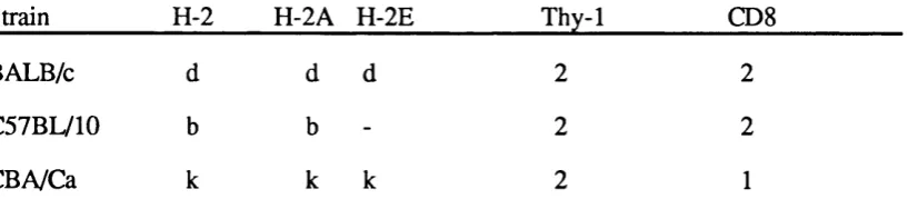
CBA/Ca (Thy 1^), C57BL/10, and BALB/c mice were obtained from colonies maintained in an SPF environment at the National Institute for Medical Research. Table ii gives a summary of the phenotypic characteristics of these mice.
Transgenic mice were generated by microinjection of DNA (Gordon etaL, 1980) into CBA/Ca oocytes obtained from superovulated mice injected with Folligon (Intervet) (Hogan et aL, 1994) at the microinjection facility at the National Institute for Medical Research. All injections were carried out by Dr. A. L. Mellor. Transgenic mice were bred and maintained in a clean breeding environment, unless required for long-term experiments, in which case they were transferred to an experimental area. Incoming mice were maintained in an isolator and offspring rederived into the clean breeding area. Table iii summarises the transgenic mice used in the course of these experiments.
T able ii Phenotypic C h aracteristics of M ouse S train s
Strain_________ H-2 H-2A H-2E__________Thy-1_________ Π8_________
BALB/c d d d 2 2
C57BL/10 b b - 2 2
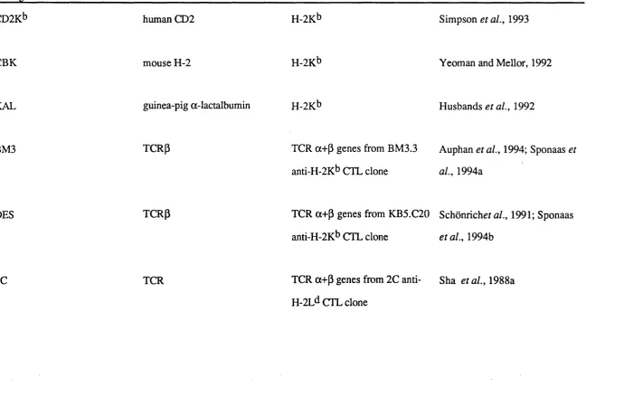
Table iii T ransgenic Mice
Transgene Code Promoter Structural Gene Reference
CD2Kb human CD2 H-2Kb Simpson etal., 1993
CBK mouse H-2 H-2Kb Yeoman and Mellor, 1992
KAL guinea-pig a-lactalbumin H-2K^ Husbands et a l, 1992
BM3 TCRP TCR a+ p genes from BM3.3 Auphan et a l, 1994; Sponaas et
anti-H-2K^ CTL clone aL, 1994a
DES TCRP TCR a+ p genes from KB5.C20 Schonricher al., 1991; Sponaas
anti-H-2K^ CTL clone et at., 1994b
2 .5 DNA P re p a ra tio n from Tail Tissue
Tissue samples from transgenic mice were obtained by tail biopsy, and digested with 0.2mg/ml Proteinase K (Boehringer, Cat. 1000144) in lysis buffer at 55°C in a shaking waterbath (Kotterman) overnight. Remaining hair was removed by centrifugation and DNA precipitated from the supernatant by with an equal volume of isopropanol. DNA was removed with a glass capillary tube, washed in 70% ethanol, and air dried before redissolving in TE.
2 . 6 G en o ty p e analysis
Every transgenic mouse used in the following experiments was tested for the presence of the transgene with one of the following techniques.
2.6.7 Southern blots
This method is similar to that adapted by Maniatis et a i, (1982), from the method of Southern (1975).
rôtisserie bottle. The probe was left to hybridize to the filter at 65®C overnight in the rôtisserie oven, then the hybridization solution was removed and the filter given 3 washes of increasing stringency (3X SSC, IX SSC, then O.IX SSC all with 0.1% SDS), to remove remaining radioactivity. The filter was incubated at -70^C in an autoradiography cassette with an X-ray film (UVP) and developed using an automated developing procedure. Exposed areas on the film indicated fragments of DNA to which the probe had hybridized.
2.6.2 Slot blots
The slot blot is similar to dot blots (Kafatos et al., 1979) and are described by Greaves et al., (1989). They are particularly useful for determining the numbers of copies of the transgene present in the genome of transgenic mice, since the DNA is localized to one area of the filter. Thus the intensity of the radioactive signal from the transgene can be compared to the signal from the same DNA sample probed for an endogenous gene.
Slot blot apparatus (Schleicher and Schuell's Minifold II, Anderman, Cat. 447 8(X)) comprises 2 perspex blocks with a nitrocellulose filter and blotting paper sandwiched between them and clamped together tightly. The upper block contains the slots, which concentrate the DNA into a small band on the filter, and the lower block contains vents and grooves and is attached to a vacuum line. As the denatured DNA is applied to the slots the liquid is drawn out of the apparatus via the vacuum line, and the DNA remains on the filter.
2 .6 J Production o f radioactive probes
Nick translation is part of the proof-reading process in the nucleus that ensures that DNA replication occurs with minimal errors. DNA polymerase I has the unique ability to extend the 3' end of single DNA strands where they have been nicked, synthesising a new DNA strand, and displacing the homologous stand in the duplex. We use nick translation to produce DNA probes labelled with radioisotopes, using a method similar to that of Rigby et al., (1977); the DNA probe is nicked with very low concentrations of DNAase I.
200ng probe DNA is added to 5|il water. In a separate tube 2|il lOx Nick mix, l^il each of O.lmM dATP, dGTP and dTTP (Pharmacia, Cat. 27-2050-01, 27-2070-01 and 27-2080-01 respectively), 0.5|ig DNAase I (Sigma, Cat. D4531), and DNA Pol I (Boehringer, Cat. 642 720) are mixed, followed by 3|il a^^P-dCTP (Amersham, Cat. AA 0085) and 5.5|il water, and this mixture is added to the probe DNA solution. Nick translation occurs at 15®C over 1 hr. Unincorporated counts were removed from the labelled probe using a Sephadex column (Pharmacia, Cat. 17-0855-02). The column was equilibrated with TE, and the probe made up to 400|il with TE. The probe was placed on the column, then eluted with up to 1ml TE, or until the number of counts from the eluant is approximately one third of the counts from the column itself. The probe was denatured at 100°C for 1 minute, and added directly to the pre-hybridization mix.
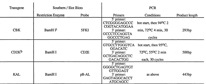
2.6.4 Polymerase Chain Reactions
This is an adaptation of the method described by Sakai et al., (1988). It is a quick method of amplifying a region of DNA in order to verify the presence of a particular DNA sequence in a DNA sample. The method described below is not quantitative, and can only be used to confirm the presence of the transgene in the sample. Oligonucleotide primers specific for each transgene were synthesised at the NIMR.
Table iv Probes and Prim ers for G enotype A nalysis o f Transgenic M ice
Transgene Southern / Slot Blots PCR
Restriction Enzyme Probe Primers Conditions Product length
CBK BamH 5FKI
5' primer: CTCGGGGAGCCC CGGTACATGGAA 3' primer: GCCCTCCAGGTA GGCCCTGAG
hot start, then 94®C 2 min, 72°C 4 min, 30
cycles
293bp
CD2Kb BamH I CD2E
5' primer: GTGCCTTGGGTCA
GGACATC 3' primer: GCTGACAGGCTC
GACACTGG
hot start, then 95®C, 72°C, 550C 2 min
each, 30 cycles
500bp
KAL BamH I pB-AL
5' primer: GGGGCTGAGTGT
GTTGGAGT 3' primer: GAGTAGGCACCT
ATGCAGCC
BM3.6 BamH I V a
5’ (Va) primer: CTGTGATGGAGA
AGACAACG 3' (Va) primer: CTTGTTGCCACTG
CCCCCAT
as above 290bp
DES Bgl IP TP900
5' (Lp2) primer: TGCCTCTGTGTAC
TCATGGC 3’ (Jp2) primer: ACAGCGTTTCTGC
ACTAGCC
as above 600bp
2C -
-5' primer: TGACACAGCCCG
ATGCTCGC 3' primer: GTCAGCGCACTTG
CAAAGCC
as above 300bp