R ES EAR CH A R T I C LE
Open Access
Pathological dislocation of the hip due to
coxotuberculosis in children: a 29-case report
Xin Jiang, Yuan Li
*, Lijun Liu, MingXin Peng, XueYang Tang, DaoXi Wang and XiaoDong Yang
Abstract
Background:The aim of this study is to evaluate the clinical outcome of various surgery methods in children suffering pathological dislocation of the hip joint due to coxotuberculosis.
Patients and methods:From January 2006 to January 2011, 29 children with coxotuberculosis hip dislocation were treated with open reduction (4 children) and acetabular reconstruction (25 children). According to degree of dislocation and age, acetabular reconstructions included Salter osteotomy (nine children), Pemberton operation (six children), Dega operation (seven children), the hip shelf arthroplasty (two children), and Chiari operation (one child). During acetabular reconstruction, 16 children underwent upper femoral rotational shortening osteotomies simultaneously, 6 children underwent the femoral head and neck reconstruction simultaneously, and 3 children were underwent corrective osteotomy. After operation, children were immobilized with plaster external fixation for 1–3 months and treated with routine anti-infective therapy for 3 days. During follow-up study (12–18 months), the hip stability was examined via X-ray. The recovery was evaluated via acetabular index (AI) and Harris hip score.
Results:Postoperative X-ray films of 29 children showed concentric reduction of all hips. Total 25 children healed I incision surgery, while 4 children with skin antrum of incision were further treated leading to 1–3 months delay of healing. Total 24 children were followed up for 2–5 years. X-ray examination revealed no redislocation after 2 years postoperative. The postoperative AI was normal (15°–20°) in 22 children and increased to 25°–30° in 2 children. After 2 years postoperative, 8 children had normal function of hip joint, 13 children had mild limitation of flexion and rotation, and 3 children had fibrous ankylosis. The average of Harris hip score was 83 (ranged, 62–90).
Conclusion:In our study, pathological hip dislocations of children attributed to coxotuberculosis were treated via open reduction and acetabular reconstruction and the outcomes were both excellent. Individual characteristic should be taken into consideration during treatment, and proper surgery approach should be adopted according to pathological changes of the hip.
Keywords:Pathological hip dislocation, Coxotuberculosis, Open reduction, Acetabular reconstruction
Introduction
Pathological dislocation of the hip joint in children is rela-tively rare occurrence and may cause lifelong disability when missed or ill-treated. The hip is a deep-seated ar-ticulation, and its dislocation may either remain hidden for some time or go undiagnosed [1]. According to the development of hip dislocation, pathological dislocation of the hip is frequently associated with heavy bone destruc-tive defect of acetabulum and femoral heads and necks as well as the residual anatomic deformity. Therefore, this
disease is difficult to treat, and several surgery operations have been explored and showed different outcomes.
Tuberculosis of the hip presents significant clinical problems, accounting for approximately 15% of osteoar-ticular tuberculosis [2]. The disease once occurred in the hip may lead to progressive destruction of the joint if untreated at an early stage, and many even proceed to pathological dislocation. Dislocated hips with infection are difficult to be managed to obtain a stable, mobile, congruous, and concentric joint. Diagnosis and effective treatment at an early stage are vital to treat the disease and save the joint [3]. The reports on the management of residual anatomic deformity of hip dislocation due to * Correspondence:[email protected]
Department of Pediatric Surgery, West China Hospital, Sichuan University, No. 37 Guoxue Road, Chengdu, Sichuan 610041, People's Republic of China
© 2014 Jiang et al.; licensee BioMed Central Ltd. This is an Open Access article distributed under the terms of the Creative Commons Attribution License (http://creativecommons.org/licenses/by/2.0), which permits unrestricted use, distribution, and reproduction in any medium, provided the original work is properly credited. The Creative Commons Public Domain Dedication waiver (http://creativecommons.org/publicdomain/zero/1.0/) applies to the data made available in this article, unless otherwise stated.
ticulation, and its dislocation may either remain hidden for some time or go undiagnosed [1]. According to the development of hip dislocation, pathological dislocation of the hip is frequently associated with heavy bone destruc-tive defect of acetabulum and femoral heads and necks as Pathological dislocation of the hip joint in children is rela-tively rare occurrence and may cause lifelong disability tively rare occurrence and may cause lifelong disability when missed or ill-treated. The hip is a deep-seated ar-ticulation, and its dislocation may either remain hidden for some time or go undiagnosed [1]. According to the Pathological dislocation of the hip joint in children is rela-tively rare occurrence and may cause lifelong disability
Pathological hip dislocation, Coxotuberculosis, Open reduction, Acetabular reconstruction
Pathological dislocation of the hip joint in children is
rela-should be taken into consideration during treatment, and proper surgery approach rela-should be adopted according to pathological changes of the hip.
Pathological hip dislocation, Coxotuberculosis, Open reduction, Acetabular reconstruction and rotation, and 3 children had fibrous ankylosis. The average of Harris hip score was 83 (ranged, 62
In our study, pathological hip dislocations of children attributed to coxotuberculosis were treated via open reduction and acetabular reconstruction and the outcomes were both excellent. Individual characteristic should be taken into consideration during treatment, and proper surgery approach should be adopted according healing. Total 24 children were followed up for 2
postoperative. The postoperative AI was normal (15°
After 2 years postoperative, 8 children had normal function of hip joint, 13 children had mild limitation of flexion and rotation, and 3 children had fibrous ankylosis. The average of Harris hip score was 83 (ranged, 62
In our study, pathological hip dislocations of children attributed to coxotuberculosis were treated via Postoperative X-ray films of 29 children showed concentric reduction of all hips. Total 25 children healed I incision surgery, while 4 children with skin antrum of incision were further treated leading to 1
healing. Total 24 children were followed up for 2–5 years. X-ray examination revealed no redislocation after 2 years postoperative. The postoperative AI was normal (15°–20°) in 22 children and increased to 25°
After 2 years postoperative, 8 children had normal function of hip joint, 13 children had mild limitation of flexion 3 months and treated with routine anti-infective therapy for 3 days. During follow-up study (12
hip stability was examined via X-ray. The recovery was evaluated via acetabular index (AI) and Harris hip score. Postoperative X-ray films of 29 children showed concentric reduction of all hips. Total 25 children healed I incision surgery, while 4 children with skin antrum of incision were further treated leading to 1
5 years. X-ray examination revealed no redislocation after 2 years child). During acetabular reconstruction, 16 children underwent upper femoral rotational shortening osteotomies simultaneously, 6 children underwent the femoral head and neck reconstruction simultaneously, and 3 children were underwent corrective osteotomy. After operation, children were immobilized with plaster external fixation for
3 months and treated with routine anti-infective therapy for 3 days. During follow-up study (12
hip stability was examined via X-ray. The recovery was evaluated via acetabular index (AI) and Harris hip score. treated with open reduction (4 children) and acetabular reconstruction (25 children). According to degree of dislocation and age, acetabular reconstructions included Salter osteotomy (nine children), Pemberton operation (six children), Dega operation (seven children), the hip shelf arthroplasty (two children), and Chiari operation (one child). During acetabular reconstruction, 16 children underwent upper femoral rotational shortening osteotomies simultaneously, 6 children underwent the femoral head and neck reconstruction simultaneously, and 3 children
From January 2006 to January 2011, 29 children with coxotuberculosis hip dislocation were treated with open reduction (4 children) and acetabular reconstruction (25 children). According to degree of dislocation and age, acetabular reconstructions included Salter osteotomy (nine children), Pemberton operation
The aim of this study is to evaluate the clinical outcome of various surgery methods in children
tuberculosis in children have rarely been documented. No comprehensive treatment protocol has been outlined.
From 2006 to 2011, we have performed surgery ap-proaches in 29 children with pathological tubercular dis-location of the hip, accounting for 3.4% of hip disdis-location in our hospital. The details of the patients' general data and clinical results are presented here.
Patients and methods
This study was approved by the Institutional Review Board of West China Hospital, Sichuan, China. Written informed consent was obtained from the patients for the publication of this report and any accompanying images. Based on the diagnosis card index at our department during the period from January 2006 to January 2011, we searched the files of patients with pathological hip dis-location caused by tuberculosis. Patients were included in the study if they met the following criteria: no previous treatment in other hospitals and radiographs available from the start of treatment to healing. Patients with slight subluxation or acetabular dysplasia were excluded.
Clinical manifestation of 29 patients with hip dislocation due to coxotuberculosis was shown in Table 1. Total 29 children (13 boys and 16 girls) with an average age of 4.3 years (ranged, 1–11 years) were included in the present study. There were 12 Han Chinese, 9 ethnic Tibetans, and 8 Yi people. Pathological tubercular hip dislocation is lo-cated in the right hip (17 children) and left hip (12 chil-dren). Four children showed subluxation of the hip joint, and 25 showed total dislocation of the hip. The mean duration of hip dislocation was 18 month (ranged, 10 days to 10 years). Besides the hip pain and lameness, other accompanied symptoms included significant local swell-ing in hip (13 children), fever (38°C–39°C) (6 children), pulmonary tuberculosis (7 children), spinal tuberculosis (2 children), tibia tuberculosis (1 child), femoral tuber-culosis (1 child), and iliac fossa abscess (1 child). The function status of injured hip joint was assessed using the Harris hip score [4,5]. The average of preoperative Harris hip score was 61 (ranged, 43–78). All children pre-sented the increased erythrocyte sedimentation rate, and the PPD skin test was positive in 23 children. Preoperative X-ray examination was performed for all children, and other examinations included computed tomography (CT) scan and three-dimensional reconstruction (23 children), color Doppler ultrasound (18 children), and magnetic resonance imaging (MRI) (12 children). The radiologic examination indicated the hip dislocation in all patients accompanied by avascular necrosis of femoral head (15 children), excalation of femoral head (6 children), total ab-sence of femoral head (2 children), and abab-sence of femoral head and neck (1 child). Additionally, eight children had increased anteversion angle (40°–60°) and three had varus deformity. The acetabular index (AI) was measured
according to Kleinberg and Lieberman [6] on the primary radiographs. It was roughly normal in 11 patients with the values of 15°–20° and increased to 25°–35° in 12 patients and to 40°–50° in 6 patients.
Surgical approaches
For the management of active tuberculosis of the hip, all patients were given routine antituberculosis therapy for 2–4 weeks before surgery. After routine preoperative preparations, patients were placed in a supine position and the contralateral hip was raised. Then, a Salter an-terior approach to the hip was performed after intub-ation and general anesthesia. Firstly, the hip lesion was removed thoroughly and histopathologically examined to make a definitive diagnosis.
Different operation methods were selected for 29 patients according to different pathological types. Four patients younger than age 2 were treated with open reduction and external fixation by plaster hip spica. Acetabular Table 1 Clinical manifestation of 29 patients with hip dislocation due to coxotuberculosis
Clinical manifestation Patients
Patients(male/female) 32 (19/13)
Age(years) 4.3 (ranged, 1–11)
Han/Tibetan/Yi nationality 12/9/8
Duration(months) 18 (ranged, 10 days–10 years) Dislocation of hip joint
Subluxation 4
Complete dislocation 25 Concomitant symptom
Hip local swelling 13
Fever (38°C–39°C) 6
Pulmonary tuberculosis 7
Spondylocace 2
Tibia tuberculosis 1
Femoral tuberculosis 1
Liac fossa abscess 1
Harris score 61 (43–78)
Acetabular index
15-20° (normal) 11
25-35° (mild) 12
40-50° (severe) 6
Preoperative examination
Damage of acetabulum 18 Ischemic necrosis of femoral head 15 Excalation of femoral head 6 Total absence of femoral head 2 Absence of femoral head and neck 1
the PPD skin test was positive in 23 children. Preoperative X-ray examination was performed for all children, and other examinations included computed tomography (CT) scan and three-dimensional reconstruction (23 children), color Doppler ultrasound (18 children), and magnetic the Harris hip score [4,5]. The average of preoperative Harris hip score was 61 (ranged, 43
sented the increased erythrocyte sedimentation rate, and the PPD skin test was positive in 23 children. Preoperative X-ray examination was performed for all children, and other examinations included computed tomography (CT) culosis (1 child), and iliac fossa abscess (1 child). The function status of injured hip joint was assessed using the Harris hip score [4,5]. The average of preoperative Harris hip score was 61 (ranged, 43
sented the increased erythrocyte sedimentation rate, and pulmonary tuberculosis (7 children), spinal tuberculosis (2 children), tibia tuberculosis (1 child), femoral tuber-culosis (1 child), and iliac fossa abscess (1 child). The function status of injured hip joint was assessed using the Harris hip score [4,5]. The average of preoperative accompanied symptoms included significant local swell-ing in hip (13 children), fever (38°C–39°C) (6 children), pulmonary tuberculosis (7 children), spinal tuberculosis (2 children), tibia tuberculosis (1 child), femoral tuber-culosis (1 child), and iliac fossa abscess (1 child). The and 25 showed total dislocation of the hip. The mean duration of hip dislocation was 18 month (ranged, 10 days to 10 years). Besides the hip pain and lameness, other accompanied symptoms included significant local swell-39°C) (6 children), 8 Yi people. Pathological tubercular hip dislocation is lo-cated in the right hip (17 children) and left hip (12 chil-dren). Four children showed subluxation of the hip joint, and 25 showed total dislocation of the hip. The mean duration of hip dislocation was 18 month (ranged, 10 days 11 years) were included in the present study. There were 12 Han Chinese, 9 ethnic Tibetans, and 8 Yi people. Pathological tubercular hip dislocation is lo-cated in the right hip (17 children) and left hip (12 chil-dren). Four children showed subluxation of the hip joint,
Preoperative examination children (13 boys and 16 girls) with an average age of 4.3
11 years) were included in the present
Acetabular index
15-20° (normal) 11
25-35° (mild) 12
40-50° (severe) 6
Liac fossa abscess 1
Harris score 61 (43
Acetabular index
15-20° (normal) 11
Spondylocace 2
Tibia tuberculosis 1
Femoral tuberculosis 1
Liac fossa abscess 1
39°C) 6
Pulmonary tuberculosis 7
Spondylocace 2
Tibia tuberculosis 1
Hip local swelling 13
39°C) 6
Pulmonary tuberculosis 7
Complete dislocation 25
Hip local swelling 13
39°C) 6
reconstruction was performed for 25 patients. Among them, nine patients aged less than 6 with the AI < 40° and femoral head congruent with the acetabula underwent Salter osteotomy. Six patients aged 3–6 with the AI > 40°, femoral head not congruent with the acetabula and un-closed Y-shaped acetabular cartilages were treated with Pemberton's operation. In addition, Dega operation was used for seven patients aged 5–14 years with total hip dis-location. The hip shelf arthroplasty was performed for two patients aged more than 14 years with hip subluxation. One patient aged more than 14 with complete hip disloca-tion was treated via Chiari operadisloca-tion.
During the acetabular reconstruction, 16 patients under-went upper femoral rotational shortening (short, 1–3 cm; mean, 2 cm) osteotomies simultaneously. Six patients also underwent the femoral head and neck reconstruction, and their greater trochanter were moved down. Corrective osteotomy was performed in three patients to correct coax vara, and the neck–shaft angle was corrected to 25° (ranged, 15°–40°).
Postoperative therapy
All affected limbs underwent external fixation postopera-tively by plaster cast for 1–3 months. To prevent add-itional infection, cephalosporins were given for 3 days after surgery, whilst all patients were given antituberculo-sis therapy for 12–18 months. Postoperative X-ray photo-graph of each patient was taken. The exercise of patients was scheduled according to the stability of the hip joints and the healing of osteotomies.
Results
Pathological examination indicated that all 29 cases were hip dislocation due to coxotuberculosis. Postoperative X-ray films showed well hip reduction of all patients. Postopera-tively, 25 patients healed I incision surgery, while 4 patients were accompanied by skin antrum, which were cut, and the healing was delayed for 1.5 months (ranged, 1–3 months).
A total of 24 patients were followed up for 3 years (ranged, 2–5 years) (Table 2). No complication was de-tected, such as hip redislocation, pathologic fracture and looseness of internal fixation during the follow-up period. X-ray examination revealed that no dislocation reoccurred at 2 years after operation. The postoperative AI was 15°–20° in 22 patients and increased mildly to 25°–30° in 2 patients. The anteversion angle was 20° (ranged, 15°–20°) and the neck shaft angle was 125° (ranged, 110°–140°), indicating normal anatomical rela-tion between the head and neck of hip (Figure 1). Among the patients associated with the femoral head and/or neck defect, 2 patients returned to normal oral morphology, 17 patients showed malunion of fem-oral head, and 3 patients with femfem-oral neck stump acted
as femoral head presented a normal relationship of for-mal head and acetabula (Figure 2).
The hip stability and weight-bearing were improved significantly. Hip pain was relieved. Twelve patients had residual mild lameness and four had moderate lameness, displaying leg length discrepancy (eight patients) with the average of 1.5 cm (ranged, 1–2 cm). After 2 years postoperatively, 8 patients had normal function of the hip joint; 13 cases had mild limitation of flexion and rotation, and 3 cases had fibrous ankylosis. The Harris hip score was 83 (ranged, 62–90).
Discussion
Pathological dislocation of a joint is a complication that directly involves the joint surfaces, which is common in the hip joint. Destructive dislocation occurs commonly in tuberculosis of the hip because of a traveling acetabu-lum (destruction of the acetabuacetabu-lum) or in septic arthritis of the hip in infancy secondary to the total destruction of femur head. Distensile dislocation occurred because of rapid distension of the joint capsule due to effusion of synovial fluid or pus [7].
Since the pathologically dislocated hips generally did not become normal spontaneously during growth, detec-tion at early stage was necessary and then appropriate remedial measures were performed [8,9]. If the abducted femoral head was hinged on the acetabular margin, which was common in subluxated and dislocated hips, spontaneous progressive cephalocotyloid improvement could not be expected without having a non-hinging hip by any measure. Johari et al. has reported the manage-ment of hip dislocations [10] and advocated a surgical Table 2 Clinical outcome of 24 patients at 2 years after surgery
Follow-up study Patients
Duration (years) 3 (ranged, 2–5) Hip joint reduction
Concentric reduction 21 Off-concentric reduction 3 Acetabular index
15°–20° 22
25°–30° 2
Femoral morphology
Normal 2
Malunion of femur head 17 Normal reduction of formal neck and acetabulum 3 Function of the hip joint
Normal 8
Mild limitation of flexion and rotation 13
Fibrous ankylosis 3
tected, such as hip redislocation, pathologic fracture and looseness of internal fixation during the follow-up period. X-ray examination revealed that no dislocation reoccurred at 2 years after operation. The postoperative AI was 15°
healing was delayed for 1.5 months (ranged, 1
A total of 24 patients were followed up for 3 years (ranged, 2–5 years) (Table 2). No complication was de-tected, such as hip redislocation, pathologic fracture and looseness of internal fixation during the follow-up period. X-ray examination revealed that no dislocation tively, 25 patients healed I incision surgery, while 4 patients were accompanied by skin antrum, which were cut, and the healing was delayed for 1.5 months (ranged, 1
A total of 24 patients were followed up for 3 years 5 years) (Table 2). No complication was de-hip dislocation due to coxotuber
films showed well hip reduction of all patients. Postopera-tively, 25 patients healed I incision surgery, while 4 patients were accompanied by skin antrum, which were cut, and the healing was delayed for 1.5 months (ranged, 1
Pathological examination indicated that all 29 cases were culosis. Postoperative X-ray n of all patients. Postopera-tively, 25 patients healed I incision surgery, while 4 patients Pathological examination indicated that all 29 cases were 18 months. Postoperative X-ray photo-graph of each patient was taken. The exercise of patients was scheduled according to the stability of the hip joints itional infection, cephalosporins were given for 3 days after surgery, whilst all patients were given antituberculo-18 months. Postoperative X-ray photo-graph of each patient was taken. The exercise of patients was scheduled according to the stability of the hip joints
residual mild lameness and four had moderate lameness, displaying leg length discrepancy (eight patients) with 3 months. To prevent
add-as femoral head presented a normal relationship of for-mal head and acetabula (Figure 2).
The hip stability and weight-bearing were improved significantly. Hip pain was relieved. Twelve patients had residual mild lameness and four had moderate lameness, as femoral head presented a normal relationship of for-mal head and acetabula (Figure 2).
Fibrous ankylosis 3
Function of the hip joint
Normal 8
Mild limitation of flexion and rotation 13
Fibrous ankylosis 3
Malunion of femur head 17
Normal reduction of formal neck and acetabulum 3
Normal 8
Normal 2
Malunion of femur head 17
Normal reduction of formal neck and acetabulum 3
30° 2
Normal 2
Malunion of femur head 17
20° 22
30° 2
intervention in failed closed reductions and in patients older than 2 years. In this study, we believed that patients even with active tuberculosis also needed aggressive treat-ment. The operation was performed after antitubercular chemotherapy for 2–4 weeks.
Davidson and Fernandez [11] have reported six cases of the dislocating type out of 31 cases of tuberculous hip (almost 20% incidence) in children younger than 14 years, and all 6 cases had poor outcomes. However, Campbell and Hoffman [12] have reported 13 cases of dislocating type of 74 hips (about 17% incidence), and the outcome was excellent/good in 46% of these cases (6 of 13). A poor outcome was found in those patients who had a narrow joint space after reduction. However, in the present study, all patients achieved satisfied out-comes which might be due to the different surgical pro-cedures we adopted based on the age and the region, range, or extent of pathological dislocation.
Zadeh et al. [13] have reported the management of developmental hip dislocations and advocated that the age at operation might be a significant prognostic factor in the long follow-up, suggesting that 94% of patients under the age of 2 years achieved an acceptable outcome, while the proportion of patients aged 2–4 years and more than 4 years was 80% and 71%, respectively. Johari et al. [10] have reported a surgical intervention in failed closed reductions in patients older than 2 years and emphasized the need of supplemental procedures such as Dega osteotomy, Staheli
shelf procedure, and femoral varus/derotational osteoto-mies combined with open reduction. Consistent with prior studies, in this study, patients under the age of 2 years were treated with open reduction associated with external fixation and the outcome was well.
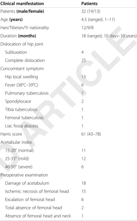
Acetabular reconstruction is essential for patients with severe destruction of acetabulum and femoral head–neck as well as low stability of articular reduction. However, young age, the extent of coverage of the acetabular com-ponent by the graft, and secure fixation and mechanical loading of the graft might be impact factors [14,15]. Salter osteotomy was only fit for children aged 1–6 years with well-developed acetabular. Meanwhile, femoral head of these patients should be generally congruent with their acetabular roof. Salter osteotomy can provide a limited correction (15°–20°) which can be considered as a dis-advantage in patients with severe dysplasia [16,17]. For children younger than 18 months, Salter osteotomy was contraindicated because satisfactory outcomes could be achieved by other measures [18]. Besides, the patients were usually too small to harvest or fix bone graft. For children older than 6 years, the prerequisites of complete reduction and reasonable congruity usually could not be met, hence it was contraindicated. Pemberton osteotomy was usually performed in patients aged 3–6 years with slightly shallower acetabulum and severe acetabular defi-ciency and was the most widely known incomplete transi-liac osteotomy. Pemberton osteotomy starts approximately Figure 1Serial radiographs of a 6-year-old boy who presented with tuberculosis of left hip dislocation.He was treated by left open reduction, Pemberton pelvis osteotomy, and corrective osteotomy in the left proximal femur.(a)Pre-operative.(b)Immediate post-operative. (c)2 years post-operative.
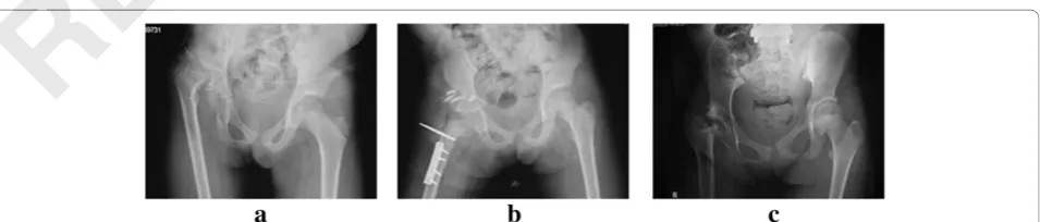
Figure 2Serial radiographs of an 11-year-old girl who presented with tuberculosis of hip dislocation, thoracolumbar kyphosis, and spinal tuberculosis.She was treated by right open reduction, Dega pelvis osteotomy, and subtrochanteric shortening osteotomy in the left proximal femur.(a)Pre-operative.(b)Immediate post-operative.(c)2 years post-operative.
reported a surgical intervention in failed closed reductions in patients older than 2 years and emphasized the need of supplemental procedures such as Dega osteotomy, Staheli proportion of patients aged 2
years was 80% and 71%, respectively. Johari et al. [10] have reported a surgical intervention in failed closed reductions in patients older than 2 years and emphasized the need of supplemental procedures such as Dega osteotomy, Staheli long follow-up, suggesting that 94% of patients under the age of 2 years achieved an acceptable outcome, while the proportion of patients aged 2–4 years and more than 4 years was 80% and 71%, respectively. Johari et al. [10] have reported a surgical intervention in failed closed reductions developmental hip dislocations and advocated that the age at operation might be a significant prognostic factor in the long follow-up, suggesting that 94% of patients under the age of 2 years achieved an acceptable outcome, while the 4 years and more than 4 cedures we adopted based on the age and the region, range, or extent of pathological dislocation.
Zadeh et al. [13] have reported the management of developmental hip dislocations and advocated that the age at operation might be a significant prognostic factor in the who had a narrow joint space after reduction. However, in the present study, all patients achieved satisfied out-comes which might be due to the different surgical pro-cedures we adopted based on the age and the region, the outcome was excellent/good in 46% of these cases (6 of 13). A poor outcome was found in those patients who had a narrow joint space after reduction. However, in the present study, all patients achieved satisfied out-comes which might be due to the different surgical
pro-these patients should be generally congruent with their acetabular roof. Salter osteotomy can provide a limited dislocating type of 74 hips (about 17% incidence), and
the outcome was excellent/good in 46% of these cases
ponent by the graft, and secure fixation and mechanical loading of the graft might be impact factors [14,15]. Salter osteotomy was only fit for children aged 1
well-developed acetabular. Meanwhile, femoral head of these patients should be generally congruent with their severe destruction of acetabulum and femoral head as well as low stability of articular reduction. However, young age, the extent of coverage of the acetabular com-ponent by the graft, and secure fixation and mechanical loading of the graft might be impact factors [14,15]. Salter were treated with open reduction associated with external fixation and the outcome was well.
Acetabular reconstruction is essential for patients with severe destruction of acetabulum and femoral head as well as low stability of articular reduction. However, shelf procedure, and femoral varus/derotational osteoto-mies combined with open reduction. Consistent with prior studies, in this study, patients under the age of 2 years were treated with open reduction associated with external fixation and the outcome was well.
shelf procedure, and femoral varus/derotational osteoto-mies combined with open reduction. Consistent with prior studies, in this study, patients under the age of 2 years shelf procedure, and femoral varus/derotational osteoto-mies combined with open reduction. Consistent with prior
He was treated by left open Immediate post-operative. He was treated by left open
10 to 15 mm above the anterior inferior iliac spine and curves gently posteriorly; the outer and inner tables of the ilium were osteotemized separately and ended at the level of the ilioischial limb of the triradiate cartilage halfway between the sciatic notch and the posterior part of the acetabular rim. Dega osteotomy was also a reshaping, in-complete osteotomy. The theoretical location of the hinge in the Dega osteotomy can involve, to varying degrees, the sciatic notch, the posterior portion of the inner pelvic cor-tex, the horizontal limb of the triradiate cartilage, and the symphysis pubis [19]. Similar to Pemberton osteotomy, Dega osteotomy also increases acetabular volume.
Subtrochanteric shortening osteotomy is a safe and pre-dictable procedure to restore the anatomic hip center and trochanteric rotation without neurologic injury in high developmental hip dislocation [20]. It allows acetabular exposure, diaphyseal shortening, and facilitates femoral derotation. In this study, although the femoral head was nearly completely absent, the joint function of patients restored to an adequate level by placing stump of femur neck inside the acetabulum. For cases with coxa vara, derotational femoral shortening was performed combined with corrective osteotomy.
Many scholars have always disputed whether internal fixator could be left in the affected hip joint with patho-logical dislocation. Some researchers believe that using internal fixator will increase numerous of risk such as infection, recrudescence, foreign body reaction, and for-mation of fistula. We hold that the internal fixator should not be adopted in chronic inflammation infection period. Our clinical data indicated that using the limited internal fixation could greatly enhance the strength of acetabular bone after reconstruction. In addition, prompt removal of the internal fixator had not precluded wound from heal up. In the present study, we did not find any case that had complications caused by internal fixator.
Conclusions
We have good results for surgical treatments in 29 patients with pathologic dislocation of tuberculous hip. Individual-ized surgical approach should be taken to treat tuberculosis hip in adolescent patients according to the pathological changes of affected acetabulum. In addition, based on this experience, the use of the early reconstructive surgery of the hip appeared effective even for patients with severely damaged acetabulum.
Competing interests
The authors declare that they have no competing interests.
Authors’contributions
XJ and YL carried out the studies, participated in the sequence alignment, and drafted the manuscript. LL carried out the immunoassays with XY. MP participated in the sequence alignment. XY participated in the design of the study and performed the statistical analysis. DW conceived of the study,
participated in its design and coordination, and helped to draft the manuscript. All authors read and approved the final manuscript.
Acknowledgement
Applied basic research programs funded projects in Sichuan province (2012JY0086): iatrogenic epiphyseal plate penetrating wound on the developmental effects of children's epiphyseal plate.
Received: 22 October 2013 Accepted: 19 February 2014 Published: 10 March 2014
References
1. Fabry G:Clinical practice.Eur J Pediatr2010,169:143–148.
2. Wang Y, Wang J, Xu Z, Li Y, Wang H:Total hip arthroplasty for active tuberculosis of the hip.Int Orthop2010,34:1111–1114.
3. Masood S:Diagnosis of tuberculosis of bone and soft tissue by fine‐
needle aspiration biopsy.Diagn Cytopathol1992,8:451–455. 4. Haddad R, Cook SD, Brinker MR:A comparison of three varieties of
noncemented porous-coated hip replacement.J Bone Joint Surg Br1990,72:2–8. 5. Harris WH:Traumatic arthritis of the hip after dislocation and acetabular
fractures: treatment by mold arthroplasty. An end-result study using a new method of result evaluation.J Bone Joint Surg1969,51:737–755. 6. Kleinberg S, LIEBERMAN HS:The acetabular index in infants in relation to
congenital dislocation of the hip.Arch Surg1936,32:1049.
7. Tahasildar N, Sudesh P, Tripathy SK, Shashidhar B:Bilateral pathological dislocation of the hip secondary to tuberculous arthritis following disseminated tuberculosis: a case report and review of the literature. J Pediatr Orthop B2012,21:567–573.
8. Moon M-S, Rhee S-K, Lee K-S, Kim S-S:A natural course of a dislocated healed tuberculous hip in a child: a case report.Clin Orthop Relat Res 1984,190:154–157.
9. Mousa H-L:Evaluation of sinus-track cultures in chronic bone infection. J Bone Joint Surg Br1997,79:567–569.
10. Johari AN, Dhawale AA, Johari RA:Management of post septic hip dislocations when the capital femoral epiphysis is present.J Pediatr Orthop B2011,20:413–421.
11. Davidson PT, Fernandez E:Bone and joint tuberculosis.Pennsylvania State University College of Medicine, Praeger Publishing: In Monograph in Tuberculosis. Edited by Schlosery D; 1983:119–131.
12. Campbell J, Hoffman E:Tuberculosis of the hip in children.J Bone Joint Surg Br1995,77:319–326.
13. Zadeh H, Catterall A, Hashemi-Nejad A, Perry R:Test of stability as an aid to decide the need for osteotomy in association with open reduction in developmental dysplasia of the hip a long-term review.J Bone Joint Surg Br2000,82:17–27.
14. Kim M, Kadowaki T:High long-term survival of bulk femoral head autograft for acetabular reconstruction in cementless THA for developmental hip dysplasia.Clin Orthop Relat Res2010,468:1611–1620.
15. Spangehl MJ, Berry DJ, Trousdale RT, Cabanela ME:Uncemented acetabular components with bulk femoral head autograft for acetabular
reconstruction in developmental dysplasia of the hip results at five to twelve years.J Bone Joint Surg Br2001,83:1484–1489.
16. Utterback TD, MacEwen GD:Comparison of pelvic osteotomies for the surgical correction of the congenital hip.Clin Orthop Relat Res1974,98:104–110. 17. Wong-Chung J, Ryan M, O’Brien TM:Movement of the femoral head after
Salter osteotomy for acetabular dysplasia.J Bone Joint Surg Br1990, 72:563–567.
18. Pekmezci M, Yazici M:Salter osteotomy: an overview.Acta Orthop Traumatol Turc2004,41:37–46.
19. Mubarak S, Valencia F, Wenger D:One-stage correction of the spastic dislocated hip. Use of pericapsular acetabuloplasty to improve coverage. J Bone Joint Surg Am1992,74:1347–1357.
20. Masonis JL, Patel JV, Miu A, Bourne RB, McCalden R, MacDonald SJ, Rorabeck CH:Subtrochanteric shortening and derotational osteotomy in primary total hip arthroplasty for patients with severe hip dysplasia: 5-year follow-up.J Arthroplasty2003,18:68–73.
doi:10.1186/1749-799X-9-16
Cite this article as:Jianget al.:Pathological dislocation of the hip due to coxotuberculosis in children: a 29-case report.Journal of Orthopaedic Surgery and Research20149:16.
hip in adolescent patients according to the pathological changes of affected acetabulum. In addition, based on this experience, the use of the early reconstructive surgery of the hip appeared effective even for patients with severely damaged acetabulum.
We have good results for surgical treatments in 29 patients with pathologic dislocation of tuberculous hip. Individual-ized surgical approach should be taken to treat tuberculosis hip in adolescent patients according to the pathological changes of affected acetabulum. In addition, based on this We have good results for surgical treatments in 29 patients with pathologic dislocation of tuberculous hip. Individual-up. In the present study, we did not find any case that had complications caused by internal fixator.
We have good results for surgical treatments in 29 patients bone after reconstruction. In addition, prompt removal of the internal fixator had not precluded wound from heal up. In the present study, we did not find any case that had complications caused by internal fixator.
not be adopted in chronic inflammation infection period. Our clinical data indicated that using the limited internal fixation could greatly enhance the strength of acetabular bone after reconstruction. In addition, prompt removal of the internal fixator had not precluded wound from heal internal fixator will increase numerous of risk such as infection, recrudescence, foreign body reaction, and for-mation of fistula. We hold that the internal fixator should not be adopted in chronic inflammation infection period. Our clinical data indicated that using the limited internal fixator could be left in the affected hip joint with patho-logical dislocation. Some researchers believe that using internal fixator will increase numerous of risk such as infection, recrudescence, foreign body reaction, and for-mation of fistula. We hold that the internal fixator should
11. Davidson PT, Fernandez E: Many scholars have always disputed whether internal
fixator could be left in the affected hip joint with
patho-1984, 9. Mousa H-L:
J Bone Joint Surg Br
10. Johari AN, Dhawale AA, Johari RA:
dislocations when the capital femoral epiphysis is present. dislocation of the hip secondary to tuberculous arthritis following disseminated tuberculosis: a case report and review of the literature. J Pediatr Orthop B2012,
8. Moon M-S, Rhee S-K, Lee K-S, Kim S-S:
healed tuberculous hip in a child: a case report. 154–
new method of result evaluation. 6. Kleinberg S, LIEBERMAN HS:
congenital dislocation of the hip.
7. Tahasildar N, Sudesh P, Tripathy SK, Shashidhar B:
dislocation of the hip secondary to tuberculous arthritis following disseminated tuberculosis: a case report and review of the literature.
A comparison of three varieties of ated hip replacement.
Traumatic arthritis of the hip after dislocation and acetabular fractures: treatment by mold arthroplasty. An end-result study using a new method of result evaluation.J Bone Joint Surg
The acetabular index in infants in relation to Diagn Cytopathol
A comparison of three varieties of ated hip replacement.
Traumatic arthritis of the hip after dislocation and acetabular fractures: treatment by mold arthroplasty. An end-result study using a
Total hip arthroplasty for active 34:1111–
Diagnosis of tuberculosis of bone and soft tissue by fine Diagn Cytopathol
Diagnosis of tuberculosis of bone and soft tissue by fine 8:451
A comparison of three varieties of ated hip replacement.J Bone Joint Surg Br
169:143
Total hip arthroplasty for active 1114.