R E S E A R C H
Open Access
Induction of antigen-specific immune responses
in mice by recombinant baculovirus expressing
premembrane and envelope proteins of West
Nile virus
Bibo Zhu
1,2, Jing Ye
1,2, Ping Lu
4, Rong Jiang
1,2, Xiaohong Yang
1,2, Zhen F Fu
1,2,3, Huanchun Chen
1,2and Shengbo Cao
1,2*Abstract
Background:West Nile Virus (WNV) is an emerging arthropod-born flavivirus with increasing distribution worldwide that is responsible for a large proportion of viral encephalitis in humans and horses. Given that there are no effective antiviral drugs available for treatment of the disease, efforts have been directed to develop vaccines to prevent WNV infection. Recently baculovirus has emerged as a novel and attractive gene delivery vehicle for mammalian cells. Results:In the present study, recombinant baculoviruses expressing WNV premembrane (prM) and envelope (E) proteins under the cytomegalovirus (CMV) promoter with or without vesicular stomatitis virus glycoprotein (VSV/G) were constructed. The recombinant baculoviruses designated Bac-G-prM/E and Bac-prM/E, efficiently express E protein in mammalian cells. Intramuscular injection of the two recombinant baculoviruses (at doses of 108or 109PFU/mouse) induced the production of WNV-specific antibodies, neutralizing antibodies as well as gamma interferon (IFN-γ) in a dose-dependent pattern. Interestingly, the recombinant baculovirus Bac-G-prM/E was found to be a more efficient immunogen than Bac-prM/E to elicit a robust immune response upon intramuscular injection. In addition, inoculation of baculovirus resulted in the secretion of inflammatory cytokines, such as TNF-α, IL-2 and IL-6.
Conclusions:These recombinant baculoviruses are capable of eliciting robust humoral and cellular immune responses in mice, and may be considered as novel vaccine candidates for West Nile Virus.
Keywords:West Nile virus, Rcombinant baculovirus, Premembrane/Envelope protein, Immune responses
Background
West Nile Virus (WNV), a mosquito-born virus, belongs to the Japanese encephalitis serogroup of flaviviruses [1] and is an outstanding example of a zoonotic pathogen as its geographic range spans North and South America, Europe, the Middle East, Africa, Western Asia and Aus-tralia [2,3]. Zoonotic transmission of WNV involves Culex mosquitos and birds as the natural reservoir hosts, with humans and horses as dead-end hosts [4]. WNV infec-tions are ranging from a sub-clinical illness, to a
self-limiting febrile syndrome or a lethal neuroinvasive disease, and the incidence of severe disease and death increases with age [5,6]. Since the emergence of WNV in north American in 1999, WNV has been responsible for over 12,000 cases of meningitis/encephalitis, and over 1,100 fatalities in humans and mass mortality of resident birds [7]. The arbovirus infection of humans and animals is be-coming a major public health and veterinary concern [4]. The WNV outbreak in North America coupled with the absence of specific therapy against WNV infection has triggered vaccine development to prevent the infection of humans and animals. It has been shown that virion enve-lope is embedded with viral enveenve-lope (E) and premem-brane/membrane (prM/M) proteins. The prM protein helps proper folding of the E protein by forming * Correspondence:[email protected]
1
State Key Laboratory of Agricultural Microbiology, Huazhong Agricultural University, Wuhan, Hubei 430070, People's Republic of China
2
Laboratory of Animal Virology, College of Veterinary Medicine, Huazhong Agricultural University, Wuhan, Hubei 430070, People's Republic of China Full list of author information is available at the end of the article
heterodimers [8] and is cleaved into M by furin [9,10]. The glycoprotein E is responsible for many properties of the virus including host range, replication, assembly, and stimulation of humoral and cellular immune responses [11]. A variety of WNV vaccine candidates based on the glycoprotein E, which evokes the majority of the neutraliz-ing antibodies, have been developed. Three equine vac-cines have been successfully commercialized in the USA, including InnovatorTM, formulated based on formalin-inactivated virus, RecombitekTM, a recombinant replica-tive canarypoxvirus vaccine, and pCBWN, a recombinant plasmid DNA vaccine [12]. Novel recombinant subunit vaccine candidates such as virus-like particles (VLPs), vec-toring vaccine candidates, DNA vaccine candidates [12], and TripliVAX JE, a chimeric vaccine expressing prM/E and NS1 gene of Japanese encephalitis virus [13], have shown protective immunity in animal model. However, there are no vaccines currently available for humans. With the growing volume of international travel and commerce, exotic pathogens can spread quickly between continents. Therefore, safe and effective vaccines of WNV are needed for humans.
In recent years, baculovirus has emerged as a vector for gene delivery and vaccine development, with promis-ing newcomer bepromis-ing Autographa californica multiple nucleopolyhedrovirus (AcMNPV), a member of baculo-viridae family. The host specificity of baculovirus was originally thought to be restricted to cells derived from a few taxonomically related insect species [14]. However, it has been shown to infect a number of mammalian cells without replication [15-18]. Additionally, accumu-lated evidence has revealed that baculovirus express for-eign proteins not only in insect cells, but also in mammalian cells under the transcriptional control of mammalian promoters [14,16,19]. Subsequently, it has been reported that a baculovirus pseudotyped with the glycoprotein (G) of the vesicular stomatitis virus (VSV/ G) appeared to help the virus escape from the endo-somes, which increases transduction efficiency in mam-malian cells [20,21]. Furthermore, the baculovirus has
advantages of being a strong adjuvant and thus can in-duce inflammatory cytokines and interferons [22-24]. To this end, several studies have demonstrated that direct vaccination with recombinant baculovirus can induce high-level humoral and cell-mediated immunity against various antigens, including pseudorabies virus glycopro-tein B (gB) [25], GP5 and M proglycopro-tein of porcine repro-ductive and respiratory syndrome virus (PRRSV) [26], capsid protein of procine circovirus type2 [27], rabies virus glycoprotein [28] and Japanese encephalitis virus (JEV) envelope protein [29].
In the present study, recombinant baculoviruses expressing WNV prM and E protein were constructed with or without pseudotyping with VSV/G. Protein ex-pression was characterized in transduced mammalian cells and its immunogenicity was investigated in a
mouse model. The results showed that direct
immunization with the recombinant baculoviruses
induced a higher level of WNV-specific antibody, neu-tralizing antibody and gamma interferon compared with E-DIII protein vaccine, suggesting that the recombinant baculoviruses expressing WNV prM and E protein could be developed as new vaccine candidates for WNV.
Results
Construction of recombinant baculoviruses expressing WNV protein
Recombinant baculovirus has been used as gene-delivery vehicles for transient expression of recombinant proteins in mammalian cells. In the present study, recombinant baculoviruses Bac-prM/E and Bac-G-prM/E expressing WNV prM and E gene under the control of the cyto-megalovirus immediate early promoter were constructed (Figure 1).
In order to investigate the transduction efficiency of the constructed recombinant baculoviruses Bac-prM/E and Bac-G-prM/E. BHK-21 cells were transduced with the constructed baculoviruses or control baculovirus (Bac-G-EGFP or Bac-EGFP) at a multiplicity of infection (MOI) of 100. An indirect immunofluorescence assay
and Western blotting were performed 48h post-transduction to measure the level of WNV protein ex-pression in mammalian cells. As shown in Figure 2A, significant fluorescence was detected in Bac-prM/E- and Bac-G-prM/E- transducd BHK-21 cells, whereas it was
not detected in the Bac-EGFP- or Bac-G-EGFP- trans-duced cells. A protein band of approximately 55 KDa was consistently observed in recombinant baculovirus-transduced BHK-21 cell lysates as detected by Western blotting (Figure 2B). BHK-21 cells were also transduced
with recombinant baculoviruses at different MOIs (10, 50 and 100) and E protein was compared by Western blot analysis. As shown in Figure 2C, the cells trans-duced with Bac-G-prM/E showed a darker band than cells transduced with Bac-prM/E and the protein expres-sion was increased in a dose-dependent pattern in cells transduced with both viruses.
Characterization of WNV-specific antibodies elicited by recombinant baculoviruses in mice
Our previous studies have demonstrated the potential of using baculovirus as a vaccination platform [29]. To deter-mine whether the recombinant baculoviruses expressing prM/E proteins could induce WNV-specific humoral im-mune responses in vivo, BALB/c mice were immunized intramuscularly with 109or 108PFU/mouse of recombinant baculovirus. Serum samples were collected at 3 and 6 weeks after the primary immunization. Then the samples were diluted 1: 500 and specific IgG responses were determined by an indirect ELISA using E-DIII protein as coating anti-gen. At 3 weeks, the antibody titers reached a detectable level in all the vaccinated groups except the group immu-nized with the Bac-G-EGFP, Bac-EGFP (control virus) and PBS, and a further increase in antibody titers were observed at 6 weeks after primary immunization (p<0.001) (Figure 3). Mice immunized with 109PFU of Bac-G-prM/E produced significantly higher antibody titer than mice received 108 PFU of Bac-G-prM/E (p<0.01). Similar result was also observed in Bac-prM/E-vaccinated group. Interestingly, mice immunized with Bac-G-prM/E developed higher anti-body titer than mice immunized with Bac-prM/E at the same dosage, but there was no statistical significance (p>0.05). Immunization with 50μg/mouse of E-DIII pro-teins induced highest antigen-specific antibody titer. As expected, all vaccinated groups produced significantly higher
WNV-specific antibody titers than mice vaccinated with control viruses (p<0.001).
In order to further study whether recombinant
baculovirus-immunized mice induce WNV-specific neutral-izing antibodies, serum samples were tested by PRNT. As shown in Table 1, mice immunized with 108or 109PFU of Bac-G-prM/E or Bac-prM/E developed mean neutralizing antibody titers of 1:16 and 1:8 or 1:11 and 1:5 at 3 weeks after primary immunization, which increased further to 1:44 and 1: 22 or 1:30 and 1:12 at 6 weeks, respectively. Although WNV-specific neutralizing antibodies were detectable in mice immunized with 50μg of E-DIII protein, the mean neutralizing antibody titer was 1:8 at 6 weeks, which was significantly lower than the groups inoculated with 109PFU of Bac-G-prM/E and Bac-prM/E (p<0.05). As expected, no neutralizing antibodies were detected in sera from mice immunized with control virus or PBS. In addition, we also determined whether the antibodies can efficiently neutralize heterologous Japanese encephalitis virus (JEV)-p3 strain. Lit-tle or none anti-JEV cross-neutralizing antibodies were detected in sera collected at 3 weeks after the first immunization. After booster immunization, anti-JEV cross-neutralizing antibody titers increased to 1:10, which were lower than those against WNV. These studies indicate that recombinant baculoviruses immunized mice developed humoral immune responses evidenced by WNV-specific neutralizing antibodies and partial anti-JEV cross-neutralizing antibodies.
WNV-specific cellular immune responses elicited by recombinant baculoviruses in mice
To further address whether the recombinant baculo-viruses can elicit cell-mediate immune responses in mice, mice were immunized with the recombinant bacu-loviruses and sacrificed at 3 weeks after the booster
immunization. Splenocytes were prepared from the immu-nized mice and restimulated in vitro with E-DIII protein. IFN-γ production was measured by ELISA. As a negative control, IFN-γproduction was also measured upon stimu-lation of splenocytes with NS3 protein of JEV. Mice immu-nized with 109PFU, 108PFU of G-prM/E and Bac-prM/E or 50μg of E-DIII protein showed significantly higher levels of IFN-γ than mice given the control virus (109PFU of Bac-G-EGFP) (Figure 4A). In particular, IFN-γ level detected in splenocytes of mice inoculated with 109 PFU of Bac-G-prM/E was significantly higher than that in splenocytes of mice immunized with E-DIII protein (p<0.01). It was also found that mice immunized with Bac-G-prM/E developed higher amount IFN-γ than mice re-ceiving Bac-prM/E, but it was not statistically significant. Moreover, IFN-γproduction in Bac-G-prM/E- or Bac-prM /E-inoculated group was significant lower when NS3 pro-tein was used as stimulation antigen and there was no sig-nificant difference among Bac-G-prM/E, Bac-prM/E, and control virus immunized group, suggesting WNV specific IFN-γproduction (Figure 4A).
To validate this observation, mRNA level of IFN-γ pro-duced in splenocytes was further analyzed by real-time PCR in parallel with the housekeeping gene. Similar to the results of IFN-γdetermined by ELISA, the mean relative level of IFN-γmRNA expression showed significant differ-ence between Bac-G-prM/E- or Bac-prM/E-immunized mice and Bac-G-EGFP- or E-DIII protein-inoculated mice
(Figure 4B). All these results demonstrate that
immunization with recombinant baculoviruses can stimu-late cellular immune responses.
Recombinant baculoviruses induce inflammatory cytokines in mice
To further understand the mechanism associated with induction of host immune response upon baculovirus
in-oculation, the expression profile of inflammatory
cytokines was monitored. Splenocytes from immunized mice were restimulated in vitro and then mRNA levels of three inflammatory cytokines, including tumor necro-sis factor alpha (TNF-α), interleukin-6 (IL-6), and interleukin-2 (IL-2), were detected by real-time PCR. As shown in Figure 5, the mean relative mRNA levels of these inflammatory cytokines in the group immunized with Bac-G-prM/E or Bac-prM/E were significantly higher than in the control group. Moreover, mice immu-nized with 109PFU of Bac-G-prM/E or Bac-prM/E pro-duced significantly higher amount of these inflammatory cytokines than mice immunized with E-DIII protein (p<0.05). These suggest that a strong inflammatory re-sponse with a complex pattern of inflammatory cyto-kines may contribute to the intrinsic immunogenic properties of the inoculated baculoviruses.
Discussion
WNV continues to present public health threat to the western hemisphere, yet, there is neither effective ther-apy nor vaccines available for humans. Thus developing a safe and effective vaccine is a priority. Several immunization strategies have been described, some of which utilize vectored vaccines expressing WNV glyco-protein E resulted in 80%-100% protective immunity in animal model [30-34]. In this present study, attempts were made by using recombinant baculovirus approach for WNV vaccine development since baculovirus exhibit low toxoxicity in mammalian cells and can induce inter-feron production [24]. Two recombinant baculoviruses, Bac-G-prM/E and Bac-prM/E, expressing the major im-munogenic protein, glycoprotein E, were constructed, and the humoral and cellular immune responses were determined in mice. WNV-specific humoral (WNV-spe-cific antibodies and neutralizing antibodies) and cellular immunity (WNV-specific IFN-γ and inflammatory cyto-kines) were induced in mice inoculated with recombin-ant baculoviruses. These data are in concordance with previous studies that showed robust humoral and cellu-lar immune responses against PRRSV, PCV, or JEV by recombinant baculoviruses [29].
It has been controversial as to the relative role of the humoral versus cellular immune responses in the WNV-protective immunity. High antibody titers are believed to protect animals against flavivirus infections through dir-ect neutralization of receptor binding, inhibition of viral fusion, and/or complement-mediated viral clearance [35,36]. Neutralizing antibody titers of >1:10 were reported to be sufficient to afford protection in humans against JE [37]. Schneeweiss et al. also reported that mice immunized once with pT-WNV-E produced neu-tralizing antibodies titer of 1:16 which can protect 100% of mice from WNV challenge [38]. However, others have argued that cellular immune responses such as robust
Table 1 Neutralizing antibody titers in mice immunized with recombinant baculoviruses (mean values of groups of 4 mice)
Vaccine Anti-WNVb Anti-JEVc
3 weeks 6 weeks 3 weeks 6 weeks
Bac-G-prM/E 109 16 44 4 10
Samples were taken three weeks and six weeks after the primary immunization.a
a
plaque-reduction neutralization tests (PRNTs);
b
WNV (NY99 strain)-specific neutralizing antibodies;c
anti-CD8+ T cell responses are essential for clearing WNV [39,40]. In our study, the antibody responses were evaluated in mice immunized with recombinant baculo-viruses. Majority of mice immunized with two dosages of Bac-G-prM/E or Bac-prM/E developed neutralizing antibodies titers of >1:16 (Table 1). Furthermore, these baculovirus-derived vaccines can also induce anti-JEV cross-neutralizing antibodies although the titers of the antibodies were lower than antibodies against WNV. These results strongly demonstrate that the antibody re-sponse induced by WNV protein could partially cross-neutralize JEV, as has been reported in previous studies
[41]. It is reasonable to anticipate that immunization with these recombinant baculoviruses expressing WNV antigens will not only provide protection against WNV infection, but also provide partial protection against JEV infection. Cellular immunity such as IFN-γ production and inflammatory cytokines responses were also deter-mined in mice after immunization with these baculo-viruses since these cytokines play a critical role in directing the cell-mediated immune responses [42]. Bac-G-prM/E- or Bac-prM/E-immunized mice produced higher levels of IFN-γ than any other group (Figure 4) and promoted the release of inflammatory cytokines, Figure 4IFN-γresponses induced by immunization with recombinant baculoviruses. (A)Production of IFN-γin the supernatant of
splenocytes harvested from immunized mice afterin vitrorestimulation was determined by ELISA. Splenocytes of immunized mice were isolated 6 weeks after the primary immunization and incubatedin vitrowith E-DIII protein or NS3 protein for 72h. Data represent the mean
including tumor necrosis factor alpha (TNF-α), interleukin-6 (IL-6), and interleukin-2 (IL-2) (Figure 5). In this case, secretion of IFN-γ, TNF-α, or IL-2 are indi-cative of a Th1 response, whereas a Th2 response is characterized by induction of IL-6 [43]. These indicate that the recombinant baculoviruses can stimulate both humoral and cellular immune responses which are essential to control dissemination of infection and to in-hibit the entry of virus into brain.
Our results also demonstrate that Bac-G-prM/E exhib-ited better immunogenicity than Bac-prM/E, as indi-cated by the higher levels of antibody titers, IFN-γ and inflammatory cytokines production. It is highly probable that the expression of VSV/G was associated with enhancement of the immune responses by augmenting transduction efficiency of baculovirus into mammalian cells [20]. In addition, the VSV/G-modified baculovirus exhibited greater resistance to animal serum inactivation than the unmodified baculovirus [20], which may con-tribute to the better immunogenicity of Bac-G-prM/E.
It has been reported that recombinant E-DIII protein induces high neutralizing antibody titers, as well as pro-tection against lethal WNV and partial propro-tection against lethal JEV [41]. Therefore, the recombinant E-DIII protein of WNV expressed inE.coliwas used as a positive control in the present study. However, intramuscular injection of mice with E-DIII protein elicited lower levels of neutralization antibody titers than mice immunized with the recombinant baculoviruses even at a low dosage (108 PFU/mouse) (Table 1), although the total IgG level was high (Figure 3). This could be due to differences in experi-mental designs such as the route of immunization, with or without adjuvant, and mouse strain. In addition, it has been shown that E-DIII protein immunization elicits low
level of neutralizing antibodies with relatively high IgG responses [38], which is consistent with our results. It is noticed that E-DIII protein also induced lower levels of cellular immune response than recombinant viruses, since E protein expressed by recombinant baculo-virus contains more T cell epitopes than its domain III [40], and the baculovirus augment cellular immunity.
Baculovirus has been shown to possess a strong adjuvant activity and to promote humoral and cellular immune responses for foreign antigens, maturation of dendritic cells, and production of inflammatory cytokines and IFN [24]. The transduction of macrophages in vitro by baculovirus led to the induction of significant levels of IL-6 and TNF-α [44]. It has been proposed that baculovirus genome, espe-cially its CpG motifs, could be recognized by macrophages and DCs. Furthermore, baculoviruses enter into the cells through mannose receptor (MR)-mediated endocytosis or phagocytosis, leading to the secretion of inflammatory cytokines through a MyD88/TLR9-dependent signaling pathway [23,24]. However, Chen et al. reported that recom-binant baculoviruses are able to induce a robust secretion of inflammatory cytokines through a TLR3-dependent pathway [45]. In our study, induction of a measurable
inflammatory responses in mice [44]. These baculovirus-induced non-specific inflammatory responses raise concerns that whether they will compromise the use of baculovirus vectors for in vivo gene therapy in humans and animals. However, Bac-G-E2 inoculated mice released inflammatory cytokines as early as 6h post-injection, and returned to back-ground levels by 48h post-injection [21]. Furthermore, there was no cytokine-induced acute toxicity or mortality in baculovirus-immunized mice, which suggests that non-specific inflammatory responses induced by baculovirus vec-tors may be minimized by host. Nevertheless, more investi-gations are needed to ensure the safety of baculovirus vectors.
The protection is an important index to evaluate the ef-ficiency of vaccine. However, the ability of our recombin-ant baculoviruses to protect against lethal challenge of WNV was not determined in consideration of the safety problem since WNV has not been found in China. Never-theless, the neutralizing antibodies and the cellular im-mune responses induced by recombinant baculoviruses may provide the basis for protection. Immunization with baculovirus expressing JEV antigens have been shown to confer protection against JEV challenge, demonstrating that cellular immunity plays a crucial role against JEV in-fection [29]. Thus it is well supported that recombinant baculoviruses expressing WNV prM/E proteins could pro-vide protective immunity against WNV challenge.
Methods
Cells, plasmids and virus
Spodoptera frugiperda 9 (Sf9) cells were propagated at 28° C in Grace’s media (Invitrogen, Carlsbad, CA, USA) sup-plemented with 10% FBS, 100 μg/ml streptomycin and 100 IU/ml penicillin. BHK- 21 cells, used for transduction and neutralization tests, were grown and maintained in Dulbecco’s modified Eagle’s medium (DMEM; Invitrogen, Carlsbad, CA, USA) supplemented with 10% heated-inactivated fetal bovine serum (FBS), 100 μg/ml strepto-mycin and 100 IU/ml penicillin, at 37°C with 5% CO2.
The prM and E genes region (GenBank accession number DQ211652) corresponding to nt 466–2469, derived from a North American NY99 strain of WNV, were synthesized and cloned into pcDNA3.1 vector backbone, to generate plasmid pcDNA3.1-prM/E. The construction of baculovirus transfer vector, pFastBac-VSV/G, in which the VSV/G gene is under the control of the polyhedrin promoter of pFast-BacTM1 (Invitrogen), was described previously [29]. Bac-G-EGFP is a pseudotype baculovirus containing a CMV pro-moter that controls the expression of EGFP [29] and was used as a control strain of the baculovirus.
Protein and monoclonal antibodies
WNV envelope protein (E protein) domain III (the sequence corresponding to nt 1858–2211) was expressed inE.colias
an inclusion body, and then purified and refolded in appro-priate buffer [46]. Protein concentrations were determined at an absorbance of 280 nm by spectrophotometry. Anti-E-DIII monoclonal antibody (MAb) was produced from the mice immunized with E-DIII protein.
Construction of recombinant baculoviruses
To generate the recombinant transfer plasmid pFastBac-G-prM/E, the DNA fragment containing the prM/E expres-sion cassette, which was controlled under the CMV pro-moter, was released from pcDNA3.1-prM/E and inserted into pFastBac-VSV/G. To generate the recombinant trans-fer plasmid pFastBac-prM/E the same prM/E expression cassette was digested and clone into pFastBac1 vector. The recombinant baculoviruses Bac-G-prM/E and Bac-prM/E were subsequently generated by using Bac-to-Bac system (Invitrogen) following the manufacturer’s instructions. The resultant viruses were further amplified by propagation in Sf9 cells and purified as described [25]. The virus pellet was resuspended in phosphate-buffer saline (PBS, PH7.4) and infectious titers were determined by a plaque assay as described in the Bac-to-Bac system, and then stored in ali-quots at−80°C until needed.
Detection of E protein in Baculovirus-transduced cells
The transduction procedure was performed as described previously with minor modifications [27,29]. BHK-21 cells were seeded into six-well plates at concentration of 1.5 × 10⁵cells/well. At approximately 70-80% confluence, culture medium was removed and cells were washed three times with PBS and incubated with baculoviruses for 4h at 28°C. After removal of the inoculum, fresh medium was added and cultures incubated at 37°C for 48h. Duplicate wells were processed in parallel for indirect immunofluorescence assay and Western blotting. At 48h after transduction, the cells were fixed with absolute methanol and processed for the indirect immunofluores-cence assay using monoclonal antibodies (MAb) against WNV E-DIII protein, followed by fluorescein isocyanate-conjugated goat anti-mouse immunoglobulin (Ig) G. For Western blot analysis, transduced cells were collected at 48h post-transduction and lysed in SDS sample buffer. Cell extracts were separated by sodium dodecyl sulfate
−10% polyacrylamide gel electrophoresis (SDS-PAGE) and electroblotted onto a nitrocellulose membrane. The non-specific antibody binding sites were blocked with 1% bo-vine serum albumin (BSA) in TBST buffer (10mM Tris– HCl PH 8.0, 150mM Nacl, and 0.05% Tween-20), and fi-nally the membrane were reacted with anti-E-DIII Mab to detect the expressed E protein.
Mouse immunization
province, China. Mouse studies were performed accord-ing to the guidelines of this institution (No. 00020502) and all experimental protocols were approved by the Re-search Ethics Committee of College of Veterinary Medi-cine, Huazhong Agricultural University, Hubei, China. Then the mice were randomly divided into eight groups (6 mice per group). Two groups were injected intramus-cularly (i.m.) with 100μl of PBS containing 1 × 10⁸ or
1 × 10⁹ PFU of Bac-G-prM/E and another two groups
were immunized with Bac-prM/E. The other three groups were injected intramuscularly with 100μl of PBS containing 1 × 10⁹ PFU of Bac-G-EGFP, Bac-EGFP, and 100μl of PBS containing 50μg of E-DIII protein, respect-ively. The last group was used as a negative control by intramuscularly injecting 100μl of PBS. Booster immuni-zations were identically performed 3 weeks later. Serum samples were collected from the tail vein of 6 individual mice before the second injection and from the retro-orbital plexus at 6 weeks after primary immunization and then store at−20°C for serological tests. At 6 weeks after the primary immunization, three mice of each group were euthanized, and splenocytes were harvested for immunological assay.
Antibody assay
Induction of E protein-specific antibody was determined by using indirect enzyme linked immunosorbent assay (ELISA). Microplates were coated with E-DIII protein (20ng/well) overnight at 4°C [38]. After blocking for 1h at 37°C with PBST (2% bovine serum albumin, PBS, 0.05% Tween 20) to avoid nonspecific binding, serum diluted in PBST (1:500) were added to each well and incubated at 37°C for 1h. After washing with PBST, bounded proteins were detected with HRP conjugated with goat mouse IgG (Boster, China). Serum anti-body titer was monitored versus optical density values. The endpoint titers of the neutralizing antibodies against WNV and JEV were determined by plaque-reduction neutralization tests (PRNTs) using WNV strain NY99 and JEV strain P3, in biosafety level–3 conditions,as described earlier [47]. Briefly, serum samples were heat-inactivated for 45min at 56°C and then serially diluted and mixed with an equal volume containing 100 PFU of
virus. The mixtures were incubated for 1h at 37°C and then inoculated onto BHK-21 cell monolayers in 12-well plates. After adsorption for 1h at 37°C, the wells were overlaid with medium containing 1.5% carboxymethyl cellulose, and the plates were incubated in a 5% CO2
in-cubator at 37°C for 3–4 days for plaque formation, then fixed with 10% formalin in PBS. Plates were washed and stained with 0.01% crystal violet. The percent plaque re-duction was calculated relative to virus controls without serum. The PRNT titers were given as the reciprocal of the highest serum dilutions which resulted in>50% re-duction of the plaques.
IFN-γdetection
Mouse splenocytes were prepared from immunized mice and resuspended in RPMI 1640 (Invitrogen, Carlsbad,
CA, USA) supplemented with 10% FBS, 2mML
-glutam-ine, 100μg/ml streptomycin and 100 IU/ml penicillin. Splenocytes (1 × 10⁶/ml) were cultured in 24-well plates at 37°C in the presence of 20μg/ml WNV E-DIII or JEV NS3 protein. After 72h incubation, culture supernatant was harvested and the presence of IFN-γwas measured with commercial mouse IFN-γimmunoassay ELISA kits (eBioscience Inc., San Diego, CA) according to the man-ufacture’s guidelines. The concentrations of IFN-γin the samples were calculated from the standard curves.
Analysis of cytokine mRNA expression by Real-time PCR
Mouse splenocytes (1 × 10⁶/ml) were maintained in 24-well plates for 20h at 37°C in the presence of 5% CO2, with or without 20μg/ml E-DIII protein. Total RNA from each immunized group was extracted using Trizol (Invitrogen) in accordance with the manufacture’s guide-lines. RNA concentrations were determined by OD260 measurements and 1μg of RNA was reverse transcribed using the Rever Tra AceTM kit (ToYoBo, China) in a 20μl reaction mixture. Afterward, the cDNA pool (0.5μl) was amplified in a 25μl reaction mixture containing
SYBRTM Green real-time Master Mix (ToYoBo, China)
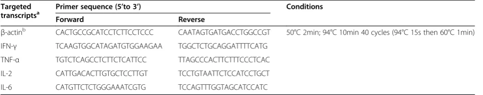
and 0.2μM each of the forward and reverse gene-specific primers (listed in Table 2), aliquoted into 96-well plates (Axygen, CA, USA), and then sealed with optical sealing tape (Axygen, CA, USA). Real-time PCR was performed
Table 2 Relative quantitative real-time PCR primers for targeted transcripts mRNAs Targeted
transcriptsa
Primer sequence (5’to 3’) Conditions
Forward Reverse
β-actinb CACTGCCGCATCCTCTTCCTCCC CAATAGTGATGACCTGGCCGT 50°C 2min; 94°C 10min 40 cycles (94°C 15s then 60°C 1min)
IFN-γ TCAAGTGGCATAGATGTGGAAGAA TGGCTCTGCAGGATTTTCATG
TNF-α TGTCTCAGCCTCTTCTCATTCC TTAGCCCACTTCTTTCCCTCAC
IL-2 CATTGACACTTGTGCTCCTTGT TCCTGTAATTCTCCATCCTGCT
IL-6 CATGTTCTCTGGGAAATCGTG TCCAGTTTGGTAGCATCCATC
a
in triplicate by using an Applied biosystems 7500 Real-time PCR System and universal cycle conditions (2min at 50°C, 10min at 94°C, 40 cycles of 15s at 94°C and then 1min at 60°C). House keeping gene β–actin was mea-sured in parallel, which was used as the internal control. In order to evaluate the expression of the target gene by relative quantity, the threshold cycle (Ct) of the sample of interest was compared to the Ct generated by a refer-ence sample referred to as the calibrator (a sample from nonstimulated splenocytes). Cytokine gene expression was normalized toβ–actin expression by the subtraction of Ct to provideΔCt values. TheΔΔCt was calculated as the difference between ΔCt values for stimulated and nonstimulated splenocytes (the calibrator). Thus, the relative difference in cytokine expression between stimu-lated and nonstimustimu-lated cells was calcustimu-lated as fold change by using equation 2-ΔΔCt.
Statistical analysis
Where specified, an unpairedt-test was used to compare the humoral and cellular immune responses between the different groups. All comparisons were made using two-tailed andP-values of 0.05 or less were considered statis-tically significant.
Competing interests
The authors declare that they have no competing interests.
Acknowledgments
This research work was supported by 863 (Grant No: 2011AA10A2) and Special Fund for Agro-scientific Research in the Public Interest (200903037). We deeply appreciate Keqian Bio Inc. (Wuhan, China) for providing experimental assistance.
Author details
1State Key Laboratory of Agricultural Microbiology, Huazhong Agricultural University, Wuhan, Hubei 430070, People's Republic of China.2Laboratory of Animal Virology, College of Veterinary Medicine, Huazhong Agricultural University, Wuhan, Hubei 430070, People's Republic of China.3Department of Pathology, University of Georgia, Athens, GA 30602, USA.4China Animal Health and Epidemiology Center, Qingdao City, Shandong Province 266032, People's Republic of China.
Authors’contributions
BZ performed the majority of experiments and involved in manuscript preparation. JY participated in editing of the manuscript. JY, PL, RJ, and XY participated part of the experiments. FZ, HC and SC conceived of the study, participated in its design and coordination, and revised the manuscript. All authors read and approved the final manuscript.
Received: 21 December 2011 Accepted: 16 July 2012 Published: 16 July 2012
References
1. Hubalek Z, Halouzka J:West Nile fever–a reemerging mosquito-borne viral disease in Europe.Emerg Infect Dis1999,5:643–650.
2. Campbell GL, Marfin AA, Lanciotti RS, Gubler DJ:West Nile virus.Lancet Infect Dis2002,2:519–529.
3. Gubler DJ:The continuing spread of West Nile virus in the western hemisphere.Clin Infect Dis2007,45:1039–1046.
4. Kulasekera VL, Kramer L, Nasci RS, Mostashari F, Cherry B, Trock SC, Glaser C, Miller JR:West Nile virus infection in mosquitoes, birds, horses, and humans, Staten Island, New York, 2000.Emerg Infect Dis2001,7:722–725.
5. Nash D, Mostashari F, Fine A, Miller J, O'Leary D, Murray K, Huang A, Rosenberg A, Greenberg A, Sherman M,et al:The outbreak of West Nile virus infection in the New York City area in 1999.N Engl J Med2001, 344:1807–1814.
6. Diamond MS:Progress on the development of therapeutics against West Nile virus.Antiviral Res2009,83:214–227.
7. Murray KO, Mertens E, Despres P:West Nile virus and its emergence in the United States of America.Vet Res2010,41:67.
8. Lorenz IC, Allison SL, Heinz FX, Helenius A:Folding and dimerization of tick-borne encephalitis virus envelope proteins prM and E in the endoplasmic reticulum.J Virol2002,76:5480–5491.
9. Zhang Y, Kaufmann B, Chipman PR, Kuhn RJ, Rossmann MG:Structure of immature West Nile virus.J Virol2007,81:6141–6145.
10. Li L, Lok SM, Yu IM, Zhang Y, Kuhn RJ, Chen J, Rossmann MG:The flavivirus precursor membrane-envelope protein complex: structure and maturation.Science2008,319:1830–1834.
11. Sanchez MD, Pierson TC, McAllister D, Hanna SL, Puffer BA, Valentine LE, Murtadha MM, Hoxie JA, Doms RW:Characterization of neutralizing antibodies to West Nile virus.Virology2005,336:70–82.
12. Dauphin G, Zientara S:West Nile virus: recent trends in diagnosis and vaccine development.Vaccine2007,25:5563–5576.
13. Ishikawa T, Wang G, Widman DG, Infante E, Winkelmann ER, Bourne N, Mason PW:Enhancing the utility of a prM/E-expressing chimeric vaccine for Japanese encephalitis by addition of the JEV NS1 gene.Vaccine2011, 29:7444–7455.
14. Kenoutis C, Efrose RC, Swevers L, Lavdas AA, Gaitanou M, Matsas R, Iatrou K: Baculovirus-mediated gene delivery into Mammalian cells does not alter their transcriptional and differentiating potential but is accompanied by early viral gene expression.J Virol2006,80:4135–4146.
15. Boyce FM, Bucher NL:Baculovirus-mediated gene transfer into mammalian cells.Proc Natl Acad Sci U S A1996,93:2348–2352. 16. Sarkis C, Serguera C, Petres S, Buchet D, Ridet JL, Edelman L, Mallet J:
Efficient transduction of neural cells in vitro and in vivo by a
baculovirus-derived vector.Proc Natl Acad Sci U S A2000,97:14638–14643. 17. Tani H, Limn CK, Yap CC, Onishi M, Nozaki M, Nishimune Y, Okahashi N,
Kitagawa Y, Watanabe R, Mochizuki R,et al:In vitro and in vivo gene delivery by recombinant baculoviruses.J Virol2003,77:9799–9808. 18. Hu YC:Baculovirus as a highly efficient expression vector in insect and
mammalian cells.Acta Pharmacol Sin2005,26:405–416.
19. Qiao M, Ashok M, Bernard KA, Palacios G, Zhou ZH, Lipkin WI, Liang TJ: Induction of sterilizing immunity against West Nile Virus (WNV), by immunization with WNV-like particles produced in insect cells.J Infect Dis
2004,190:2104–2108.
20. Barsoum J, Brown R, McKee M, Boyce FM:Efficient transduction of mammalian cells by a recombinant baculovirus having the vesicular stomatitis virus G glycoprotein.Hum Gene Ther1997,8:2011–2018. 21. Facciabene A, Aurisicchio L, La Monica N:Baculovirus vectors elicit
antigen-specific immune responses in mice.J Virol2004,78:8663–8672. 22. Condreay JP, Witherspoon SM, Clay WC, Kost TA:Transient and stable
gene expression in mammalian cells transduced with a recombinant baculovirus vector.Proc Natl Acad Sci U S A1999,96:127–132. 23. Abe T, Hemmi H, Miyamoto H, Moriishi K, Tamura S, Takaku H, Akira S,
Matsuura Y:Involvement of the Toll-like receptor 9 signaling pathway in the induction of innate immunity by baculovirus.J Virol2005,79:2847–2858. 24. Abe T, Matsuura Y:Host innate immune responses induced by
baculovirus in mammals.Curr Gene Ther2010,10:226–231.
25. Aoki H, Sakoda Y, Jukuroki K, Takada A, Kida H, Fukusho A:Induction of antibodies in mice by a recombinant baculovirus expressing
pseudorabies virus glycoprotein B in mammalian cells.Vet Microbiol1999, 68:197–207.
26. Wang S, Fang L, Fan H, Jiang Y, Pan Y, Luo R, Zhao Q, Chen H, Xiao S: Construction and immunogenicity of pseudotype baculovirus expressing GP5 and M protein of porcine reproductive and respiratory syndrome virus.Vaccine2007,25:8220–8227.
27. Fan H, Pan Y, Fang L, Wang D, Wang S, Jiang Y, Chen H, Xiao S: Construction and immunogenicity of recombinant pseudotype baculovirus expressing the capsid protein of porcine circovirus type 2 in mice.J Virol Methods2008,150:21–26.
baculovirus expressing the glycoprotein of rabies virus in mice.Arch Virol
2011,156:753–758.
29. Li Y, Ye J, Cao S, Xiao S, Zhao Q, Liu X, Jin M, Chen H:Immunization with pseudotype baculovirus expressing envelope protein of Japanese encephalitis virus elicits protective immunity in mice.J Gene Med2009,11:57–65. 30. Pletnev AG, Putnak R, Speicher J, Wagar EJ, Vaughn DW:West Nile virus/
dengue type 4 virus chimeras that are reduced in neurovirulence and peripheral virulence without loss of immunogenicity or protective efficacy.Proc Natl Acad Sci U S A2002,99:3036–3041.
31. Despres P, Combredet C, Frenkiel MP, Lorin C, Brahic M, Tangy F:Live measles vaccine expressing the secreted form of the West Nile virus envelope glycoprotein protects against West Nile virus encephalitis.
J Infect Dis2005,191:207–214.
32. Iglesias MC, Frenkiel MP, Mollier K, Souque P, Despres P, Charneau P:A single immunization with a minute dose of a lentiviral vector-based vaccine is highly effective at eliciting protective humoral immunity against West Nile virus.J Gene Med2006,8:265–274.
33. Iyer AV, Pahar B, Boudreaux MJ, Wakamatsu N, Roy AF, Chouljenko VN, Baghian A, Apetrei C, Marx PA, Kousoulas KG:Recombinant vesicular stomatitis virus-based west Nile vaccine elicits strong humoral and cellular immune responses and protects mice against lethal challenge with the virulent west Nile virus strain LSU-AR01.Vaccine2009,27:893–903.
34. Martina BEE, van den Doel P, Koraka P, van Amerongen G, Spohn G, Haagmans BL, Provacia LBV, Osterhaus ADME, Rimmelzwaan GF:A Recombinant Influenza A Virus Expressing Domain III of West Nile Virus Induces Protective Immune Responses against Influenza and West Nile Virus.PLoS One2011,6:e18995.
35. Pierson TC, Fremont DH, Kuhn RJ, Diamond MS:Structural insights into the mechanisms of antibody-mediated neutralization of flavivirus infection: implications for vaccine development.Cell Host Microbe2008,4:229–238. 36. Vogt MR, Moesker B, Goudsmit J, Jongeneelen M, Austin SK, Oliphant T,
Nelson S, Pierson TC, Wilschut J, Throsby M, Diamond MS:Human monoclonal antibodies against West Nile virus induced by natural infection neutralize at a postattachment step.J Virol2009,83:6494–6507. 37. Hoke CH, Nisalak A, Sangawhipa N, Jatanasen S, Laorakapongse T, Innis BL, Kotchasenee S, Gingrich JB, Latendresse J, Fukai K,et al:Protection against Japanese encephalitis by inactivated vaccines.N Engl J Med1988, 319:608–614.
38. Schneeweiss A, Chabierski S, Salomo M, Delaroque N, Al-Robaiy S, Grunwald T, Bürki K, Liebert UG, Ulbert S:A DNA vaccine encoding the E protein of West Nile Virus is protective and can be boosted by recombinant domain DIII.Vaccine2011,29:6352–6357.
39. Shrestha B, Diamond MS:Role of CD8+ T cells in control of West Nile virus infection.J Virol2004,78:8312–8321.
40. Parsons R, Lelic A, Hayes L, Carter A, Marshall L, Evelegh C, Drebot M, Andonova M, McMurtrey C, Hildebrand W,et al:The memory T cell response to West Nile virus in symptomatic humans following natural infection is not influenced by age and is dominated by a restricted set of CD8+ T cell epitopes.J Immunol2008,181:1563–1572.
41. Martina BE, Koraka P, van den Doel P, van Amerongen G, Rimmelzwaan GF, Osterhaus AD:Immunization with West Nile virus envelope domain III protects mice against lethal infection with homologous and heterologous virus.Vaccine2008,26:153–157.
42. Estcourt MJ, Ramshaw A, Ramsay AJ:Cytokine responses in virus infections: effects on pathogenesis, recovery and persistence.Curr Opin Microbiol1998,1:411–418.
43. Spellberg B, Edwards JE Jr:Type 1/Type 2 immunity in infectious diseases.
Clin Infect Dis2001,32:76–102.
44. Abe T, Takahashi H, Hamazaki H, Miyano-Kurosaki N, Matsuura Y, Takaku H: Baculovirus induces an innate immune response and confers protection from lethal influenza virus infection in mice.J Immunol2003,171:1133–1139. 45. Chen GY, Shiah HC, Su HJ, Chen CY, Chuang YJ, Lo WH, Huang JL, Chuang CK,
Hwang SM, Hu YC:Baculovirus transduction of mesenchymal stem cells triggers the toll-like receptor 3 pathway.J Virol2009,83:10548–10556. 46. Liu J, Liu B, Cao Z, Inoue S, Morita K, Tian K, Zhu Q, Gao GF:
Characterization and application of monoclonal antibodies specific to West Nile virus envelope protein.J Virol Methods2008,154:20–26. 47. Li Y, Xu M, Chen L, Zhu J, Ye J, Liu X, Sun Y, Chen H, Cao S:Evaluation of
murine bone marrow-derived dendritic cells loaded with inactivated virus as a vaccine against Japanese encephalitis virus.Vaccine2009, 27:6004–6010.
doi:10.1186/1743-422X-9-132
Cite this article as:Zhuet al.:Induction of antigen-specific immune responses in mice by recombinant baculovirus expressing
premembrane and envelope proteins of West Nile virus.Virology Journal 20129:132.
Submit your next manuscript to BioMed Central and take full advantage of:
• Convenient online submission
• Thorough peer review
• No space constraints or color figure charges
• Immediate publication on acceptance
• Inclusion in PubMed, CAS, Scopus and Google Scholar
• Research which is freely available for redistribution