Structural Insight into the Binding
Interactions of Native and Mutant Structures
of Dihydrofolate Reductases
Vinod Kumar Yata, Anant Saxena, Vinita Sharma
Assistant Professor, Dept. of Biotechnology, Dr B R Ambedkar National Institute of Technology Jalandhar, Punjab,
India
B.Tech Student, Dept. of Biotechnology, Dr B R Ambedkar National Institute of Technology Jalandhar, Punjab, India
B.Tech Student, Dept. of Biotechnology, Dr B R Ambedkar National Institute of Technology Jalandhar, Punjab, India
ABSTRACT: Dihydrofolate reductase (DHFR) is an important enzyme in the cell and is the target for antifolate drugs such as methotrexate, pyrimethamine and trimethoprim. In this study, we compared the binding interactions of trimethoprim with native and mutant structure of DHFR by computational methods. We generated four mutant structures of DHFR by using UCSF chimera. Binding energies of trimethoprim with native and mutant structures of DHFR were computed by using Autodock. Finally, one mutant structure (1YHO_Q35I) which has been generated by computational methods, has shown a high affinity binding affinity towards the trimethoprim the native other mutant structures.
KEYWORDS: Affinity, Dihydrofolate reductase, Docking, Trimethoprim
.
1. INTRODUCTION
Dihydrofolate reductase, DHFR, is an enzyme that has a significant role in regulating the amount of Tetrahydrofolate in the cell [1]. It reduces dihydrofolic acid to tetrahydrofolic acid by using NADPH which can be converted to the Tetrahydrofolate cofactors. Trimethoprim is a bacteriostatic antibiotic and it inhibits the reduction of dihydrofolic acid (DHF) to tetrahydrofolic acid (THF). THF is an essential precursor in the thymidine synthe-sis pathway and interference with this pathway inhibits bacterial DNA synthesis. The mechanism of action of trimethoprim (TMP) is the inhibition of DHFR, an en-zyme that participates in the recycling of folates by reducing dihydrofolate to Tetrahydro-folate in bacteria, pneumocystis and mammalian cells [2].
To carry out the computational analysis, the three-dimensional (3D) structures of native and mutant Dihydrofolate Reductase were taken from the crystal structures of Protein Data Bank [3,4]. Non-covalent molecular interaction between the face of an electron-rich π system and an adjacent cation were calculated for both the native
and mutant structures by protein interaction calculator (PIC). We generated four mutant structures 1YHO_D21V, 1YHO_K18V, 1YHO_Q35I and 1YHO_R91Y by using UCSF chimera. Finally, binding energies of trimethoprim with the native and five mu-tants were calculated by autodock to identify the high affinity structure.
II. EXPERIMENTAL APPROACHES
2.1. Cation- π Interactions
2.2. Generation of mutants
The best mutating residues were selected on the basis of the results of CUPSAT server, whether they were stabilizing or destabilizing, on the basis of torsion angles and ΔΔG values. These residues were replaced with the potentially
important ones, obtained from HotSpot Wizard, in UCSF Chimera 1.10.1 [6].
2.3. Molecular Docking:
Docking of native and mutant structures of DHFR and five model structures with trimethoprim ligand were performed using AUTODOCK version 4.2. [7, 8] Preparation of protein structures: Protein molecules were added to Molecular Graphics laboratory (MGL) Tools Version 1.5.6 and these tools were used to remove water molecules from these molecules and hydrogen’s were also added with Kollman charges to the protein molecule. Finally all the files were saved in PDB format.
2.4. Ligand preparation: Tripmethoprim molecule is taken from RCSB, protein data bank, with DrugBank ID- DB03125. The three dimensional structure was retrieved in SDF format after that UCSF Chimera was used to convert in inot PDB file.
2.5. Docking procedure: Protein and ligand molecules were loaded in Autodock tools win-dow and Grid box was built with respect to urea binding sites of that particular protein structures. Default parameters were set for all the docking. The resultant Grid files were saved in .gpf file format. Docking file was prepared with the help of loaded molecules in .dpf file format. Autogrid4.exe file was used for preparing grid log file and map files. Autodock4.exe file was used for preparing docking log file. Resultant (protein-TMP complex) files were saved in PDB format for further analysis.
Table.1.Cation-pi interactions
Donor Residue in Native type (PDB code- 1YHO)
Acceptor Residue in Native type (PDB code- 1YHO) Distanc e ( A°) Angle (in Degrees) Donor Residue in Native type (PDB code- 3S3V)
Acceptor Residue in Native type (PDB code- 3S3V) Distanc e ( A°) Angle (in Degrees)
PHE34 ARG70 5.90 95.18 TYR33 ARG36 5.48 110.07
TRP57 ARG65 3.78 31.81 PHE34 LYS35 4.82 94.28
PHE58 LYS54 3.94 166.69 TRP57 ARG65 5.73 51.16
III. RESULTS AND DISCUSSION
3.1.Cation-Pi interactions
Cation-Pi interactions of TPM-DHFR complexes were calculated by Protein Interac-tions Calculator (PIC) is a server. Naitve and mutant structures of DHFR which are avi-alble in pdb database were chosen for this study. Distance between the two aminoacids and angles were computed for both the structure and values are compared in Table.1.a total of four Cation-Pi interactions were observed in native DHFR-TMP complex. Among four TRP57 –ARG65 is shortest distance Cation-Pi interaction with a distance of 3.75 A° and PHE34-ARG70 is the longest distance with a distance of 5.9 A°.PHE-LYS132 is found to be highest bond angle cation-Pi interaction with a bond angle of 166.69º and TRP57-ARG65 found to be smallest bond angle cation-Pi interaction with a bond angle of 31.81º. Eight Cation-Pi interactions were observed in mutant DHFR-TMP complex. PHE34 –LYS35 is shortest distance Cation-Pi interaction with a dis-tance of 4.82 A° and TYR156-LYS155 is the longest distance with a distance of 5.9 A°. PHE64-LYS63 is found to be highest bond angle cation-Pi interaction with a bond angle of 136.08º and TRP57-ARG65 found to be smallest bond angle cation-Pi interac-tion with a bond angle of 51.16º. in conclusion, mutant structure with TMP complex has shown comparativevely better Cation-Pi interactions than native structure with TMP complex.
Generation of mutants
In our previous study, the following residues were obtained as potentially important sites for mutagenesis Pro26, Asp21, Arg91, Lys18, Gln35 with a mutability score of 9, 9, 9, 6 and 6. These residues were then checked for stabilizing and destabilizing alter-natives in the CUPSAT server and the following results were obtained on the basis of
of torsion angles and ΔΔG values. On the basis of torsion angle’s stability and the value of ΔΔG values the best amino
acid residues were selected and then mutated using UCSF Chimera. Five model structures were obtained by mutating the following resi-dues 1YHO_D21V, 1YHO_K18V, 1YHO_P26K, 1YHO_Q35I and 1YHO_R91Y.
Table.2. Binding energy scores obtained by AutoDock for different mutants of native DHFR
Mutants generated by Chimera
Binding Energy (kcal/mol)
Interacting amino acids Distance (in angstrom)
1YHO_D21V -5.19 GLU30
LEU22 VAL8
2.19 2.24 2.6
1YHO_K18V -7.59 GLU30
THR136 VAL8
2.13 2.30 2.17
1YHO_Q35I -7.81 GLU30
VAL8 PHE31
2.20 2.11 2.18
1YHO_R91Y -7.39 GLU30
PHE31 VAL8
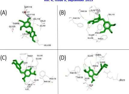
Figure 1. Mutant structures of native dihydrofolate reductase visualized by Discovery Studio. (A) 1YHO_D21V (B) 1YHO_K18V (C) 1YHO_Q35I (D) 1YHO_R91Y. TMP shown in Green color. Hydrogen bonds are
indicated as Green dotted lines.
3.2. Molecular docking
The native and the mutant structures of DHFR were docked in our previous study and the binding energies were -5.31 and -1.57 kcal/mol respectively [9]. Mutant structures of DHFR which are generated by chimera were docked with TMP in the same binding pocket where native structure was docked. The binding energies were shown in Table.2. The results of molecular docking shows that the binding energy of mutant DHFR (3S3V) is more than that of native DHFR (1YHO) and hence the entire focus shifted on molecular docking of the mutants generated for the native DHFR structure. From the Table2, the model structure 1YHO_Q35I is the most stable mutant with the most negative binding energy and will possibly bind best with the ligand (trimethoprim). Docking poses of 1YHO_D21V, 1YHO_K18V, 1YHO_P26K, 1YHO_Q35I and 1YHO_R91Y with TMP were visualized in discovery studio (Figure 1). TMP has shown has major interaction with GLU30,VAL8, PHE31of 1YHO_Q35I with a binding energy of 7.81 kcal/mol.GLU30 plays important role in interaction of TMP with all the mutated structures of DHFR generated by chimera.
human DHFR protein with a novel binding site using PdbSum, when mu-tated with Isoleucine at 35th position of Glutamine, resulting in high binding affinity because of lower binding energy.
REFERENCES
[1] Del Santo, Molly, and Mamoru Nozawa. "Human Dihydrofolate Reductase."Structure and Function of Proteins (2006): 12.
[2] Chen, Mann-Jy, et al. "The functional human dihydrofolate reductase gene."Journal of Bio-logical Chemistry 259.6 (1984): 3933-3943.
[3] Kovalevskaya, Nadezhda V., et al. "Solution structure of human dihydrofolate reductase in its complex with trimethoprim and NADPH." Journal of biomolecular NMR 33.1 (2005): 69-72
[4] Cody, Vivian, et al. "Crystallographic analysis reveals a novel second binding site for trimethoprim in active site double mutants of human dihydrofolate reductase." Journal of struc-tural biology 176.1 (2011): 52-59.
[5] Tina, K. G., Rana Bhadra, and Narayanaswamy Srinivasan. "PIC: protein interactions calcu-lator." Nucleic acids research 35.suppl 2 (2007): W473-W476.
[6] Pettersen, Eric F., et al. "UCSF Chimera—a visualization system for exploratory research and analysis." Journal of computational chemistry 25.13 (2004): 1605-1612.
[7] Morris, G. M., Huey, R., Lindstrom, W., Sanner, M. F., Belew, R. K., Goodsell, D. S. and Olson, A. J, Autodock4 and AutoDockTools4: automated docking with selective receptor flexiblity. J. Compu-tational Chemistry 2009, 16: 2785-91.
[8] Goodsell, D. S., Morris, G. M. and Olson, A. J. (1996), Automated Docking of Flexible Ligands: Applications of AutoDock J. Mol. Recognition, 9: 1-5.