_____________________________________________________________________________________________________
*Corresponding author: E-mail: [email protected], [email protected];
www.sciencedomain.org
Rhinal Snuff: A Possible Gateway for Sinonasal
Malignancy
Pratiksha Hada
1*, Vikram Singh
1, Sakshi Sharma
1and Shivam Dubey
11
Department of Oral Medicine and Radiology, RKDF Dental College and Research Centre, NH – 12, Bhopal, M.P., India.
Authors’ contributions
This work was carried out in collaboration between all authors. All authors read and approved the final manuscript.
Article Information
DOI: 10.9734/BJMMR/2017/31300 Editor(s): (1) Emad Tawfik Mahmoud Daif, Professor of Oral & Maxillofacial Surgery, Cairo University, Egypt. (2) Ibrahim El-Sayed M. El-Hakim, Ain Shams University, Egypt and Riyadh College of Dentistry and Pharmacy, Riyadh,
Saudi Arabia. (3)Salomone Di Saverio, Emergency Surgery Unit, Department of General and Transplant Surgery, S. Orsola Malpighi University Hospital, Bologna, Italy. Reviewers: (1) Marwa Mokbel ElShafei, Misr International Univeristy, Egypt. (2)Sucheta Bansal, Himachal Institute of Dental Sciences, Paonta Sahib, India. (3)Mehmet Zahit Adisen, Kirikkale University, Turkey. (4)Sameep S. Shetty, Manipal College of Dental Sciences, Manipal University, Manipal, India. Complete Peer review History:http://www.sciencedomain.org/review-history/17999
Received 30th December 2016 Accepted 15th February 2017 Published 1st March 2017
ABSTRACT
Aim: To present a case report of malignancy of maxillary antrum invading the oral cavity
associated with the habit of nasal snuff.
Presentation of Case: We describe here a case of a 40-year-old man who developed malignancy
of maxillary antrum invading the hard palate after 20 years of snuff usage. He reported to our Department with the chief complaint of swelling and nasal discharge from right nostril since 4 months. On further questioning, patient revealed that he noticed swelling on the right middle 1/3rd of the face along with clear watery discharge from right nostril. Simultaneously, he also noticed a small pea sized swelling on the surface of hard palate which was gradual in onset and progressed to attain the present size.
Discussion: Use of smokeless tobacco products is common worldwide, with its increasing
consumption in the developing countries and nasal snuff being the primeval form. Malignancy of the maxillary antrum is an uncommon neoplasm with a small percentage of occurrences. Different studies revealed that continuous use of nasal snuff proved to be carcinogenic.
Conclusion: Malignancy of maxillary antrum is relatively uncommon and its treatment poses
several challenges to the head and neck surgeons, radiotherapists and medical oncologists. Nasal snuff is a deleterious habit and its prolonged usage proves to be fatal to the patient’s life, as happened in this case. Hence the use of tobacco in any form should be condemned in the society.
Keywords: Nasal snuff; maxillary antrum; sinonasal malignancy; smokeless tobacco; nicotine; carcinogenic.
1. INTRODUCTION
Through the ages, use of tobacco is a great health problem and has been used extensively by communities in various forms, the most common being the smoking and smokeless tobacco. The major difference between smoking and the smokeless forms of tobacco is the absence of carbon monoxide and oxides of nitrogen and tar. Smokeless tobacco is consumed without burning the product and is ingested either orally or nasally. Among them, nasal snuff being the oldest form [1]. The inhalation of nasal snuff is a common addiction in the Indian subcontinent because a large variety of homemade snuff is easily available. Snuff is made from ground or pulverized tobacco leaves. It is insufflated or “snuffed” into the nasal cavity, delivering a swift hit of nicotine and a lasting flavored scent. According to various literatures, some people consider nasal snuff as a safe alternative of smoking as well as chewing tobacco, but this rare habit of nasal snuff proves to be carcinogenic for paranasal sinuses [2].
Majority of the paranasal sinus malignancies originate from maxillary antrum, of which squamous cell carcinoma is the commonest histological variety [2]. Malignancy of maxillary antrum is a relatively rare neoplasm, representing a small percentage (0.2%) of human malignant tumors [2,3]. The incidence seems to vary in different parts of the world, with highest number of cases being reported in the developing countries [3].
Publications on the incidence of malignancy of maxillary antrum and its relation to taking nasal snuff have been reported very rarely because of the limited spread of this form of tobacco abuse today [3]. We now report a case of malignancy of maxillary antrum invading hard palate with special emphasis laid on usage of nasal snuff.
2. CASE REPORT
In October 2015, a 40-year-old male patient presented to the Department of Oral Medicine
and Radiology with the chief complaint of swelling and nasal discharge from right nostril since 4 months. On further anamnesis, patient revealed that he noticed swelling on the right middle 1/3rd of the face along with clear watery discharge from right nostril. Simultaneously, he also noticed a small pea sized swelling on the surface of hard palate which was gradual in onset and progressed to attain the present size.
Further questioning revealed that the patient had habit of using nasal snuff since 20 years, with a frequency of 3 to 4 times per day. His medical as well as dental history was irrelevant and vital signs were normal.
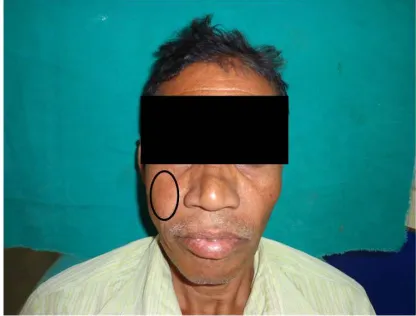
Clinical examination showed slight swelling on the right middle 1/3rd of the face [Fig. 1]. Two right submandibular lymph nodes were palpable, approximately 1x1.5 cm in size, firm in consistency, non-tender, non-indurated and mobile. Intraorally, a single ulcerative growth was present on right posterior slope of hard palate, roughly oval in shape measuring approximately 4×4 cm crossing the midline. Its medio-lateral extension was 1.5 cm away from 25, 26 to alveolar bone of right maxilla and further extending upwards involving upper right vestibular region. Surrounding mucosa appeared normal [Fig. 2]. On palpation, inspectory findings with respect to site, shape, size and extent were confirmed. Margins were irregular and the ulcer was tender and bleeding on palpation was also evident. An initial impression of malignancy of maxillary antrum was made.
Differential diagnoses of mucoepidermoid carcinoma, acinic cell carcinoma and adenoid cystic carcinoma were considered.
carcinoma was also ruled out as no facial nerve paralysis was seen and mostly it is not associated with the habit of nasal snuff.
Fig. 1. Front profile of the patient showing swelling on the right side of middle 1/3rd of
the face
Fig. 2. Intra oral ulcerative growth present on the hard palate and right alveolus
Panoramic radiograph revealed a radiolucent shadow measuring 3×3 cm on right half of maxilla involving the palate and the maxillary antrum. There was destruction of right maxillary alveolus [Fig. 3].
Water’s view showed a well-defined radio opacity extending into the right maxillary antrum with obliteration of the sinus space and destruction of lateral wall of the nose and medial wall of the orbit [Fig. 4].
The Chest X-ray showed multiple enlarged hilar lymph nodes suggestive of lung metastasis [Fig. 5]. T4N2aM1 staging was given.
CT Scan showed a hypo dense lobulated soft tissue mass of approximately 6×6 cm in size in the right maxillary cavity causing significant surrounding bony destruction with destroyed bony maxillary walls, right lateral nasal wall, right maxillary alveolus, right nasal cavity and masticator space [Fig. 6].
Biopsy specimen (10x) revealed stratified squamous epithelial cells invading the deeper connective tissues showing numerous keratin pearls [Fig. 7].
Based upon the above investigations, final diagnosis of well differentiated squamous cell carcinoma of maxillary antrum involving hard palate was reached. Patient was referred to Jawaharlal Nehru Cancer Hospital where 6 cycles of chemotherapy were advised for the patient followed by palliative care for his terminal illness. Later, patient succumbed within 6 months time.
3. DISCUSSION
Sinonasal malignancies are very difficult to diagnose and are associated with a poor prognosis. Although, majority of these malignancies are locally advanced because its symptoms are nonspecific [4]. They are often confused with allergies or sinus infection and are neglected for a long time. They tend to remain localized to maxillary antrum and during evolution they invade adjacent structures such as bone, base of the skull, facial soft tissue, oral cavity, and orbits, hence the first line of treatment remains to be radiotherapy followed by surgical intervention and reconstruction [4,5]. Sinonasal malignancy has a predilection for males with a ratio of 2.3:1 and the mean age of occurrence being 64 years [6].
relatively higher amounts in snuff as compared to other forms of tobacco [10,11].
Nicotine is the main psychoactive substance in tobacco. It is absorbed very rapidly through the
nasal mucosa into the maxillary sinus and within 10 minutes, blood nicotine concentrations are higher than that of smoked tobacco [12]. The variation of nicotine consumption depends on the strength of snuff, the size of the pinch, and the
Fig. 3. Orthopantomograph showing a radiolucent shadow on right half of maxilla involving the palate and the maxillary antrum with the destruction of right maxillary alveolus
Fig. 4. Water’s view showing a well-defined radio opacity extending into the right maxillary antrum with obliteration of the sinus space and destruction of lateral wall of the nose and
way it is snuffed. The nasal absorption of nicotine is influenced strongly by the pH of the snuff, if alkaline the nicotine is completely protonated and the rate of absorption is very high [13].
For some people snuff may be a satisfactory and acceptable substitute for cigarette smoking, but it will not decrease the number of cancers
seemingly caused by tobacco usage, it will merely change the site of occurrence [14,15]. The close proximity of usage of nasal snuff and it being the most important rationale of sinonasal malignancies, makes it life threatening and fatal for many people as happened in the above reported case. “Early detected is well cured” is the key to success for such cases.
Fig. 5. The chest X-ray showing multiple enlarged hilar lymph nodes suggestive of lung metastasis
Fig. 6. CT Scan (transverse section) showing significant bony destruction with destroyed bony maxillary walls, right lateral nasal wall, right maxillary alveolus, right nasal cavity and
Fig. 7. Biopsy specimen (10x) revealed stratified squamous epithelial cells and numerous keratin pearls
4. CONCLUSION
Malignancy of the maxillary antrum is relatively uncommon and its treatment poses several challenges to the head and neck surgeons, radiotherapists and medical oncologists. The reasons for the poor outcomes are that they often present in advanced stages, the complex anatomy and the close proximity of critical structures compromise effectual surgical excision and radiation deliverance, and substantial uncertainty surrounds fundamental aspects of treatment, hence the optimal therapy remains to be defined. Conflicting ideas are found concerning the uses of snuff, a substitute for other forms of tobacco as snuff does not contaminate the atmosphere for non-users but the long term use of nasal snuff induces malignant changes and its usage should not be promoted in the society.
CONSENT
As per international standard or university standard written patient consent has been collected and preserved by the authors.
ETHICAL APPROVAL
It is not applicable.
COMPETING INTERESTS
Authors have declared that no competing interests exist.
REFERENCES
1. Temple DJ. The absorption of nicotine from tobacco snuff through the nasal mucosa. Arch Pharm. 1976;304(12):745-9.
2. Evens G, Treyer F. The old snuff. London: Nabu Press; 1920.
3. Sapundzheiv N, Werner JA. Nasal snuff: Historical review and health related aspects. J Laryngol Otology. 2003;117(9): 686-91.
4. Nunez F, Suarez C, Alvarez I, Losa JL, Barthe P, Fresno M. Sino – nasal adenocarcinoma: Epidemiological and clinic-pathological study of 34 cases. J Otolaryngol. 1993;22(2):86-90.
the maxillary antrum mimicking invasive fungal sinusitis: The diagnostic dilemma of an extensive paranasal sinus mass. J Health Res Rev. 2015;2:112-5.
6. Sharma S, Sharma SC, Singhal S. Carcinoma of the maxillary antrum. A 10-year experience. Ind J Otolaryngol. 1991; 43:191-4.
7. Holm H, Jarvis M, Russel M, Feyerabend C. Nicotine intake and dependence in Swedish snuff takers. Psycho- pharmacology (Berl). 1992;108:507-11. 8. Boffetta P, Hecht S, Gray N, Gupta P,
Straif K. Smokeless tobacco and cancer. Lancet Oncol. 2008;9(7):667-75.
9. Asplund H. Snuff – how dangerous it is? The controversy continues. J Intern Med. 2001;250:457-61.
10. Mehanna P, Smith G. Maxillary Carcinoma- A wolf in sheep’s clothing. Can Fam Physician. 2009;55(3):262-4.
11. Russell MA, Jarvis MJ, Feyerabend C. A new age for snuff. The Lancet. 1980; 1(8166):474-5.
12. Vasudevan V, Kailasam S, Radhika MB, Venkatappa M, Devaiah D, Shrihari TG, et al. Well differentiated squamous cell carcinoma of maxillary sinus. J Indian Aca Oral Med Radiol. 2012;24(3):250-4. 13. Popovic D, Milisavljevic D. Malignant
tumors of the maxillary sinus. A ten – year experience. Med Biol. 2004;11(1):31-4. 14. Qureshi SS, Devendra AC, Talole Sanjay
DT, Dcruz AK. Squamous cell carcinoma of the maxillary sinus: A Tata Memorial Hospital experience. Indian J Cancer. 2006;43(1):26-9.
15. Sreedharan S, Hedge MC, Pai R, Rhodrigues S, Kumar R, Rasheed A. Snuff- induced malignancy of the nasal vestibule: A case report. American J Otolaryngol. 2007;28:353-6.
_________________________________________________________________________________
© 2017 Hada et al.; This is an Open Access article distributed under the terms of the Creative Commons Attribution License (http://creativecommons.org/licenses/by/4.0), which permits unrestricted use, distribution, and reproduction in any medium, provided the original work is properly cited.
Peer-review history: