Title: How Cadaver Bone Transplants Mineralize and Sclerotic Bone Fails
Author: Gregory Gene Steiner DDS MS
Abstract
Background: Cadaver bone is possibly the most common transplant material used today. Common types of cadaver bone transplants are freeze dried bone allografts and xenografts. In the case of freeze dried bone allograft transplants, it was theorized that these materials mineralize by way of osteoinduction and stimulating osteogenesis. However, these theories have been proven false. It has been proposed that these materials mineralize via osteoconduction however, there are no studies to support this hypothesis. This study was undertaken to determine how these transplants mineralize and what type of bone they produce.
Materials and Methods: This study is a histological analysis of human cadaver bone graft healing from the incipient stages of mineralization through completed mineralization. All cadaver bone grafts used for evaluation in this study were particulate bone graft materials in the maxilla or mandible. No block grafts were evaluated.
Results: The mineralization of cadaver bone transplants was produced by an inflammatory response to the transplanted tissue. The histologic findings of the mineralized bone produced by this process was sclerotic bone. No resorption of cadaver bone graft particles was found. When loaded the sclerotic bone was found to fail through an accumulation of microfractures.
Conclusions: Particulate freeze-dried bone allografts and xenografts do not heal via the normal processes of mineralization. Cadaver bone grafts produce significant inflammation and are hypothesized to mineralize by a process termed antigenic ossification. The process of antigenic ossification produces sclerotic bone that is not capable of self-repair which can ultimately lead to bone failure.
Keywords: allograft, xenograft, inflammation, sclerotic bone, implant failure, antigenic ossification.
Corresponding author: Gregory Gene Steiner Email: [email protected]
Affiliation: CEO SteinerBio 1051 Olsen Street Building 3611 Henderson, Nevada USA 89011
Preprints (www.preprints.org) | NOT PEER-REVIEWED | Posted: 15 October 2018 doi:10.20944/preprints201810.0300.v1
Introduction
The popularity of freeze dried cadaver bone transplants for use in the treatment of skeletal defects coincided with the findings of Urist et al. (1), which identified the presence and function of bone morphogenic proteins (BMPs). With the discovery that freeze-dried cadaver bone contained proteins that have the potential for osteoinduction and stimulating osteogenesis, it was assumed that the growth factors in cadaver bone also stimulated bone growth when grafted into humans. However, beginning in the 1990’s, research showed that autografts and freeze-dried allografts are not osteoinductive in humans (2-7). Likewise, studies proved that freeze-dried cadaver bone does not stimulate osteogenesis in humans (8-12). Most current publications now claim that allografts are osteoconductive, presuming that this is the only other way bone can mineralize. However, again, there is no scientific support for the assumption that freeze-dried allografts or xenografts are osteoconductive.
Since cadaver bone is not osteoinductive, does not stimulate osteogenesis, and there is no scientific support showing that cadaver bone is osteoconductive, the question remains as to the process by which these transplants produce mineralization. Irrespective of the extensive use of cadaver bone for bone grafting over decades of use and hundreds of histologic studies, there is no published histologic research that can be found on how these materials produce mineralization. This article will present the early histology of cadaver bone mineralization and discuss the type of tissue that these materials produce. With the assessment of the histology of the mineralization produced by these materials a new process of mineralization will be proposed.
Materials and Methods
All the transplants evaluated were particulate bone grafts. Two types of bone grafts were evaluated: particulate mineralized freeze-dried bone allograft and particulate low temperature bovine xenograft. Human histologic samples were taken at various time points. The early histologic samples were taken from healing extraction sites during the mineralization period. The samples were acquired from patients who requested that the cadaver bone be removed from their jaw and replaced with synthetic resorbable graft materials. The removal of the cadaver bone grafts was at the request of the patients due to fear of the patient that the presence of the cadaver bone graft material might pose a future health threat. The histologic samples taken of mineralized freeze-dried allografts in the mineralizing phase, were from patients who were clinically asymptomatic and had no signs of pathology. Histologic samples of fully healed cadaver bone graft sites were taken via a trephine at the time of implant placement. The sites had no clinical signs of pathology. Histology and clinical cases of sclerotic bone were obtained from patients who were treated for implant failure after grafting with cadaver bone. None of these patients were enrolled in a controlled clinical trial. All cases were retrospective. All patients gave written permission consenting that any tissue removed during routine therapy could be used for scientific evaluation. All trephine bone core samples were demineralized and stained with H and E.
Results
Early particulate freeze-dried allograft mineralization
histologically was found to be infected with actinomycetes with no bone formation (data not shown).
In Figure 1A, the radiograph of extraction socket #7 at seven weeks shows a normal appearance. However, there is a clear distinction between the extraction socket wall and the bone graft in the socket. There is no evidence that bone is growing from the socket wall into the extraction socket. In Figure 1B, the cadaver bone particles are identified and the black arrow points to newly formed mineralized tissue on the surface of an allograft particle. New mineralization is only present on the surface of the allograft particles. There is no growth of mineralized tissue from the periphery which indicates that this bony defect is not healing via osteoconduction. Figure 1C is a high-power view of an allograft particle with new mineralization forming on the surface as identified by the black arrow. What is also evident in image 1C is intense inflammation with the presence of cytotoxic T cells. The identification of the cells as cytotoxic T cells was made by the pathology department at Queens Hospital, Honolulu, Hi. This finding is understandable because these cells are involved in organ rejection during organ transplantation (13). By comparison figure 1D is from a site grafted with a synthetic biocompatible graft material six weeks prior to this histology. The histology shows robust bone growth and no inflammation.
Figure 1
Figure 1. Mineralized freeze-dried bone allograft. (A) Radiograph of extraction socket. (B and C) Hematoxylin and Eosin staining of mineralized allograft site containing intense inflammatory infiltrate. The black arrows indicate incipient mineralization. (D) Hematoxylin and Eosin staining of biocompatible synthetic bone graft (Socket Graft) showing robust osteogenesis and no inflammation.
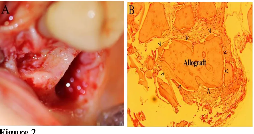
Figure 2 is of a molar extraction site, 8 weeks after extraction and grafting with particulate mineralized freeze-dried bone allograft and covered with a collagen membrane. The patient requested that the cadaver bone be removed and the socket grafted with a synthetic biocompatible resorbable bone graft. Figure 2A is a photograph of the molar extraction site after the cadaver bone had been removed. Note, that no bone formation has occurred on the extraction socket wall at eight weeks indicating that osteoconduction is not occurring. Figure 2B is a high-power magnification showing an allograft particle covered with new mineralization and a decrease in inflammation. There is no evidence of mineralization proceeding from the periphery, which again rules out osteoconduction. At this time frame, the only mineralization present is a thin layer of mineralization covering the allograft particle.
In the histology of the early mineralization process of allograft particles, the amount of mineralization present is inversely related to the amount of inflammation present in the tissue. As the allograft particles become covered by mineralized tissue this isolates the foreign antigenic graft material from the hosts’ immune system and inflammation subsides. Normal bone formation does not occur in the presence of inflammation. The theories of allograft particles stimulating osteogenesis through the release of osteoinductive factors such as BMP-2 requires the resorption of the allograft bone first in order to release these molecules from the mineralized matrix of the allograft particle. There were no osteoclasts present in these samples.
The histology of allograft mineralization rules out mineralization via osteoinduction or the stimulation of osteogeneses in addition to ruling out mineralization via osteoconduction.
Figure 2
Figure 2. Mineralized freeze-dried bone allograft. (A) Photograph of molar extraction site after allograft was removed. (B) Hematoxylin and Eosin staining of mineralized freeze-dried allograft particle. The black arrows identify the new mineralization that is covering the surface of the allograft particle.
composed of allograft particles covered in a thin layer of mineralization. When a non-biocompatible bone graft material such as mineralized freeze-dried bone allograft is used, the particles on the surface are either completely covered in mineralized tissue or encased in granulation tissue. In figure 3B both situations are occurring. Where the granules are encased in mineralization there is no inflammation. However, where the bone graft granules are loose in the soft connective tissue, bleeding and inflamed granulation tissue is present. When a biocompatible bone graft material is used, the surface graft particles are not covered in mineralized tissue and no inflammation is present. This finding supports the concept that transplanted tissue produces mineralization for the purpose of encapsulating the antigenic graft material and isolating it from the host. Figure 3C is a core sample taken from the second molar site at the time of implant placement. The histology shows remaining allograft particles encased in sclerotic bone. Sclerotic bone is found in other bony lesions such as arthritic joints and calcified arteries and is formed in response to inflammation (14,15). Figure 3D is a site that was grafted with mineralized allograft prior to implant placement. The Implant was in function for a number of years before it failed abruptly with the diagnosis of sclerotic bone failure.
Figure 3
Figure 3. Sclerotic bone from mineralized freeze-dried bone allograft. (A) Radiograph of extraction site 6 months after grafting with mineralized freeze-dried bone allograft. Black arrows indicate radiolucent boarder around graft site. (B) Photograph of the ridge 6 months after grafting. (C) Hematoxylin and Eosin staining showing sclerotic bone. (D) Radiograph of site that was grafted with allograft prior to implant placement showing bone failure of grafted bone.
Figure 4 shows a site grafted with Bio-Oss. Bio-Oss is a low temperature bovine xenograft that retains bovine matrix proteins. The bone graft was in place for 10 years. The radiograph in figure 4A shows a very dense mineralized area mesial to the molar. The image in figure 4B is the trephine immediately after taking a core sample from the densely mineralized tissue. Particles of Bio-Oss graft material are obvious but what is most striking, is the lack a vascularity with essentially no bleeding. Image 4C is the demineralized histology stained with H and E. With the mineralization removed from the Bio-Oss particles, the histology shows the collagen matrix that was inside the mineralized bovine bone. The bone in the histologic sample is amorphous with no cement lines and no lamellar bone. No resorption is occurring in association with either the Bio-Oss particles or the mineralization produced by the patient. There is no blood supply is evident. This histology is pathognomonic for sclerotic bone.
Sclerotic bone failure
Mineralized cadaver bone that contains proteins, irrespective of whether it is of animal or human origin, produces sclerotic bone (16). While sclerotic bone is pathologic, the question remains about how this affects the ability of this tissue to support load. A study published in the Journal of Periodontology found that the most significant factor associated with marginal bone loss was implants placed in sites grafted with cadaver bone (17,18). Most dentists think of implant failure as a result of infection, referring to the process of bone loss as periimplantitis. In periimplantitis, as is periodontal disease, the bone loss is a result of an infection and the bone is resorbed in advance of the invading bacteria. Therefore, the granulation tissue never contains any bone particles. However, sclerotic bone in other parts of the human body fails by way of microfracture; not infection. This process was well documented by studying the formation of
sclerotic bone in the joints of race horses (19)
Joint inflammation resulting from trauma or disease converts normal bone into sclerotic bone (19). Once sclerotic bone is formed, it is unable to be reversed and with continual loading microfractures develop in the sclerotic bone. Microfractures are a common occurrence in normal bone as the microfracture allows bone to absorb load without resulting in a complete fracture of the bone (20). However, normal bone microfractures can be repaired because normal bone contains basic multicellular units that are comprised of both osteoblasts and osteoclasts that remodel the microfracture to repair the damaged bone. In sclerotic bone produced by cadaver bone grafts, there are no functioning basic multicellular units, and therefore this bone lacks the ability to repair microfractures. As shown in the horse sclerotic bone model, the microfractures are unable to be repaired and the microfractures accumulate until the bone collapses (19). It is proposed that this same process occurs in sites that are grafted with cadaver bone.
Figure 5
The radiograph in Figure 6A is of a mobile implant that was placed approximately 4 years prior to implant loss. Upon communication with the surgeon that removed the molar, the site was grafted with a combination of Bio-Oss and mineralized freeze-dried bone allograft in 1998. Figure 6B shows removal of the implant, which contained a portion of the bone that was prepared for histology. Figure 6C shows the histology of the grafted site which shows allograft particles and Bio-Oss particles encased in sclerotic bone. The differing size of the osteocyte lacuna allows the determination of what particles are from the allograft and what particles are bovine. After approximately 18 years, there was no resorption of the cadaver bone graft particles and the implant was lost due to bone graft failure after being functional for approximately 4 years. Figure 6 shows a failed combination allograft/Bio-Oss bone graft after 1.5 years in function. The images of these two cases of bone failure are very similar.
Figure 6
Figure 6. Failure of combination Allograft/Bio-Oss. (A) Radiograph of failed implants approximately 4 years after implant placement. (B) Photograph of the removed implant with attached bone used for histology. (C) Hematoxylin and Eosin staining showing sclerotic bone. (D) Radiograph of a failed combination allograft/Bio-Oss bone graft.
Discussion
Normal bone formation is produced by the induction of bone stem cells, stimulating osteogenesis or osteoconduction. Contrary to current dogma normal bone formation is free of inflammation. Our entire skeleton is formed free of inflammation and therefore a regenerative process that uses the same mechanism of bone formation will be fee of inflammation as shown by the histology of the biocompatible bone graft materials. The concept that bone regeneration requires an inflammatory process comes from studying how long bone fractures heal. Of course, when a bone is fractured inflammation will be present, but it subsides before osteogenesis begins. Another difference in fractured long bone healing is that it is endochondral bone that goes through a cartilaginous phase (callous) prior to osteogenesis. However, the maxilla and mandible are intramembranous bones and do not have a cartilaginous phase. When holes are created in the maxilla or mandible they proceed directly with osteogenesis with no callous formation. It is well known that inflammation inhibits bone formation and induces bone resorption. The immune response to cadaver bone transplants produces inflammation that blocks the normal processes of bone formation. The presence of inflammation can induce cells other than osteoblasts to become mineralizing cells. In cardiovascular disease, it is known that the cells that form the mineralized tissue in our arteries are reprogramed smooth muscle cells (21). The mineralized tissue that forms in our arteries is bone but is not produced by cells of osteogenic origin. The inflammation caused by cardiovascular disease prompts smooth muscle cells to be deprogramed and these cells are then reprogramed to produce mineralized tissue. In the presence of inflammation, endothelial cells can also become reprogramed into mineralizing cells that produces the calcified tissue found in atherosclerosis (22).
Bone that forms in soft tissues outside the skeleton is called as heterotopic ossification. In the presence of inflammation caused by trauma, bone forms in muscle through an inflammatory process that stimulates endothelial cells to be reprogramed into mineralizing cells. The process of endothelial-mesenchymal transition in the presence of inflammation is postulated as the process that produces ectopic mineralization (23).
The conversion of smooth muscle cells in arteries into mineralizing cells is considered a protective mechanism. The bone produced in arteries surrounds the soft plaques and prevents them from breaking loose and producing an infarct. The mineralization process on cadaver bone particles occurs on the surface of the graft particles and it is proposed that this process is also protective. Mineralized freeze-dried bone allograft particles cannot be resorbed and the mineralization on the surface of the allograft particles isolates the foreign inflammatory tissue from the host. Therefore, it is postulated that cells that produce mineralization in sites grafted with cadaver bone may be reprogramed endothelial cells.
It is proposed that in order to protect the host, the same process of mineralization found in heterotopic ossification is used in the mineralization of particulate cadaver bone grafts. There are two significant differences between heterotopic ossification and the mineralization produced by particulate cadaver bone transplants. The mineralization produced by cadaver bone transplants is in bone and the inflammation is not cause by trauma, but by an immune response. The process of mineralization of cadaver bone transplants is therefore given a specific term called antigenic ossification.
The term sclerotic is defined as the inability of a tissue to respond or adapt to change. There are over 200 histologic studies on allograft cadaver bone in the literature. The studies of mineralized freeze-dried bone allografts report that an approximate average of 30% of the area is comprised of retained graft particles. Studies have shown that the percentage of retained graft particles does not change over time (24). Mineralized freeze-dried bone allograft particles have never been reported to be in the process of resorption and there is no histology in the literature showing an osteoclast in a resorption lacuna on the surface of an allograft particle. There are no osteoclasts in Figure 3C and no remodeling has occurred. Basic multicellular units are required for bone remodeling and there are no basic multicellular units are found in this histology. Therefore, the bone is termed sclerotic.
Different types of cadaver bone heal through the same process, but with varying mineral density related to the degree of inflammation produced. The antigenic response would be expected to be greater to bovine proteins than human proteins. A common low-temperature xenograft made from cow bone is an example of this process. Low-temperature bovine bone xenografts contain animal proteins and produce significant inflammation when grafted into humans. The difference in immune response to animal and human transplanted proteins can be seen in the degree of mineralization produced by the bone graft. The more antigenic the material, the higher the level inflammation and mineralization.
Sclerotic bone produced by particulate cadaver bone transplants lack a number of features found in normal bone. Because sclerotic bone never remodels into lamellar bone, sclerotic bone has no cement lines, which are exclusive to lamellar bone. The cement lines found in lamellar bone contain an elevated amount of elastic fibers that function to stop propagation of microfractures by absorbing the shock so the fracture is stopped at the cement line (25). Sclerotic bone has no cement lines and no osteoclasts to repair the microfractures, so the microfractures increase until the bone collapses. Since sclerotic bone is a homogeneous material without the ability to repair, the point at which sclerotic bone fails is simply a function of the amount of load and the frequency of loading.
In periodontal disease and periimplantitis, bacterial invasion produces inflammation and bone is resorbed ahead of the infection. This is proposed to be a protective mechanism to prevent osteomyelitis. There is no bone found in the granulation tissue of periodontal disease or periimplantitis because it is fully resorbed ahead of the infection. However, because sclerotic bone lacks osteoclasts and is unable to be resorbed, the bone fails by a breaking up and the granulation tissue contains fragments of bone that are not found in periodontitis or periimplantitis.
The granulation tissue in sites of failed sclerotic bone is commonly infected. However, the infection is considered to be secondary to the breakup of the sclerotic bone. Sclerotic bone failure is commonly misdiagnosed as periimplantitis because the clinical picture of sclerotic bone failure and periimplantitis can appear to be very similar radiographically. In the United States where cadaver bone grafts are common, it is estimated that approximately 50% of lost implants are a result of sclerotic bone failure and with the continued use of cadaver bone grafts this percentage is likely to increase.
Conclusions
It is proposed that particulate mineralized freeze-dried allografts and xenografts are non-biocompatible and heal through an inflammatory process called antigenic ossification. The mineralization process produces sclerotic bone which never resorbs and fails by the accumulation of microfractures. Prosthetic failure is a significant issue in both medicine and dentistry. The increasing use of dental implants is also associated with an increasing wave of unexplained failures. Understanding that particulate cadaver bone grafts do not produce normal bone and that the bone produced is subject to failure provides the clinician with the ability to better diagnose prosthetic failure and avoid the large burden these failures place on the clinician and patient.
Compliance with Ethical Standards There was no research done on human subjects. The tissue samples were collected during routine surgical therapy.
Conflict of Interest: The author declares no conflict of interest.
Funding: The study was privately funded by the author.
Ethical Approval: There was no research done to acquire the tissue samples. The tissue samples were acquired during routine dental treatment where the tissue needed to be removed in order to treat the patient. Therefore, no ethical approval was needed.
Informed Consent: Informed consent was obtained from all individual participants to use discarded tissue obtained during routine surgery for scientific evaluation.
References
1. Urist MR, Strates BS. Bone morphogenetic protein.J Dent Res. 1971;50:1392–1406
2. Groeneveld EH, van den Bergh JP, Holzmann P, ten Bruggenkate CM, Tuinzing DB, Burger EH. Mineralization processes in demineralized bone matrix grafts in human maxillary sinus floor elevations. J Biomed Mater Res. 1999;48(4):393-402.
3. Piattelli A, Scarano A, Corigliano M, Piattelli M. Comparison of bone regeneration with the use of mineralized and demineralized freeze-dried bone allografts: a histological and
4. Becker W, Urist M, Becker BE, Jackson W, Parry DA, Bartold M, Vincenzzi G, De Georges D, Niederwanger M. Clinical and histologic observations of sites implanted with intraoral autologous bone grafts or allografts. 15 human case reports. J Periodontol. 1996
Oct;67(10):1025-33.
5. Intini G, Andreana S, Buhite RJ, Bobek LA. A comparative analysis of bone formation induced by human demineralized freeze-dried bone and enamel matrix derivative in rat calvaria critical-size bone defects. J Periodontol. 2008 Jul;79(7):1217-24.
6. Paul BF, Horning GM, Hellstein JW, Schafer DR. The osteoinductive potential of demineralized freeze-dried bone allograft in human non-orthotopic sites: a pilot study. J Periodontol. 2001 Aug;72(8):1064-8.
7. Becker W, Schenk R, Higuchi K, Lekholm U, Becker BE. Variations in bone regeneration adjacent to implants augmented with barrier membranes alone or with demineralized freeze-dried bone or autologous grafts: a study in dogs.Int J Oral Maxillofac Implants. 1995 Mar-Apr;10(2):143-54.
8. Xiao Y, Parry DA, Li H, Arnold R, Jackson WJ, Bartold PM. Expression of extracellular matrix macromolecules around demineralized freeze-dried bone allografts. J Periodontol. 1996 Nov;67(11):1233-44.
9. Becker W, Clokie C, Sennerby L, Urist MR, Becker BE. Histologic findings after implantation and evaluation of different grafting materials and titanium micro screws into extraction sockets: case reports. J Periodontol. 1998 Apr;69(4):414-21.
10. Froum S, Cho SC, Rosenberg E, Rohrer M, Tarnow D. Histological comparison of healing extraction sockets implanted with bioactive glass or demineralized freeze-dried bone allograft: a pilot study. J Periodontol. 2002 Jan;73(1):94-102.
11. Simon BI, Zatcoff AL, Kong JJ, O'Connell SM. Clinical and Histological Comparison of Extraction Socket Healing Following the Use of Autologous Platelet-Rich Fibrin Matrix (PRFM) to Ridge Preservation Procedures Employing Demineralized Freeze Dried Bone Allograft
Material and Membrane. Open Dent J. 2009 May 20;3:92-9.
12. Thompson DM, Rohrer MD, Prasad HS. Comparison of bone grafting materials in human extraction sockets: clinical, histologic, and histomorphometric evaluations. Implant Dent. 2006 Mar;15(1):89-96.
13. Manca F, Ferry B, Jaakkola M, Halttunen J, Horsmanheimo L, Häyry P. Frequency and functional characterization of specific T-helper cells infiltrating rat kidney allografts during acute rejection. Scand J Immunol. 1987 Mar;25(3):255-64.
14. Sokolove J, Christin ML. Role of inflammation in the pathogenesis of osteoarthritis: latest findings and interpretations. Ther Adv Musculoskelet Dis. 2013 Apr; 5(2): 77–94.
15. Woollard KJ, Geissmann F. Monocytes in atherosclerosis: subsets and function. Nat Rev Cardiol. 2010 Feb; 7(2): 77–86.
16. Tolstunov L, Chi J Alveolar ridge augmentation: comparison of two socket graft materials in implant cases. Compend Contin Educ Dent. 2011 Nov-Dec;32(9):E146-55.
17. Corcuera-Flores JR, Alonso-Domínguez AM, Serrera-Figallo MÁ, Torres-Lagares D, Castellanos-Cosano L, Machuca-Portillo G. Relationship Between Osteoporosis and Marginal
Jan;87(1):14-20.
18. Huang HY, Ogata Y, Hanley J, Finkelman M, Hur Y. Crestal bone resorption in augmented bone using mineralized freeze-dried bone allograft or pristine bone during submerged implant healing: a prospective study in humans. Clin Oral Implants Res. 2016 Feb;27(2):e25-30.
19. Norrdin RW, Stover SM. Subchondral bone failure in overload arthrosis: a scanning electron microscopic study in horses. J Musculoskelet Neuronal Interact. 2006 Jul-Sep;6(3):251-7.
20. Dittmer KE, Firth EC. Mechanisms of bone response to injury. J Vet Diagn Invest. 2017 Jul;29(4):385-395.
21. Leem J, Lee IK. Mechanisms of Vascular Calcification: The Pivotal Role of Pyruvate Dehydrogenase Kinase 4. Endocrinol Metab. 2016 Mar;31(1):52-61.
22. Cheng SL, Shao JS, Behrmann A, Krchma K, Towler DA. Dkk1 and MSX2-Wnt7b signaling reciprocally regulate the endothelial-mesenchymal transition in aortic endothelial cells.
Arterioscler Thromb Vasc Biol. 2013 Jul;33(7):1679-89.
23. Medici D, Shore EM, Lounev VY, Kaplan FS, Kalluri R, Olsen BR. Conversion of vascular endothelial cells into multipotent stem-like cells. Nat Med. 2010 Dec;16(12):1400-6.