A Review on Deep Learning Technique and Its
Application in Medical Image Processing
Sumit Nirankari1, Shalini Vashisth2
M.Tech, Department of Computer Science and Engineering, Shri Ram College of Engineering and Management
(Palwal), J. C. Bose University of Science & Technology, YMCA, Faridabad, Haryana, India1
Assistant Professor, Department of Computer Science and Engineering, Shri Ram College of Engineering and
Management(Palwal), J. C. Bose University of Science & Technology, YMCA, Faridabad, Haryana, India.2
ABSTRACT:-This review paper design to introduces the machine learning algorithms as applied to medical image
analysis, focusing on convolution neural networks, and emphasizing clinical aspects of the field. The advantage of machine learning in an era of medical big data is that significant hierarchal relationships within the data can be discovered algorithmically without laborious hand-crafting of featuresWe propose and evaluate the convolution neural network designed for classification of ILD patterns The 7outputs of ILD patterns: healthy, ground glass opacity (GGO), micro nodules, consolidation, reticulation, honeycombing and a combination of GGO/reticulation. To train and evaluate the CNN, we used first deep CNN designed for the specific problem. Finally we classify the performance (85.5%) demonstrated the potential of CNNs in analyzing lung patterns.
KEYWORDS : machine learning ,deep learning ,medical images, Convolution neural networks
I. INTRODUCTION
The diagnosis of an ILD involves questioning the patient about their clinical history, a thorough physical examination, pulmonary function testing, a chest X-ray and a CT scan. The imaging data are interpreted by assessing the extent and distribution of the various ILD textural patterns in the lung CT scan. Typical ILD patterns in CT images are, honeycombing, ground glass opacity(GGO),consolidation and micro nodules. The low diagnostic accuracy and the high inter-and intra-observer variability, which has been reported to be as great as 50%.In ambiguous cases, additional invasive procedures are required, such as bronchoalveolar lavage and histological conurbation Current medical imaging systems are capable of providing large amounts of images, which needs in - depth analysis. Normally, experts perform certain evaluation procedures upon the medical images that may lead to operational errors which require hugeamount of time for making evaluations. MR images are qualitatively and quantitatively analyzed by experts based on Their professional experience, but it is certainly limited by the human vision system, where human eye vision is restricted to analyze 8 bits of grey level. Nowadays, MRI systems are capable of offering images of the organs up to 65,535 gray levels. Certain vital information acquired through an MRI scanner cannot be analyzed using a normal human eye, which has visual constraints. Machine learning algorithms have the potential to be invested deeply in all fields of medicine, from drug discovery to clinical decision making, significantly altering the way medicine is
practiced. The success of machine learning
II LITERATURE SURVEY
Syed MS Islam [1]In this work, we examine the strength of deep learning approaches for pathology detection in chest
radiographs. Convolution neural networks (CNN) deep architecture classification approaches have gained popularity due to their ability to learn mid and high level image representations. We explore the ability of CNN learned from a non-medical dataset to identify different types of pathologies in chest x-rays. We tested our algorithm on a 433 image dataset. The best performance was achieved using CNN and GIST features. We obtained an area under curve (AUC) of 0.87-0.94 for the different pathologies. The results demonstrate the feasibility of detecting pathology in chest x-rays using deep learning approaches based on non-medical learning. This is a first-of-its-kind experiment that shows that Deep learning with ImageNet, a large scale non-medical image database may be a good substitute to domain specific representations, which are yet to be available, for general medical image recognition tasks.
Yaniv Bar[2] ; In this work, we examine the strength of deep learning approaches for pathology detection in chest
radiographs. Convolution neural networks (CNN) deep architecture classification approaches have gained popularity due to their ability to learn mid and high level image representations. We explore the ability of CNN learned from a non-medical dataset to identify different types of pathologies in chest x-rays. We tested our algorithm on a 433 image dataset. The best performance was achieved using CNN and GIST features. We obtained an area under curve (AUC) of 0.87-0.94 for the different pathologies. The results demonstrate the feasibility of detecting pathology in chest x-rays using deep learning approaches based on non-medical learning. This is a first-of-its-kind experiment that shows that Deep learning with ImageNet, a large scale non-medical image database may be a good substitute to domain specific representations, which are yet to be available, for general medical image recognition tasks. 2
Kevin Ho-Shon[3]Medical image classification and concept detection are two important tasks for efficient and robust
medical retrieval systems and also help with downstream tasks such as knowledge discovery, medical report generation, medical question answering, and clinical decision making. We investigate the effectiveness of transfer learning on the modality classification task using state-of-the-art deep convolution neural networks pretrained on generic images. We also compare the performance of the traditional pipeline of handcrafted features with multi-label learning algorithms with end-to-end deep learning features for the concept detection task. Experimental results on the modality classification task show that transfer learning can leverage the patterns learned from large training data to the medical domain where little labeled data is available. Moreover, results on the concept detection task show that the deep learning approach provides better and more powerful feature representations compared to handcrafted feature extraction methods. The results on both tasks suggest that deep transfer learning methods are effective in the medical domain where data is scarce.
Jahanzaib Latif [4]Machine and deep learning algorithms are rapidly growing in dynamic research of medical
Nirmala Singh [5]Over 300 million diagnostic radiology images are taken every year in the United States [1]. The pressure on healthcare providers will increase considerably to operate more efficiently and accurately with growing demand for diagnostic services. The National Institute of Medicine estimates that 12 million Americans are misdiagnosed every year [2]. More accurate and efficient decision support tools for doctors could greatly reduce that number. Deep learning is a technology inspired by the workings of the human brain. Networks of artificial neurons analyze large datasets to automatically discover underlying patterns, without human intervention. Deep learning excels at identifying patterns in unstructured data, which most people know as media such as medical images, sound, video, and text. This paper talks about using deep learning networks to analyze medical imaging data such as X-rays and Magnetic Resonance Imaging (MRI) to increase diagnostic accuracy in less time and at reduced cost compared to traditional diagnostic methods.
III. EXISTING SYSTEM
In existing system presegmented lungs field, an ILD quantification map of entire lung is generated. The CAD system to attempt the final diagnosis. The first CAD systems for ILDs proposed classical feature extraction methods to describe 2D texture, gray level co-occurrence matrices (GLCM), run (RLM) and fractal analysis.The CNN learned feature train on ANN classifier and input size is 224*224.Very little has been done on the problems of texture recognition and medical image analysis.
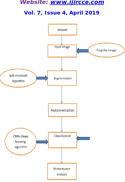
IV. PROPOSED SYSTEM
Fig 1 proposed flow chart
The proposed work will also take input from the output of this application and integrate them with the concept of ontology. Fig 1 shows a steps for the proposed flow Following are the steps of Pre-processingSegmentation Feature extraction Classification Performance measure Detection Segmentation partitions the image into set of semantically meaningful, homogenous, and non -overlapping regions of similar attributes such as
A pre-processing of an image is done before applying the threshold algorithm. Pre-processing involves enhancement of a given MRI image to improve the interpretability of the information present in images for human viewers. A filtering approach is used to enhance the given MRI image . It involves a combination of median filter for the reduction of salt and pepper noise and high pass filtering performs image sharpening. The enhanced image is then segmented using maximum entropy based thresholding method which selects a threshold value that corresponds to maximum entropy between the lower and higher frequency bands. The frequency bands correspond to intensity levels of the object and background respectively.
Thresholding Operations Thresholding is a non-linear operation that converts a gray-scale image into a binary image
Feature Extraction: is the combined process of feature extraction and selection which plays a vital role in brain image processing. The initial feature selection process minimized the dimensionality of the feature set, by doing this it takes minimum time for detecting as well as computational costs. Then to extract the best set of features from the raw dataset
Pre-Processing Stage: In this phase image is enhanced in the way that finer details are improved and noise is removed
from the image. Most commonly used enhancement and noise reduction techniques are implemented that can give best possible results. Enhancement will result in more prominent edges and a sharpened image is obtained, noise will be reduced thus reducing the blurring effect from the image. In addition to enhancement, image segmentation will also be applied. This improved and enhanced image will help in detecting edges and improving the quality of the overall image. Edge detection will lead to finding the exact location of tumor.[5]
Image To Gray Scale:-An MRI image is chosen from the file to be processed. This image is converted to gray scale
image. These images have shades of gray between 0 to 255, where 0 corresponds to black and 255 to white for instance
Post-Processing Stage: In processing segmentation is done using following methods. 1) Threshold Segmentation:
Threshold segmentation is one of the simplest segmentation methods. The input gray scale image is converted into a binary format. The method is based on a threshold value which will convert gray scale image into a binary image format. The main logic is the selection of a threshold value. [7],[9]
Morphological Operators: After converting the image in the binary format, some morphological operations are
applied on the converted binary image. The purpose of the morphological operators is to separate the tumor part of the image. Now only the tumor portion of the image is visible, shown as white color. This portion has the highest intensity than other regions of the image. Morphological operators are applied after the watershed segmentation. Some of the commands used in morphing ow, we are going to talk about all steps for the proposed system for
Performance Analysis: the works of deep learning CNN based classification techniques The factors involved in
performance comparison AccuracyAccuracy: Accuracy is calculating the ratio of number of correct assessment to the total number of assessments. In the entire dataset initially the number of relevant images were extracted and compared to entire dataset by applying the belowmentioned formula in which data quality and errors were the important factors which are measure in terms of percentages(%).
Accuracy=TN+TP/TN+TP+FN+FP
V. CONCLUSION
In general computer vision tasks, attempts have been made to circumvent limited data by using smaller filters on deeper layers [47],with novel CNN architecture combinations [86], or hyper parameter optimization In adding up to deep learning algorithms, their applications are reviewed and compared by means of other machine learning methods. although deep neural networks achieve good performance on many tasksTo train and evaluate the CNN, we used first deep CNN designed for the specific problem. Finally classify the performance (85.5%) demonstrated the potential of CNNs in analyzing lung patterns in this design.
REFERENCES
1. Syed MS Islam Hassan Mahmood Adel Ali Al-Jumaily Scott Claxton Deep Learning of Facial Depth Maps for Obstructive Sleep Apnea
Prediction IEEE 12th International Symposium on Biomedical Imaging (ISBI) DOI: 10.1109/iCMLDE.2018.00036 Sydney, pp. 110 –115 Australia 2015
2. Yaniv Bar Idit Diamant Lior Wolf Sivan LiebermanChest pathology detection using deep learning with non-medical training
doi.org/10.1080/21681163.2016.1138324 Volume -2 16 Pages 259-263 May 2016
3. Ho-Shon ; Sarvnaz Karim Len Hamey Modality Classification and Concept Detection in Medical Images Using Deep Transfer Learning
International Conference on Image and Vision Computing New Zealan(IVCNZ)DOI: 10.1109/IVCNZ.2018.8634803 pp. 147-152 –
13Teleconference Location: Auckland, New Zealand, 2018
5. Nirmala Singh Sachchidanand Singh Object classification to analyze medical imaging data using deep learningInternational Conference on Innovations in Information, Embedded and Communication Systems (ICIIECS)DOI: 10.1109/ICIIECS.2017.8276099Coimbatore, India2017
6. J.selvakumar , A.Lakshmi, T.Arivoli, “Brain Tumor Segmentation and Its Area Calculation in Brain MR images using K-Mean Clustering and
Fuzzy C-Mean Algorithm” , IEEE-International Conference On Advances In Engineering, Science And Management March Volume 7 30, 31, 2017
7. Sudipta Roy, Samir K.Bandyopadhyay, “Detection and Quantification of Brain Tumor from MRI of Brain and it’s Symmetric Analysis”,
IJICTR, Volume 2 No. 6, June 2012.
8. Krishna Kant Singh , Akansha Singh,“A Study Of Image Segmentation Algorithms For Different Types Of Images”, IJCSI International
Journal of Computer Science Issues, Vol. 7, Issue 5,September 2018
9. Qurat-Ul-Ain, L.G., Kaz mi, S.B., Jaffar, M.A ., Mirza, A.M.: ‘ Classifi cation andsegment ation of brain tumor using texture analysis’ , Recent
Adv. Artif. Intell.Knowl. Eng. Data Bases, 10, pp. 147 –155,2015
10. 2 Khalid, N.E.A., Ibrahim, S., Haniff, P.N.M.M.: ‘MRI brain abnormalitiessegmentation using K-nearest neighbors (k-NN) ’, Int. J. Comput.
Sci. Eng.(IJCSE), , 3, (2), pp. 980 –990,2011
11. 3 Aslam, H.A., Ramashri, T., Ahsan, M.I.A.: ‘A new approach to imagesegmentation for brain tumor detection using pillar K-means algorithm
’,Int. J. Adv. Res. Comput. Commun. Eng., , 2, (3), pp. 1429 –1436, 2013
12. Maity, A., Pruitt, A.A., Judy, K.D.: ‘Cancer of the central nervous system’ (ClinicalOncology, 2008, 4th edn.)
13. Ricci, P.E., Dungan, D.H.: ‘Imaging of low and intermediate-grade gliomas ’,Semradonc, , 11, (2), pp. 103 –112,2001
14. Rajini, N.H., Narmatha, T., Bhavani, R.: ‘Automatic classification of MR braintumor images using decision tree ’. Special Issue of Int. J. of ComputerApplications on Int. Conf. on Electronics, Communication and InformationSystems (ICECI 12), , pp. 10 –13,2012
15. Armstrong, T.S., Cohen, M.Z., Weinbrg, J., Gilbert, M.R.: ‘Imaging techniques inneuro oncology ’, Semoncnur, 20, (4), pp. 231 –239,2004,
16. Bandyopadhyay, S.K.: ‘Detection of brain tumor-a proposed method ’, J. GlobalRes. Comput. Sci., , pp. 55 –63 /2011
17. Anbeek, P., Vincken, K.L., Viergever, M.A.: ‘Automated MS-lesion segmentationby K-nearest neighbor classification’, Midas J. MS Lesion
Segmentation (MICCAI2008 Workshop), 2008