D
E
V
E
LO
P
M
E
N
T
INTRODUCTION
Postembryonic development of metazoans follows a series of genetically programmed events. However, developmental timing can be modified by environmental conditions. For example, in amphibians, an increase of thyroid hormone triggers metamorphosis. However, conditions unfavorable to tadpole survival such as high conspecific density, low water level or the presence of predators can significantly advance the timing of metamorphosis (reviewed by Boorse, 2004). Similarly in humans, the onset of puberty is reproducible among individuals but, in addition to genetic mutations, environmental conditions can advance or delay puberty by mechanisms that mostly have yet to be elucidated (Ebling, 2005). In the nematode Caenorhabditis elegans, analysis of mutants displaying abnormal postembryonic development has revealed molecular mechanisms of developmental timing control. C. elegans goes from hatching to reproductive adulthood through four larval stages (L1 to L4), all terminated by a molt. Each larval stage is associated with a developmental program characterized by specific patterns of cell division, differentiation and migration. Mutants have been isolated that bypass or repeat some of these stage-specific developmental programs. Genes whose mutation causes such temporal transformations are called heterochronic genes (reviewed by Ambros, 2000; Pasquinelli and Ruvkun, 2002; Rougvie, 2001; Thummel, 2001). They define an intrinsic developmental timer that specifies the identity of each larval stage. However, in addition to genetic constraints, C. elegans development is modulated by the environment. The best characterized example is the choice between reproductive development and a facultative L3 diapause (reviewed by
Riddle and Albert, 1997). Under favorable conditions, eggs develop into reproductive adults within 3 days. If L1 larvae are exposed to adverse conditions, including limited food, high temperature and high population density, animals can enter a facultative L3 diapause stage called the dauer larva (Cassada and Russell, 1975). Dauer larvae survive for several months without feeding and are able to resume development to fertile adults when conditions are favorable again. A complex genetic network involving a TGF, an insulin-like and a nuclear hormone receptor (NHR) pathway controls dauer entry (reviewed by Riddle and Albert, 1997). These three pathways integrate environmental cues perceived by several classes of sensory neurons, thus defining converging neuroendocrine pathways (Bargmann and Horvitz, 1991; Li et al., 2003; Ren et al., 1996).
In addition to dauer-inducing conditions, C. elegans developmental rate can also vary according to food quantity and quality (Houthoofd et al., 2002) or population density (Golden and Riddle, 1984a). The cellular and molecular bases of these controls are poorly characterized. To investigate a potential role of the nervous system in the temporal regulation of development, we pharmacologically manipulated acetylcholine (ACh)-mediated transmission at early stages of C. elegans development. ACh is the prominent excitatory neurotransmitter in this species: of the 302 neurons that compose the nervous system of an adult hermaphrodite, one-third are cholinergic (Rand, 1997). The two classes of receptors mediating ACh-mediated transmission in the mammalian nervous system – muscarinic G-protein-coupled receptors and nicotinic ligand-gated ion channels – are expressed in C. elegans (Bargmann, 1998). Genome sequence analysis detected up to 42 genes potentially encoding nicotinic acetylcholine receptor (nAChR) subunits (Bargmann, 1998). Some of these subunits have been involved in defined functions such as feeding (McKay et al., 2004), egg laying (Kim et al., 2001), locomotion (Fleming et al., 1997; Lewis et al., 1980a; Lewis et al., 1980b) and copulation (Garcia et al., 2001). However, the function of most C. elegansnAChRs is unknown.
Activation of nicotinic receptors uncouples a developmental
timer from the molting timer in
C. elegans
Anne-Françoise Ruaud and Jean-Louis Bessereau*
C. elegansdevelops through four larval stages (L1 to L4) separated by molts. The identity of larval stages is mostly determined by stage-specific expression of heterochronic genes, which constitute an intrinsic genetic timer. However, extrinsic cues such as food availability or population density also modulate the developmental timing of C. elegans by mechanisms that remain largely unknown. To investigate a potential role of the nervous system in the temporal regulation of C. elegansdevelopment, we pharmacologically manipulated nicotinic neurotransmission, which represents a prominent signaling component in C. elegans nervous system. Exposure to the nicotinic agonist DMPP during post-embryonic development is lethal at the L2/L3 molt. Specifically, it delays cell divisions and differentiation during the L2 stage but does not affect the timing of the molt cycle, hence causing exposure of a defective L3 cuticle to the environment after the L2/L3 molt. Forcing development through a previously
uncharacterized L2 diapause resynchronizes these events and suppresses DMPP-induced lethality. Nicotinic acetylcholine receptors (nAChRs) containing the UNC-63 subunit are required, probably in neurons, to trigger the action of DMPP. Using a forward genetic screen, we further demonstrated that the nuclear hormone receptor (NHR) DAF-12 is necessary to implement the developmental effects of DMPP. Therefore, a novel neuroendocrine pathway involving nAChRs and the NHR DAF-12 can control the speed of stage-specific developmental events in C. elegans. Activation of DMPP-sensitive nAChRs during the second larval stage uncouples a molting timer and a developmental timer, thus causing a heterochronic phenotype that is lethal at the subsequent molt.
KEY WORDS: Developmental timing, Nicotinic acetylcholine receptor, DAF-12 Development 133, 2211-2222 (2006) doi:10.1242/dev.02392
ENS, Biologie cellulaire de la synapse, Paris, 75005 France; Inserm, U789, Paris, F-75005, France.
*Author for correspondence (e-mail: [email protected])
D
E
V
E
LO
P
M
E
N
T
We manipulated nicotinic neurotransmission by exposing animals to the nicotinic agonist 1,1-dimethyl-4-phenylpiperazinium (DMPP). We observed that chronic exposure to DMPP slowed development at the second larval stage without affecting the molt timing, thus causing a lethal heterochronic phenotype at the L2/L3 molt. Genetic analysis of the sensitivity to DMPP identified nAChRs and the nuclear hormone receptor DAF-12 as components of a novel neuroendocrine pathway that controls developmental timing in C. elegans.
MATERIALS AND METHODS General methods and strains
Unless noted otherwise, worms were cultured at 20°C on NG agar plates inoculated with Escherichia coli OP50 (Sulston and Hodgkin, 1988). Synchronous developing populations were obtained by allowing young gravid adults to lay eggs for 30 minutes. Eggs were then collected and transferred onto new plates. The wild strain N2, and the following mutant alleles and transgenic markers were used: daf-9(dh6) X, daf-12(kr16,kr32,
rh61,rh273,rh284,rh285,rh61rh411,rh62rh157) X,din-1(dh127) II,
eat-6(ad467, ad792) V,lin-15(n765ts) X,pep-2(lg601) X,unc-29(e1072) I,
unc-38(x20) I,unc-63(kr13, x37) I,krEx200 andkrEx201 [Punc-63::unc-63;
Pmyo-3::GFP; Prab-3::GFP],krEx198 andkrEx199
[Punc-63::unc-63-SL2-GFP; lin-15(+)], krEx164and krEx165[3::unc-63;
Pmyo-3::GFP]and syIs50[Pcdh-3::GFP].
Mutants scored DMPP sensitive were: acr-5(ok180, ok182, ok205) III,
acr-7(tm863) II, acr-8(ok1240) X, acr-9(ok933) X, acr-11(ok1345) I,
acr-12(ok367) X, acr-14(ok1155) II, acr-15(ok1214) V, acr-16(ok789) V,
acr-18(ok1285) V, acr-19(ad1674) I, acr-21(ok1314) III, acr-22(tm627) X,
des-2(u695) deg-3(u662) V, eat-2(ad453) IIand lev-1(kr6) IV.
RNAi clones used in DMPP sensitivity tests were: C02C2.3, R13A5.4, F48E3.7 (acr-22), C04C3.2, F17E9.7, F17E9.8, T05B4.1, T01H10.7, T01H10.2, T01H10.1, T01H10.5, F11C7.1, F28F8.1 (acr-18), F53E10.2
(acr-17), K03F8.2 (acr-5), K03B8.9 (deg-3), T26H10.1 (des-2), R06A4.10
(acr-20), C31H5.3 (acr-19), F25G6.4 (acr-15), T09A5.3 (acr-7),
Y48B6A.4, F25G6.3 (acr-16), D2092.3 (acr-11), C40C9.2 (acr-9), R01E6.4
(acr-12), C35C5.5 (acr-13), F21F3.5 (unc-38), T08G11.5 (unc-29) and
F09E8.7 (lev-1).
Nicotinic agonist resistance assays and dauer pheromone
1,1-Dimethyl-4-phenylpiperazinium (DMPP) (Sigma) was dissolved in water and added to 55°C-equilibrated NG agar just before plates were poured. Gravid adult worms were allowed to lay eggs for several hours on standard plates. Eggs were then carefully transferred on DMPP-containing plates and counted. Surviving L4, adult and dauer larvae were scored after 2.5 (25°C), 3 (20°C) or 5 (15°C) days of development. Dauer pheromone was purified as described (Golden and Riddle, 1984b) and added to peptone-free DMPP plates when mentioned.
Levamisole (Sigma) was dissolved in water and added to standard NG agar. Levamisole resistance was scored as the fraction of adult worms moving after 2 hours on 1 mM levamisole.
DMPP resistance screen and daf-12allele identification
N2 worms were mutagenized by germline mobilization of the Drosophila
transposon Mos1(Williams et al., 2005). Young-adult F1 worms were transferred on 0.75 mM DMPP plates and allowed to lay eggs for 1 day. Three days later, plates were screened for healthy living adult animals. In EN16 [daf-12(kr16::Mos1)] and in EN32 [daf-12(kr32::Mos1)], Mos1
insertions were localized, respectively, at positions 10,665,405 and 10,664,945 of chromosome X by inverse PCR (WormBase website: http://www.wormbase.org, release WS140, date 06/2005).
Plasmid constructions pAF54 Pmyo-3::unc-63
unc-63 full-length cDNA was amplified by PCR from a pMT3-UNC-63 plasmid given by E. Culetto using Phusion Taq (FINNZYME) (primers GGGCCATGGTACCAGAAAAAATGGGACCAAATGACC and GGG-GGGCTCGAGCTAAGCAAGAGCCGGCGTGTT) and sequenced. It was then digested by KpnI and NaeI, and cloned into the pPD115.62 plasmid using KpnI and EcoRI sites.
pAF66 Punc-63::unc-63-SL2-GFP
Two PCR fragments were independently amplified with Phusion polymerase on wild-type (N2) genomic DNA using primers ACGTTAGT-GCACATTCTGAAAATTTTATTTTTTAAGTTG and GTAGGGTAACT-GTAGTTCAGG (fragment 1), and ACGTTAGTGACATATCGATCCC-CAACAACAC and AATGTCGATGCAATAATCACAACGGTTCC (fragment 2). Fragment 1 was digested by XhoI andApaLI, and fragment 2 by ApaLI. The digested fragments were then ligated together into pBS KSII+ digested by XhoI and EcoRV to generate pVR10. The 5⬘part of the
unc-63 genomic region was PCR amplified with Phusion polymerase
(primers GCGGTACCTAGGTTAGAGCCCCAACAGG and AAAACT-CACCGCTTGGAATG) and cloned into pVR10 using KpnI and XhoI to generate pAF67. We PCR amplified an SL2-GFP fragment from pEXPR gcy-32-egl-2(gf) (kind gift from M. de Bono) with GCGGATCCATCGAT-GCTGTCTCATCCTACTTTCA and ATCGATGTACGGCCGACTAG-TAGGAA, both containing a ClaI site, and cloned it into pVR10 using ClaI (pAF65). We then generated pAF66 by subcloning a NaeI BlpI fragment from pAF67 into pAF65.
pAF68 Punc-63::unc-63
The 3⬘end of unc-63cDNA together with unc-543⬘UTR was subcloned from pAF54 to pVR10 using ClaI and SpeI. We then subcloned the unc-63
5⬘region from pAF67 into this plasmid using KpnI and XhoI.
Germline transformation
Transformation was performed by microinjection of plasmid DNA into the gonad (Mello et al., 1991). unc-63(kr13) worms were injected with a DNA mixture containing pAF54 (Pmyo-3::unc-63) (10 ng/l), 1 kb+ DNA ladder (INVITROGEN) (85 ng/l) and pPD115.62 (Pmyo-3::GFP) (5 ng/l) as a co-transformation marker or pAF68 (Punc-63::unc-63) (20 ng/l), pHU4
(Prab-3::GFP) (20 ng/l), pPD115.62 (5 ng/l) and 1kb+ (55 ng/l).
pAF66 (Punc-63::unc-63-SL2-GFP)was injected in lin-15(n765ts)at 20 ng/l with EKL15 [lin-15(+)] (80 ng/l) as a co-injection marker.
Light microscopy
Animals were anesthetized with M9 buffer containing 20 mM sodium azide, mounted on 2% agarose in M9 pads and examined by epifluorescence and/or DIC optics using an Axioskop compound microscope (Zeiss). For confocal microscopy, 3.8 mM tricaine and 0.42 mM tetramisole were added to the anesthetic. Animals were examined using a Leica (Nussloch, Germany) TCS SP2 AOBS confocal microscope. Confocal image reconstructions were obtained with ImageJ.
Electron microscopy Chemical fixation
Just-molted L3 larvae grown on standard or 0.75 mM DMPP plates were fixed in 2.5% glutaraldehyde, 1% paraformaldehyde, 0.1 M cacodylate (pH 7.2) at 4°C and cut preferentially at the head and tail. Samples were then mounted into agar blocks, postfixed in 1% OsO4, 0.1 M cacodylate buffer,
dehydrated in a series of alcohols and embedded in Araldite (Ernest F. Fullam; Latham, NY).
High-pressure freezing
Young L3 animals were immobilized by high-pressure freezing just after L2 cuticle shedding, then dehydrated by freeze substitution and embedded in Araldite as previously described (Rostaing et al., 2004).
RESULTS
Chronic exposure to DMPP is lethal at the L2/L3 molt
D
E
V
E
LO
P
M
E
N
T
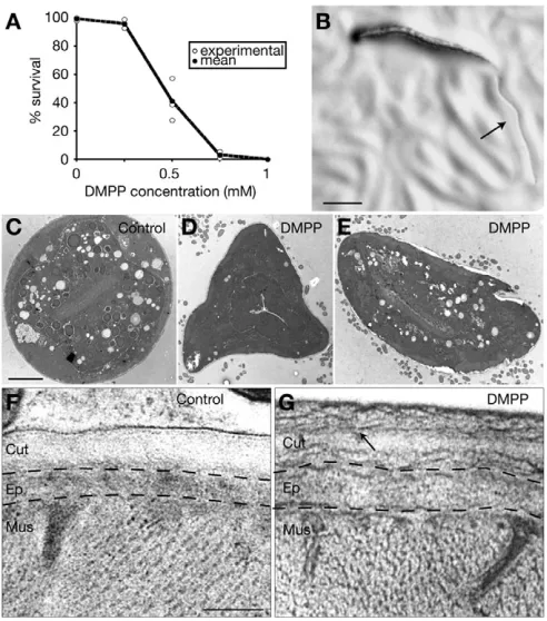
transient increase of locomotory activity with moderate muscle hypercontraction (data not shown), consistently with activation followed by desensitization of nAChRs in the locomotory system. By contrast, treating C. elegansanimals with DMPP during development had a more dramatic phenotype. When larvae hatched and developed in the presence of the drug, DMPP caused a concentration-dependent lethality (Fig. 1A). Fully penetrant lethality was obtained for concentrations above 0.75 mM. This concentration is very similar to the 1 mM required in vivo to achieve a lethal effect with levamisole, a nematode-specific nicotinic agonist that activates nAChRs present at neuromuscular junctions (Lewis et al., 1980a; Lewis et al., 1980b; Richmond and Jorgensen, 1999). Therefore, DMPP-induced lethality is compatible with a specific action of the drug on nAChRs in the animal. By following the development of worms grown on DMPP, we showed that most worms exposed to high DMPP concentrations died immediately after the L2/L3 molt (Fig. 1B). The death phenotype was stereotyped: shedding of the old cuticle was rapidly followed by complete arrest of locomotion, vacuolization of most tissues, and corpses dissolved within a few hours. This result is not in favor of a cumulative toxicity but indicates that DMPP could specifically disrupt worm physiology at the L2/L3 molt.
Chronic exposure to DMPP induces aberrant L3 cuticle synthesis
The temporal coincidence between molt and death suggested that lethality could be due to defects of the L3 cuticle, which starts being exposed to the surrounding environment at the L2/L3 molt. C. elegans cuticle is a complex protein matrix that serves as an exoskeleton and as a diffusion barrier that insulates the animal from the outside (Johnstone, 2000). Cuticle misfunction would explain the inability to maintain fluid homeostasis and subsequent cell vacuolization caused by osmotic unbalance. To test the existence of cuticle defects, we analyzed just-molted L3 larvae grown on DMPP using either classical electron microscopy (EM) or EM after high-pressure freezing (HPF) (Rostaing et al., 2004). At low magnification, we observed an irregular shape of the animals, suggesting defects in the mechanical properties of the cuticle and the maintenance of internal hydrostatic pressure (Fig. 1C-E). At higher magnification, DMPP-treated L3 larvae displayed abnormal fibrillar structures, pseudo-layers and thickening of the underlying epidermal cells, in contrast to wild-type L3 cuticle, which is uniform in thickness and density (Fig. 1F,G). Together, these results indicate that continuous exposure to the nicotinic agonist DMPP during postembryonic development causes L3 cuticle defects that may be responsible for lethality at the L2/L3 molt.
DMPP is toxic during L2 stage by uncoupling developmental speed from the molt cycle
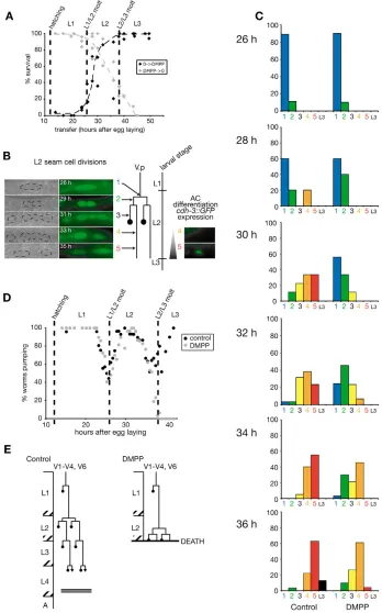
Although animals are exposed to DMPP from the beginning of the first larval stage until they die at the L2/L3 molt, no phenotype is observed at the L1/L2 molt. As the L3 cuticle is synthesized during late L2 stage, the effects of DMPP on development might be restricted to the second larval stage. To establish DMPP sensitivity period, we performed transfer experiments (Fig. 2A). When larvae hatched on DMPP were removed from drug-containing plates before the L1/L2 molt, they developed to adulthood. Conversely, larvae grown on standard plates placed on DMPP after the L2/L3 molt were resistant to the drug. From these two experiments, we concluded that DMPP toxicity is restricted to the L2 stage.
[image:3.612.52.298.304.583.2]C. eleganscuticle is synthesized by the underlying epidermis and disrupting epidermis development has been shown to affect the properties of the cuticle being synthesized (Singh and Sulston, 1978). Lateral cells of the epidermis (‘seam cells’) undergo stereotyped cell divisions at each larval stage. To analyze epidermis development in DMPP-treated individuals, we followed seam cell divisions using differential interference contrast (DIC) microscopy and the green fluorescent protein (GFP) reporter Pcdh-3::GFP(Kirouac and Sternberg, 2003; Pettitt et al., 1996). This reporter is also expressed in the anchor cell (AC), a specialized cell from the somatic gonad which differentiates during late L2 stage. Based on seam cell divisions and anchor cell differentiation, we divided the L2 stage into five successive temporal stages (Fig. 2B). In the wild type, seam cells underwent an equational division immediately followed by a stem cell division in which the anterior daughter cell fuses with the epidermal syncytium hyp7. Later during L2 stage, GFP was detected in the anchor cell (Fig. 2B). On DMPP, the two seam cell divisions were delayed by several hours and most animals had just completed the second division by the end of the second larval stage. In addition, the anchor cell precursor was observed under DIC but none of the animals expressed GFP (Fig. 2C). These data indicated that DMPP delayed the development of at least two different tissues during the L2 larval stage.
Fig. 1. DMPP is lethal at the L2/L3 molt and induces aberrant L3 cuticle synthesis.(A) Fraction of wild-type larvae reaching adulthood
when grown on increasing concentrations of DMPP (n⭓50 worms).
Experiments were performed at 20°C. (B) Representative example of an animal exposed to 0.75 mM DMPP which dies as an early L3 larvae, just after shedding the L2 cuticle (arrow). Scale bar: 0.1 mm. (C-E) Electron microscopy (EM) pictures after chemical fixation of young L3 larvae.
Scale bar: 5 m. Worms grown on DMPP (D,E) have irregular shapes
D
E
V
E
LO
P
M
E
N
T
The end of the L2 larval stage is defined by the L2/L3 molt. Does DMPP also delay this molt? The lethargus period, which corresponds to the initiation of the molting process, is marked by behavioral changes, including cessation of rhythmic pharyngeal contractions (also called pharyngeal pumping) (Singh and Sulston, 1978). Pumping rate was monitored in a synchronized developing population (Golden and Riddle, 1984a). In the absence of DMPP, L1/L2 and L2/L3 molts occurred at 26 and 36 hours after egg laying, respectively. Exposure to DMPP did not affect the timing of L1/L2 or L2/L3 molts (Fig. 2D). Therefore, DMPP uncouples the L2
developmental timer from the molting timer (Fig. 2E) and causes a lethal heterochronic phenotype characterized by molt triggering while epidermal cell are undergoing mid-L2 developmental events.
Resynchronization of the molt cycle with other developmental events suppresses DMPP-induced lethality
[image:4.612.49.397.181.739.2]If the lack of synchronization between L2 seam cell divisions and the L2/L3 molt on DMPP accounts for the lethal cuticular defects, delaying molting on DMPP might allow the completion of epidermis
Fig. 2. DMPP is toxic during the L2 stage by uncoupling cell divisions and differentiation events from the molt cycle.(A) Toxicity period assayed by transfer experiments of wild-type animals. Each dot represents the surviving fraction of a synchronous
population (n⭓25 worms) transferred at
a given time from standard to DMPP plates (black) or from DMPP to standard plates (gray). (B,C) Exposure to DMPP delays L2 development. (B) L2 development was divided in five stages based on seam cell (SC) divisions and anchor cell (AC) differentiation (1, undivided SC; 2, SC divided once; 3, SC divided twice; 4, anterior SC fused to
hyp7, no Pcdh-3::GFPexpressed in AC;
5, Pcdh-3::GFPexpressed in AC). (C) The
proportion of worms belonging to these five stages was scored every 2 hours during the L2 stage (n>20). Bars represent the mean of two independent experiments (exp 1: 30, 32, 34, 36 hours; exp 2: 26, 28, 32, 34, 36 hours). (D) DMPP does not affect the timing of L1/L2 and L2/L3 molts. Each dot represents the percentage of worms pumping at a given time (n>25 individuals). (E) Schematic representation of worm development at high DMPP
concentration (⭓0.75 mM). L2
D
E
V
E
LO
P
M
E
N
T
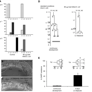
development, hence suppressing DMPP toxicity. To test this prediction, we had to find a means to regulate development timing at the L2 larval stage. Environmentally triggered diapauses are well described at the L1 and L3 larval stages (Riddle and Albert, 1997; Rougvie, 2005), but not at the L2 stage. However, we were intrigued by the results of Jeong et al. who reported that animals grown on a limited amount of heat-killed E. coli‘remained at L1 or L2 stages and did not grow to adult’ (Jeong et al., 2005). This developmental arrest was not further investigated. We therefore monitored development of worms grown in the same conditions as used in this study. Most individuals arrested at the L2 stage (Fig. 3A) and survived up to 9 days (data not shown). Specifically, arrested larvae kept moving and pumping. Seam cell development arrested after the second division and storage granules accumulated in the epidermis (Fig. 3B-D). When transferred onto an unlimited amount of standard food (live E. coli), arrested L2 larvae resumed development to adulthood (data not shown). We reasoned that forcing development through this L2 arrest might resynchronize molting and other developmental events on DMPP. We therefore grew animals in the presence of DMPP on limited amounts of heat-killed bacteria. Entry in L2 arrest did not differ between DMPP and control conditions. Arrested larvae survived on DMPP without molting. After 7 days, we transferred L2 arrested larvae onto DMPP-containing plates with standard food (live E. coli). Forty-four percent of the animals resumed development and reached adulthood (Fig. 3E), compared with 2% of control worms. These results support the hypothesis that lethality on DMPP is caused by drug-induced heterochrony.
UNC-63-containing nAChRs are required to implement the DMPP signal
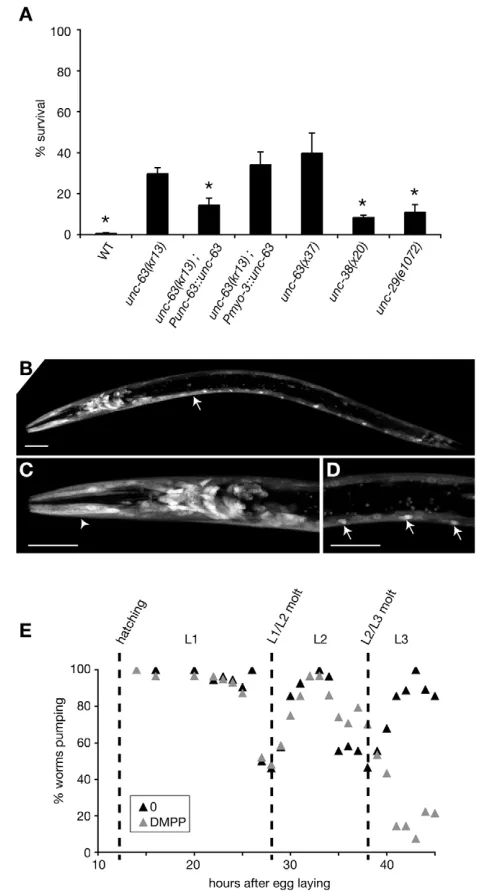
DMPP is a broad-spectrum nicotinic agonist that can activate both vertebrate (Galligan, 1999; Manetti et al., 1999; Vizi and Lendvai, 1999) and C. elegans nAChRs (Janet Richmond, personal communication). To identify which nAChRs might be targeted by DMPP and control L2 development in C. elegans, we performed RNAi (Kamath et al., 2001) against 30 nAChRs subunit candidate genes in the wild-type and RNAi-hypersensitive rrf-3 strain (Simmer et al., 2002) and tested 19 available loss-of-function mutants in nAChR subunit-coding genes for sensitivity to DMPP (see Materials and methods). Among these genes, unc-63 loss-of-function mutations conferred partial resistance to DMPP (Fig. 4A). Partial resistance might be explained by functional redundancy between nAChR subunits, another subunit substituting for UNC-63 in unc-63mutant animals. Alternatively, DMPP might target distinct nAChRs, among which UNC-63-containing nAChRs are major DMPP effectors.
[image:5.612.58.420.376.740.2]Previous reports indicate that unc-63is expressed in body-wall muscles and in neurons (Culetto et al., 2004). However, recent gene predictions indicate that the unc-63 promoter fragment used to characterize unc-63expression pattern included an open reading frame upstream of unc-63. To refine this analysis, we used a shorter unc-63promoter fragment. The unc-63-coding sequence expressed under the control of this promoter was able to rescue DMPP resistance of unc-63(0)mutants (Fig. 4A). GFP expression driven by the Punc-63 promoter was detected in body-wall
Fig. 3. Forcing development through an L2 arrest suppresses DMPP-induced lethality.(A) Worms
grown on a low amount (80 g) of
heat-killed bacteria arrest development as L2 larvae. Bars represent the % of larvae at each developmental stage, mean of two independent experiments. (B,C) DIC pictures of a mid-L2 larva
grown on live E. coli(B) and an arrested
L2 larva grown on 80 g heat-killed E.
coli(C). Arrested L2 larvae show a
seam cell pattern typical of a worm having completed the two successive L2 divisions without extensive anterior sister cell migration. There is high storage droplet content in the
epidermis (C). Scale bar: 10 m.
(D) Schematic representation of worm development on low amount of
heat-killed E. colias schematized in Fig. 2D.
(E) DMPP resistance of animals after L2 arrest. Both groups were grown on 0.75 mM DMPP. Worms transferred on standard plates after a 3-day arrest in L2 were partially resistant to DMPP (‘3 days L2 diapause’) when compared with control (‘continuous
D
E
V
E
LO
P
M
E
N
T
muscles, in many head and tail neurons and in motoneurons (Fig. 4B-D). In muscle, UNC-63 is part of a well-characterized receptor present at neuromuscular junctions that is pharmacologically
identified based on its sensitivity to the nicotinic agonist levamisole. The levamisole-sensitive receptor consists of four obligatory subunits (LEV-1, UNC-29, UNC-38 and UNC-63) that assemble with an unknown stoichiometry to form a heteropentameric receptor. Mutating one of these subunits is sufficient to inactivate fully the levamisole receptor (Culetto et al., 2004; Richmond and Jorgensen, 1999). Two lines of evidence suggest that this muscle receptor is not the molecular target responsible for DMPP-induced L2 heterochrony. First, null mutations in unc-29and unc-38are as levamisole resistant as unc-63(0)(see Fig. S1 in the supplementary material) but far less DMPP resistant (Fig. 4A). Second, muscle-specific expression of UNC-63 in an unc-63(0)background rescued levamisole but not DMPP sensitivity (Fig. 4A and see Fig. S1 in the supplementary material). Therefore, illegitimate activation of an UNC-63-containing AChR expressed in neurons is likely the cause of heterochronic L2 development. Unfortunately, we could not rescue unc-63(0)DMPP resistance by using the pan-neuronal promoter Prab-3(Nonet et al., 1997) to express UNC-63 in neurons (data not shown), but this negative result might reflect a tight regulation of UNC-63 expression in DMPP-responsive neurons that is not achievable with Prab-3.
We have previously shown that delaying the L2/L3 molt by the L2 arrest renders wild-type worms partially DMPP resistant. Therefore, partial DMPP resistance of unc-63(0)mutants could reflect an alteration of the molt cycle timing. Alternatively, unc-63 might be required to trigger DMPP-induced developmental delay. To distinguish between these two hypotheses, we monitored the unc-63(0)molt cycle with and without DMPP. The timing of both L1/L2 and L2/L3 molts was not affected in unc-63(0) mutants when compared with the wild type (Fig. 4C). Together, these results indicate that illegitimate activation of a neuronal UNC-63 containing nAChR by DMPP is, at least in part, causing a developmental delay at the second larval stage.
The DAF-12 nuclear hormone receptor is necessary for efficient DMPP-induced L2 developmental delay
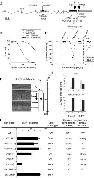
[image:6.612.53.294.117.565.2]To identify the molecules required to implement DMPP effect, we performed a forward genetic screen for mutants that can develop on DMPP. To speed up the cloning of such DMPP-resistance genes, we used an insertional mutagenesis technique based on the mobilization of the Drosophilatransposon Mos1in the C. elegansgermline (Bessereau et al., 2001; Williams et al., 2005). Among seven resistant mutants, we identified two strains carrying a Mos1 insertion in the daf-12gene. DAF-12 encodes a nuclear hormone receptor (NHR) involved in many processes in C. elegans, including temporal patterning, dauer formation and aging (Antebi et al., 1998; Antebi et al., 2000). One of the Mos1 insertions isolated introduced a late stop in the DAF-12 open reading frame, and the second one was in the stop codon (Fig. 5A). As these two Mos1alleles are not predicted to be null, we tested the DMPP sensitivity of the null allele daf-12(rh61rh411) and demonstrated that mutant animals were resistant to high DMPP concentrations (Fig. 5B). As discussed for unc-63, resistance to DMPP might have reflected alterations of the molt cycle timing. We observed that the L2/L3 molt was not delayed indaf-12(0)mutants (Fig. 5C). However, analysis of seam cell divisions showed that DMPP was no longer inducing L2 developmental delay in daf-12(0)mutants (Fig. 5D). Therefore the DAF-12 protein is required to implement DMPP-induced developmental delay.
Fig. 4. UNC-63-containing nAChRs are required to implement the DMPP signal.(A) Survival on 0.75 mM DMPP. Error bars represent
s.e.m. (n⭓3). Punc-63::unc-63: extrachromosomal array carrying an
unc-63genomic fragment; Pmyo-3::unc-63: muscle specific promoter
driving the expression of an unc-63cDNA.
unc-63(kr13);Punc-63::unc-63, unc-38(x38) and unc-29(e1072) are significantly less DMPP resistant than unc-63(kr13) (*: P< 0.05, Mann-Whitney test). Pmyo-3::unc-63
does not rescue unc-63(kr13)DMPP resistance while it restores
wild-type locomotion and levamisole sensitivity (see Fig. S1 in the
supplementary material). (B-D) unc-63expression profile. Confocal
picture of a transgenic larva expressing P63::63-SL2-GFP.
unc-63is expressed in body-wall muscles (B, arrow), head muscles (C,
arrowhead), and in many neurons in the head (C) and the ventral cord
(D, arrow). Scale bar: 20 m. (E) unc-63(kr13) does not alter the timing
D
E
V
E
LO
P
M
E
N
T
Inactivating non-dauer daf-12 activity confers DMPP resistance
Genetic analysis has separated two daf-12activities (Antebi et al., 1998; Antebi et al., 2000). One activity, probably corresponding to the non-liganded form of this nuclear receptor, is promoting L3 dauer diapause via the activation of genes required for dauer formation. Null alleles of daf-12are unable to form dauer larvae
[image:7.612.51.364.147.740.2]in any environmental conditions (dauer formation-deficient Daf-d phenotype). A seconDaf-d activity, probably generateDaf-d by the DAF-12 receptor bound to a steroid hormone, acts in non-dauer development. Mutations that are predicted to impair hormone binding or interactions with co-activators or co-repressors cause heterochronic phenotypes, including reiteration of the L2 seam cell division program at the L3 stage and reiteration of the L3
Fig. 5. DMPP resistance of daf-12and daf-9
mutants. (A) daf-12gene structure. Mutation
sites of newMos1alleles and alleles tested for
DMPP resistance are indicated. *, STOP codon. (B) Dose-response sensitivity to DMPP of wild-type and 12(rh61rh411) mutants. (C) daf-12(rh61rh411) does not alter the timing of L1/L2 and L2/L3 molts. Each dot represents the percentage of worms pumping at a given time (n>25 individuals). broken lines indicate the timing of wild-type events. (D)
daf-12(rh61rh411)is insensitive to DMPP-induced developmental delay. L2 development was divided into five stages based on seam cell (SC) divisions and anchor cell (AC) differentiation (see Fig. 2B). Developmental stage was monitored using DIC optics, which did not allow the discrimination between classes 3 and 4. The proportion of worms belonging to each class
was scored 32 hours after egg laying (n⭓12).
Data presented are from one representative experiment out of three independent trials.
(E) DMPP resistance of daf-12and daf-9 mutants
(0.75 mM DMPP). Error bars represent s.e.m.
(n⭓3). Table presenting dauer and heterochronic
D
E
V
E
LO
P
M
E
N
T
gonadal migration program at the L4 stage. Based on heterochronic and dauer-formation phenotypes, daf-12alleles were grouped into six classes (Antebi et al., 1998) (Fig. 5E). To test whether the function of DAF-12 in the response to DMPP corresponds to a previously defined activity, we evaluated the DMPP sensitivity of the strongest available daf-12 allele of each class. We showed that class 1 to 4 mutants are strongly resistant to DMPP, whereas class 5 are sensitive and class 6 are only weakly resistant (Fig. 5E). Therefore, resistance to DMPP is unrelated to dauer formation activity but is qualitatively correlated with extra-gonadal heterochronic phenotypes. These phenotypes are thought to reveal the loss of DAF-12 non-dauer activity, which induces the progression from L2 to L3 developmental programs. To confirm that DAF-12 non-dauer activity is required for the DMPP effect, we analyzed din-1S and daf-9 mutants. DIN-1S encodes a transcriptional co-repressor required only for the dauer-inducing activity of DAF-12 (Ludewig et al., 2004). din-1Snull mutants, which are Daf-d but have no heterochronic phenotype, are still sensitive to DMPP (Fig. 5E). DAF-9 is a P450 cytochrome supposed to be a key component of the sterol-derivative DAF-12 ligand biosynthetic pathway (Gerisch et al., 2001; Jia et al., 2002). In the absence of DAF-9, DAF-12 probably exists as a non-liganded form and causes dauer formation. We showed that daf-9null mutants are DMPP resistant (Fig. 5E). These results strongly suggest that the liganded form of DAF-12 is necessary for DMPP toxicity and reveal a previously undescribed role of DAF-12 in coordinating cell divisions with molts at the second larval stage.
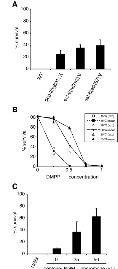
Environmental cues modulate DMPP sensitivity
Three major environmental parameters can affect DAF-12 activity and trigger dauer formation in the wild type: food availability, temperature and population density. Do these cues also modulate DMPP sensitivity? First, we restricted food availability throughout development using pep-2 and eat-6 mutants. pep-2encodes an intestinal peptide transporter (Meissner et al., 2004) that is essential for the uptake of intact peptides from the gut lumen. eat-6encodes a Na+/K+ATPase expressed specifically in the worm pharynx that is necessary for efficient pharynx muscle contractions (Davis et al., 1995). pep-2(0)animals and strong eat-6loss-of-function mutants are starved and develop slowly. We observed that a significant fraction of pep-2(0)and eat-6(lf)mutants can develop on DMPP (Fig. 6A), thus suggesting that restricting food availability leads to partial DMPP resistance. Second, we tested the effect of temperature on DMPP sensitivity. In wild-type N2 strain, raising the temperature increased the survival of animals on DMPP (Fig. 6B). Third, DAF-12 activity is modulated by population density that is monitored via the concentration of a constitutively secreted pheromone (Golden and Riddle, 1982; Golden and Riddle, 1984b; Jeong et al., 2005). High concentration of this pheromone induces dauer formation. We monitored the effect of DMPP in the presence of increasing concentrations of pheromone. The experiments were performed at 20°C so that none of the animals entered dauer diapause. Exposing worms to dauer pheromone increased survival on DMPP in a concentration-dependent manner (Fig. 6C). Modulation of the sensitivity to DMPP by these three environmental parameters cannot be explained by an increase of the L2 stage duration: in our conditions, reduced food availability increased generation time; high temperature speeded up development; and pheromone had no effect on overall developmental speed (data not shown). Therefore, analysis of DMPP sensitivity unmasks developmental effects of dauer-inducing stimuli in non-dauer development, possibly by modulating the ratio between distinct DAF-12 activities.
DISCUSSION
Chronic exposure ofC. elegansto the nicotinic agonist DMPP unmasked a novel function of nAChRs in the control of postembryonic development. We demonstrated that illegitimate activation of nAChRs during the second larval stage induced a lethal heterochronic phenotype by disconnecting developmental speed
A
B
10080
60
40
20
0
% sur
viv
al
DMPP concentration 15°C (exp) 15°C 20°C (exp) 20°C 25°C (exp) 25°C
(mean)
(mean) (mean)
NGM
0 25 50
peptone- NGM + pheromone (μL) 100
80
60
40
20
0
% sur
viv
al
C
10080
60
40
20
0
% sur
viv
al
WT
pep-2(lg601) X eat-6(ad792) V eat-6(ad467) V
[image:8.612.325.524.146.599.2]0 0.5 1
Fig. 6. Environmental modulation of the sensitivity to DMPP. (A)
DMPP resistance of ‘genetically starved’ worms. Surviving eat-6and
pep-2mutants on DMPP were scored 4 and 5 days after egg laying, respectively. By this time, most mutants grown on control plates had reached adulthood. (B) DMPP toxicity dose-response curves at 15, 20 and 25°C (wild-type N2 strain). (C) High pheromone concentrations induce partial DMPP resistance at 20°C. As pheromone signaling is antagonized by a food signal, animals were grown on plates lacking peptone, which slightly decreases the quantity of available food (Golden and Riddle, 1984a). Error bar represents s.e.m., n=3 independent experiments. Dauer larvae are not induced under these experimental conditions but pheromone activity was tested
D
E
V
E
LO
P
M
E
N
T
from the molting timer, hence resulting in deadly exposure of a defective cuticle to the surrounding environment. The DMPP primary target is probably neuronal as loss of expression of the nAChR subunit UNC-63 in neurons partially protects the animals from DMPP toxicity. Using a forward genetic screen, we further demonstrated that the nuclear hormone receptor DAF-12 is required to implement the developmental effects of DMPP. These results probably define a previously undescribed neuroendocrine pathway that is able to modulate the timing of developmental events in response to environmental parameters.
A novel function of nAChRs in C. elegans
Analysis of the C. elegans genome identified up to 42 genes encoding putative nAChR subunits (Bargmann, 1998), a high number in comparison with the 17 genes identified in humans (Corringer et al., 2000). In nAChRs, a given subunit can associate with different partners to build heteropentameric receptors with distinct pharmacological and electrophysiological properties (McGehee and Role, 1995). Therefore, the potential repertoire of nAChRs expressed in C. elegansis surprisingly large, especially with respect to the small number of neurons that constitute the C. elegansnervous system. Targeted inactivation of nAChR-subunit encoding genes was undertaken to understand the functions fulfilled by this large number of receptors, but several of these mutants do not display phenotypes that would enable a function to be associated with a given subunit. In this work, we used the nicotinic agonist DMPP to activate nAChRs and unmasked a novel function of nicotinic signaling in C. elegans. As for any chemical, formal identification of the in vivo drug target is challenging. In this study, several lines of evidence substantiate the hypothesis that DMPP causes developmental defects by activating nAChRs. First, DMPP is a well-characterized specific nicotinic agonist in other systems in vitro and in vivo (Galligan, 1999; Manetti et al., 1999; Vizi and Lendvai, 1999). Second, DMPP is able to activate C. elegans nAChRs at neuromuscular junctions (Janet Richmond, personal communication). Third, and most importantly, mutants that do not express the nAChR subunit UNC-63 are partially resistant to DMPP-induced developmental delay. Partial resistance is probably explained by functional redundancy among the nAChR subunit family. Such redundancy exists in the well-characterized C. elegans levamisole-sensitive nAChR present at the neuromuscular junction: the LEV-8 subunit is part of the receptor but lev-8(0)mutants are only weakly resistant to levamisole, suggesting that additional subunits or additional nAChRs can compensate for the loss of LEV-8 (Towers et al., 2005). UNC-63 is part of a heteromeric nAChR at neuromuscular junctions and is highly divergent from subunits that form homopentameric receptors (Jones and Sattelle, 2004). Therefore, it is predicted that additional nAChR subunits assemble with UNC-63 to form DMPP targets. RNAi experiments performed against other nAChR-subunit-encoding genes failed to identify these subunits. It is highly probable that, despite the use of the RNAi hypersensitive strain rrf-3, this technique was not efficient enough to inactivate fully nAChR expression in neurons. Alternatively, redundancy might preclude the identification of DMPP targets among the nAChR gene family by a loss-of-function strategy.
DAF-12 is required to implement nAChR stimulation in the control of development
In a forward genetic screen for animals that can reach adulthood on DMPP, we isolated two novel mutant alleles of daf-12. DAF-12 is a nuclear hormone receptor that belongs to the vitamin D and pregnane X receptor family (Antebi et al., 2000). It binds DNA and
regulates gene transcription by interacting with transcriptional co-activators and co-repressors (Ludewig et al., 2004). daf-12null mutants are unable to form dauer larvae and suppress most mutants that constitutively develop into dauer. Conversely, some daf-12 mutants are constitutive and are epistatic to most dauer-defective mutants. Therefore, DAF-12 is considered to be the main transcriptional output of a complex genetic network that controls the dauer decision. DAF-12 remains an orphan receptor, but genetic and biochemical evidence indicates that a steroid hormone regulates DAF-12 activity (Gerisch et al., 2001; Jia et al., 2002; Matyash et al., 2004). Schematically, DAF-12 could act as a developmental switch that alternates between a hormone-free form necessary for dauer formation and a hormone-bound form that promotes non-dauer development. Disrupting this non-dauer activity results in heterochronic phenotypes. Analysis of DMPP resistance unmasked a novel function of DAF-12 at the second larval stage, which is independent of DAF-12 function in dauer formation. However, DMPP resistance of daf-12alleles qualitatively correlates with extra-gonadal heterochronic phenotypes. This suggested that the non-dauer activity of DAF-12, mediated by its hormone-bound form (Antebi et al., 2000), is required to implement DMPP developmental phenotypes. This hypothesis was further supported by the DMPP-resistance of daf-9mutants, which presumably abrogate DAF-12 ligand synthesis (Gerisch et al., 2001; Jia et al., 2002). Therefore, we concluded that the ability of nAChRs to affect L2 development depends on DAF-12 non-dauer activity mediated by the hormone-bound form of DAF-12.
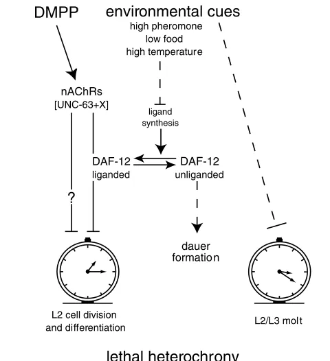
DAF-12 is able to regulate the transcription of a wide range of targets (Ao et al., 2004; Shostak et al., 2004). Liganded-DAF-12 might directly or indirectly control the L2-stage expression of one gene or a subset of genes participating in the predicted signaling cascade triggered by exposure to DMPP. Obvious candidates would be nAChRs. However, we do not favor this hypothesis because unc-63expression remains unchanged in daf-12null mutants (data not shown). Alternatively, UNC-63-containing nAChRs might regulate DAF-12 activity that, in turn, would control L2 development speed. However, analysis of unc-63; daf-12double-null mutants indicates that UNC-63 and DAF-12 do not function in a simple linear pathway (data not shown). In addition, UNC-63 is not expressed in the epidermal syncytium hyp7 or in the XXX cells (see Fig. S2 in the supplementary material), the two DAF-9 expressing tissues involved in DAF-12 ligand synthesis. As L2 development speed is unchanged in daf-12null mutants, a likely hypothesis would be that the non-dauer DAF-12 activity regulates components of the DMPP-triggered signaling cascade that control L2 development speed (Fig. 7).
D
E
V
E
LO
P
M
E
N
T
Description of a novel L2 diapause stage
Over the course of manipulating C. elegans development timing, we identified a previously undescribed developmental arrest. Under favorable conditions, C. elegans eggs develop into reproductive adults within 3 days. However, in adverse conditions, development can pause at specific steps. Newly hatched larvae deprived of food do not initiate postembryonic cell divisions. Animals can survive several weeks in this state of L1 diapause and resume development when placed on food. Similarly, the dauer larva represents a long-lived form of L3 diapause. Here, we identified a previously undescribed developmental arrest at the mid-L2 stage. When grown on low amount of heat-killed E. coli, animals developed to the second larval stage and then arrested development immediately after completing the second seam cell division. They accumulated storage granules in the epidermis but remained sensitive to stress such as desiccation (data not shown). Individuals could survive for up to 9 days and then resume development on food. This arrest probably reflects a specific control of L2 development rather than a starvation process caused by food restriction. First, worms arrested before all heat-killed bacteria had been eaten. Second, all animals arrested at exactly the same stage. Third, young L1 larvae placed on the same amount of heat-killed bacteria, but in the presence of pheromone, passed the L2 arrest stage and formed L3 dauer larvae (Jeong et al., 2005). Therefore, we propose that this previously undescribed stage corresponds to a regulated developmental pause at the L2 stage, i.e. an L2 diapause.
Independent timers control development and molting
In C. elegans, the combination of invariant cell lineage and discontinuous postembryonic development provided a means to identify heterochronic genes that temporally specify cell identity. Because mutating these genes causes entire stage-specific programs to occur earlier or later, or be skipped (reviewed by Ambros, 2000; Pasquinelli and Ruvkun, 2002; Rougvie, 2001; Thummel, 2001), the temporal regulation of C. eleganspostembryonic development is viewed as the successive selection of stage-specific developmental modules. However, whether a single timer is launched at each larval stage and how events are coordinated within each program remains largely unknown. The present results show that stimulation of nicotinic receptors can uncouple the timing of cell division and differentiation from the molt timing at the second larval stage. This suggests that at least two timers exist at the L2 stage: one to control molting and one to regulate cell divisions and differentiation (Fig. 7). The mechanisms that control C. elegansmolt timing are still poorly understood (Frand et al., 2005) in contrast to those in insects where each molt is triggered by a pulse of ecdysteroid. The regulation of developmental speed and especially the mechanisms that control the timing of cell divisions within each larval stage are also poorly characterized. Specifically, heterochronic mutants were not reported to display a change in developmental speed within a given larval stage [for a discussion, see Kipreos (Kipreos, 2005)].
Our data suggest that nicotinic signaling in the nervous system represents one of the upstream regulators of developmental speed and might provide a way to connect environmental signals to animal development. We showed that DAF-12 plays a central role in this regulation and interacts in a non-linear pathway with nicotinic signaling. As DAF-12 functions in complex genetic and molecular networks to control temporal patterning, dauer formation and aging, DMPP-sensitive nAChRs could modulate one branch of these networks. The insulin-like pathway is an interesting candidate as it has been shown to control developmental timing in Drosophila (Bateman and McNeill, 2004) and to interact with the ecdysone NHR to regulate animal growth (Colombani et al., 2005). Further analysis of the developmental effects of nAChR stimulation during C. elegans post-embryonic development might represent an interesting paradigm to analyze how the nervous system can modulate a genetically encoded timer in response to extrinsic factors, and how multiple timers are coordinated during the post-embryonic development of a multicellular organism.
We thank M. de Bono, E. Culetto, A. Fire, I. Katsura, D. Sattelle, V. Robert for plasmids; and the Caenorhabditis Genetic Center, the International C. elegans
Gene Knockout Consortium and the Japanese National BioResources Project for strains. We also thank A. Antebi for sharing unpublished results and reagents; C. Braendle and M.-A. Felix for drawing our attention to the L2 diapause; B. Matthieu for help with confocal microscopy; M.-A. Felix, M. Labouesse and P. Léopold for discussion and critical reading of the manuscript; O. Meyrignac for his help with electron microscopy; and H. Gendrot for technical help. A.-F.R. was supported by a fellowship from the Ministère de la Recherche and by the Association pour la Recherche contre le Cancer. This work was funded by an AVENIR grant from the Institut National de la Santé et de la Recherche Médicale and by the ACI BCMS from the Ministère de la Recherche.
Supplementary material
Supplementary material for this article is available at http://dev.biologists.org/cgi/content/full/133/11/2211/DC1
References
Ambros, V.(2000). Control of developmental timing in Caenorhabditis elegans.
Curr. Opin. Genet. Dev. 10, 428-433.
Antebi, A., Culotti, J. G. and Hedgecock, E. M.(1998). daf-12 regulates
environmental cues
high pheromone low food high temperature
lethal heterochrony
L2 cell division
and differentiation L2/L3 mol t
DMPP
nAChRs [UNC-63+X]
DAF-12 liganded
DAF-12 unliganded ligand
synthesis
dauer formation
[image:10.612.52.292.58.321.2]?
D
E
V
E
LO
P
M
E
N
T
developmental age and the dauer alternative in Caenorhabditis elegans.
Development125, 1191-1205.
Antebi, A., Yeh, W. H., Tait, D., Hedgecock, E. M. and Riddle, D. L.(2000). daf-12 encodes a nuclear receptor that regulates the dauer diapause and developmental age in C. elegans. Genes Dev.14, 1512-1527.
Ao, W., Gaudet, J., Kent, W. J., Muttumu, S. and Mango, S. E.(2004). Environmentally induced foregut remodeling by PHA-4/FoxA and DAF-12/NHR.
Science305, 1743-1746.
Bargmann, C. I.(1998). Neurobiology of the Caenorhabditis elegans genome.
Science282, 2028-2033.
Bargmann, C. I. and Horvitz, H. R.(1991). Control of larval development by chemosensory neurons in Caenorhabditis elegans. Science251, 1243-1246.
Bateman, J. M. and McNeill, H.(2004). Temporal control of differentiation by the insulin receptor/tor pathway in Drosophila. Cell119, 87-96.
Bessereau, J. L., Wright, A., Williams, D. C., Schuske, K., Davis, M. W. and Jorgensen, E. M.(2001). Mobilization of a Drosophila transposon in the Caenorhabditis elegans germ line. Nature413, 70-74.
Boorse, G. C.(2004). Endocrine mechanisms underlying plasticity in metamorphic timing in spadefoot toads. Integr. Comp. Biol. 43, 646-657.
Cassada, R. C. and Russell, R. L.(1975). The dauerlarva, a post-embryonic developmental variant of the nematode Caenorhabditis elegans. Dev. Biol.46, 326-342.
Colombani, J., Bianchini, L., Layalle, S., Pondeville, E., Dauphin-Villemant, C., Antoniewski, C., Carre, C., Noselli, S. and Leopold, P.(2005).
Antagonistic actions of ecdysone and insulins determine final size in Drosophila.
Science310, 667-670.
Corringer, P. J., Le Novere, N. and Changeux, J. P.(2000). Nicotinic receptors at the amino acid level. Annu. Rev. Pharmacol. Toxicol.40, 431-458.
Culetto, E., Baylis, H. A., Richmond, J. E., Jones, A. K., Fleming, J. T., Squire, M. D., Lewis, J. A. and Sattelle, D. B.(2004). The Caenorhabditis elegans unc-63 gene encodes a levamisole-sensitive nicotinic acetylcholine receptor alpha subunit. J. Biol. Chem.279, 42476-42483.
Davis, M. W., Somerville, D., Lee, R. Y., Lockery, S., Avery, L. and Fambrough, D. M.(1995). Mutations in the Caenorhabditis elegans Na,K-ATPase alpha-subunit gene, eat-6, disrupt excitable cell function. J. Neurosci. 15, 8408-8418.
Ebling, F. J.(2005). The neuroendocrine timing of puberty. Reproduction129, 675-683.
Fleming, J. T., Squire, M. D., Barnes, T. M., Tornoe, C., Matsuda, K., Ahnn, J., Fire, A., Sulston, J. E., Barnard, E. A., Sattelle, D. B. et al.(1997). Caenorhabditis elegans levamisole resistance genes lev-1, unc-29, and unc-38 encode functional nicotinic acetylcholine receptor subunits. J. Neurosci. 17, 5843-5857.
Frand, A. R., Russel, S. and Ruvkun, G.(2005). Functional genomic analysis of C. elegans molting. PLoS Biol.3, e312.
Galligan, J. J.(1999). Nerve terminal nicotinic cholinergic receptors on excitatory motoneurons in the myenteric plexus of guinea pig intestine. J. Pharmacol. Exp. Ther.291, 92-98.
Garcia, L. R., Mehta, P. and Sternberg, P. W.(2001). Regulation of distinct muscle behaviors controls the C. elegans male’s copulatory spicules during mating. Cell107, 777-788.
Gerisch, B., Weitzel, C., Kober-Eisermann, C., Rottiers, V. and Antebi, A.
(2001). A hormonal signaling pathway influencing C. elegans metabolism, reproductive development, and life span. Dev. Cell1, 841-851.
Golden, J. W. and Riddle, D. L.(1982). A pheromone influences larval development in the nematode Caenorhabditis elegans. Science218, 578-580.
Golden, J. W. and Riddle, D. L.(1984a). The Caenorhabditis elegans dauer larva: developmental effects of pheromone, food, and temperature. Dev. Biol.102, 368-378.
Golden, J. W. and Riddle, D. L.(1984b). A pheromone-induced developmental switch in Caenorhabditis elegans: temperature-sensitive mutants reveal a wild-type temperature-dependent process. Proc. Natl. Acad. Sci. USA81, 819-823.
Houthoofd, K., Braeckman, B. P., Lenaerts, I., Brys, K., De Vreese, A., Van Eygen, S. and Vanfleteren, J. R.(2002). Axenic growth up-regulates mass-specific metabolic rate, stress resistance, and extends life span in Caenorhabditis elegans. Exp. Gerontol.37, 1371-1378.
Jeong, P. Y., Jung, M., Yim, Y. H., Kim, H., Park, M., Hong, E., Lee, W., Kim, Y. H., Kim, K. and Paik, Y. K.(2005). Chemical structure and biological activity of the Caenorhabditis elegans dauer-inducing pheromone. Nature433, 541-545.
Jia, K., Albert, P. S. and Riddle, D. L.(2002). DAF-9, a cytochrome P450 regulating C. elegans larval development and adult longevity. Development129, 221-231.
Johnstone, I. L.(2000). Cuticle collagen genes. Expression in Caenorhabditis elegans. Trends Genet.16, 21-27.
Jones, A. K. and Sattelle, D. B.(2004). Functional genomics of the nicotinic acetylcholine receptor gene family of the nematode, Caenorhabditis elegans.
BioEssays 26, 39-49.
Kamath, R. S., Martinez-Campos, M., Zipperlen, P., Fraser, A. G. and Ahringer, J.(2001). Effectiveness of specific RNA-mediated interference
through ingested double-stranded RNA in Caenorhabditis elegans. Genome Biol.2, research0002.1-10.
Kim, J., Poole, D. S., Waggoner, L. E., Kempf, A., Ramirez, D. S., Treschow, P. A. and Schafer, W. R.(2001). Genes affecting the activity of nicotinic receptors involved in Caenorhabditis elegans egg-laying behavior. Genetics157, 1599-1610.
Kipreos, E. T.(2005). C. eleganscell cycles: invariance and stem cell divisions. Nat. Rev. Mol. Cell Biol.6, 766-776.
Kirouac, M. and Sternberg, P. W.(2003). cis-Regulatory control of three cell fate-specific genes in vulval organogenesis of Caenorhabditis elegans and C. briggsae. Dev. Biol.257, 85-103.
Lewis, J. A., Wu, C. H., Berg, H. and Levine, J. H.(1980a). The genetics of levamisole resistance in the nematode Caenorhabditis elegans. Genetics95, 905-928.
Lewis, J. A., Wu, C. H., Levine, J. H. and Berg, H.(1980b). Levamisole-resistant mutants of the nematode Caenorhabditis elegans appear to lack
pharmacological acetylcholine receptors. Neuroscience5, 967-989.
Li, W., Kennedy, S. G. and Ruvkun, G.(2003). daf-28 encodes a C. elegans insulin superfamily member that is regulated by environmental cues and acts in the DAF-2 signaling pathway. Genes Dev.17, 844-858.
Ludewig, A. H., Kober-Eisermann, C., Weitzel, C., Bethke, A., Neubert, K., Gerisch, B., Hutter, H. and Antebi, A.(2004). A novel nuclear
receptor/coregulator complex controls C. elegans lipid metabolism, larval development, and aging. Genes Dev.18, 2120-2133.
Manetti, D., Bartolini, A., Borea, P. A., Bellucci, C., Dei, S., Ghelardini, C., Gualtieri, F., Romanelli, M. N., Scapecchi, S., Teodori, E. et al.(1999). Hybridized and isosteric analogues of N1-acetyl-N4-dimethyl-piperazinium iodide (ADMP) and N1-phenyl-N4-dimethyl-piperazinium iodide (DMPP) with central nicotinic action. Bioorg. Med. Chem.7, 457-465.
Matyash, V., Entchev, E. V., Mende, F., Wilsch-Brauninger, M., Thiele, C., Schmidt, A. W., Knolker, H. J., Ward, S. and Kurzchalia, T. V.(2004). Sterol-derived hormone(s) controls entry into diapause in Caenorhabditis elegans by consecutive activation of DAF-12 and DAF-16. PLoS Biol.2, e280.
McGehee, D. S. and Role, L. W.(1995). Physiological diversity of nicotinic acetylcholine receptors expressed by vertebrate neurons. Annu. Rev. Physiol.57, 521-546.
McKay, J. P., Raizen, D. M., Gottschalk, A., Schafer, W. R. and Avery, L.
(2004). eat-2 and eat-18 are required for nicotinic neurotransmission in the Caenorhabditis elegans pharynx. Genetics166, 161-169.
Meissner, B., Boll, M., Daniel, H. and Baumeister, R.(2004). Deletion of the intestinal peptide transporter affects insulin and TOR signaling in Caenorhabditis elegans. J. Biol. Chem.279, 36739-36745.
Mello, C. C., Kramer, J. M., Stinchcomb, D. and Ambros, V.(1991). Efficient gene transfer in C.elegans: extrachromosomal maintenance and integration of transforming sequences. EMBO J.10, 3959-3970.
Nonet, M. L., Staunton, J. E., Kilgard, M. P., Fergestad, T., Hartwieg, E., Horvitz, H. R., Jorgensen, E. M. and Meyer, B. J.(1997). Caenorhabditis elegans rab-3 mutant synapses exhibit impaired function and are partially depleted of vesicles. J. Neurosci. 17, 8061-8073.
Pasquinelli, A. E. and Ruvkun, G.(2002). Control of developmental timing by micrornas and their targets. Annu. Rev. Cell Dev. Biol.18, 495-513.
Peckol, E. L., Zallen, J. A., Yarrow, J. C. and Bargmann, C. I.(1999). Sensory activity affects sensory axon development in C. elegans. Development126, 1891-1902.
Pettitt, J., Wood, W. B. and Plasterk, R. H.(1996). cdh-3, a gene encoding a member of the cadherin superfamily, functions in epithelial cell morphogenesis in Caenorhabditis elegans. Development122, 4149-4157.
Rand, J. B. and Nonet, M. L.(1997). Synaptic transmission. In C. Elegans II(ed. D. L. Riddle, T. Blumenthal, B. J. Meyer and J. R. Priess), pp. 611-644. Cold Spring Harbor, NY: Cold Spring Harbor Laboratory Press.
Ren, P., Lim, C. S., Johnsen, R., Albert, P. S., Pilgrim, D. and Riddle, D. L.
(1996). Control of C. elegans larval development by neuronal expression of a TGF-beta homolog. Science274, 1389-1391.
Richmond, J. E. and Jorgensen, E. M.(1999). One GABA and two acetylcholine receptors function at the C. elegans neuromuscular junction. Nat. Neurosci.2, 791-797.
Riddle, D. L. and Albert, P. S.(1997). Genetic and environmental regulation of dauer larva development. In C. Elegans II(ed. D. L. Riddle, T. Blumenthal, B. J. Meyer and J. R. Priess), pp. 739-768. Cold Spring Harbor, NY: Cold Spring Harbor Laboratory Press.
Rostaing, P., Weimer, R. M., Jorgensen, E. M., Triller, A. and Bessereau, J. L.
(2004). Preservation of immunoreactivity and fine structure of adult C. elegans tissues using high-pressure freezing. J. Histochem. Cytochem.52, 1-12.
Rougvie, A. E.(2001). Control of developmental timing in animals. Nat. Rev. Genet.2, 690-701.
Rougvie, A. E.(2005). Intrinsic and extrinsic regulators of developmental timing: from miRNAs to nutritional cues. Development132, 3787-3798.
Shostak, Y., Van Gilst, M. R., Antebi, A. and Yamamoto, K. R.(2004). Identification of C. elegans DAF-12-binding sites, response elements, and target genes. Genes Dev.18, 2529-2544.
D
E
V
E
LO
P
M
E
N
T
Ahringer, J. and Plasterk, R. H.(2002). Loss of the putative RNA-directed RNA polymerase RRF-3 makes C. elegans hypersensitive to RNAi. Curr. Biol.12, 1317-1319.
Singh, R. N. and Sulston, J. E.(1978). Some observations on moulting in
Caenorhabditis elegans. Nematologica24, 63-71.
Sulston, J. and Hodgkin, J.(1988). Methods. In The Nematode Caenorhabditis Elegans(ed. W. B. Wood), pp. 587-606. Cold Spring Harbor, NY: Cold Spring Harbor Laboratory Press.
Thummel, C. S.(2001). Molecular mechanisms of developmental timing in C. elegans and Drosophila. Dev. Cell1, 453-465.
Towers, P. R., Edwards, B., Richmond, J. E. and Sattelle, D. B.(2005). The Caenorhabditis elegans lev-8 gene encodes a novel type of nicotinic acetylcholine receptor alpha subunit. J. Neurochem.93, 1-9.
Vizi, E. S. and Lendvai, B.(1999). Modulatory role of presynaptic nicotinic receptors in synaptic and non-synaptic chemical communication in the central nervous system. Brain Res. Brain Res. Rev.30, 219-235.
Williams, D. C., Boulin, T., Ruaud, A. F., Jorgensen, E. M. and Bessereau, J. L.