C A S E R E P O R T
Open Access
Simultaneous and sequential hemorrhage
of multiple cerebral cavernous
malformations: a case report
Nundia Louis and Robert Marsh
*Abstract
Background:The etiology of cerebral cavernous malformation hemorrhage is not well understood. Causative physiologic parameters preceding hemorrhagic cavernous malformation events are often not reported. We present a case of an individual with sequential simultaneous hemorrhages in multiple cerebral cavernous malformations with a new onset diagnosis of hypertension.
Case presentation:A 42-year-old white man was admitted to our facility with worsening headache, left facial and tongue numbness, dizziness, diplopia, and elevated blood pressure. His past medical history was significant for new onset diagnosis of hypertension and chronic seasonal allergies. Serial imaging over the ensuing 8 days revealed sequential hemorrhagic lesions. He underwent suboccipital craniotomy for resection of the lesions located in the fourth ventricle and right cerebellum. One month after surgery, he had near complete resolution of his symptoms with mild residual vertigo but symptomatic chronic hypertension.
Conclusions:Many studies have focused on genetic and inflammatory mechanisms contributing to cerebral cavernous malformation rupture, but few have reported on the potential of hemodynamic changes contributing to cerebral cavernous malformation rupture. Systemic blood pressure changes clearly have an effect on angioma pressures. When considering the histopathological features of cerebral cavernous malformation architecture, changes in arterial pressure could cause meaningful alterations in hemorrhage propensity and patterns.
Keywords:Angioma, Cavernoma, Cavernous malformation, Hemorrhage, Hypertension
Background
Cerebral cavernous malformations (CCMs), also known as cavernomas or cavernous angiomas, are classically de-fined as low pressure hamartomatous berrylike vascular lesions with minimal to no intervening brain paren-chyma composed of thin-walled endothelial-lined sinus-oidal spaces devoid of smooth muscle [1–4]. It has been suggested that CCMs arise from failure of vascular stabilization in angiogenesis which promotes the devel-opment of capillary dysplasia, weak intercellular junc-tions, and defective smooth muscle recruitment [5]. An estimated 0.5 % of the population has CCMs [3, 6]. While many patients remain asymptomatic, others tend to develop epilepsy, neurological deficits, or hemorrhage
[1, 4, 6]. Sporadic and inherited forms of CCM have been described. The sporadic form often results in single isolated lesions while the inherited form is associated with multiple lesions and mutations of the endothelial genesCCM1,CCM2orCCM3[5, 7].
The etiology of CCM rupture is not well understood. It has been demonstrated that CCM lesions elicit inflam-matory responses that involve tumor necrosis factor alpha (TNF-α) and interleukins (ILs). Upregulation of angiogenic factors such as vascular endothelial growth factor (VEGF) have also been described. These processes are implicated in the promotion of angiogenesis and breakdown of the blood–brain barrier (BBB) leading to the progression and rupture of CCM [5]. CCM2 and
CCM3 mutations have also been linked to higher hemorrhage rates [1, 8]. Simultaneous hemorrhages of multiple CCM lesions are anecdotally common but few have been reported [7, 9, 10]. Causative physiologic * Correspondence:[email protected]
Department of Neurosurgery, Cabell Huntington Hospital, Marshall University, 1600 Medical Center Drive, Huntington, WV25701USA
parameters preceding hemorrhagic CCM events are often not described even in case reports. We present a case of an individual with simultaneous and sequential hemorrhages in multiple CCMs with a new onset diag-nosis of hypertension.
Case presentation
History and examination
We describe the case of a 42-year-old white man who was transferred to our facility due to worsening headache of 6 months evolution and new onset left facial and tongue numbness with dizziness. A head computed tomography indicated two areas suspicious for acute hemorrhage: one within the fourth ventricle and the other adjacent to the calvarium in his right cerebellum (Fig. 1a). His past med-ical history was significant for acute sinusitis and recent onset of hypertension. A physical examination at the time of presentation revealed blood pressure of 161/98 mmHg, ataxia, dysmetria, vertigo, and a positive Romberg’s test. Magnetic resonance imaging (MRI) was obtained with and without contrast.
The MRI showed two additional lesions: one in his left lateral cerebellar hemisphere and the other in his medial posterior left temporal lobe (Fig. 1b). All lesions exhibited significant signal dropout on gradient echo sequences. Heterogeneous enhancement was noted in the right cere-bellar mass (Fig. 1c). The differential diagnosis included multiple cavernous malformations or hemorrhagic meta-static lesions.
Erythrocyte sedimentation rate, C-reactive protein, carbohydrate antigen 19-9, carcinoembryonic antigen, and a chest X-ray were ordered and found to be within normal limits. A computed tomography (CT) scan per-formed the next day showed increased hemorrhage size within the ventricular lesion and a new hemorrhagic hyperdensity within the left medial temporal location (Fig. 1d). The patient symptomatically improved over his 2-day hospital course with complete resolution of his dizziness and ataxia. He was discharged with orders for a repeat MRI after hemorrhage resolution and further testing. Surgical resection was delayed due to his pre-senting symptoms, uncertainty of etiology, and specific reports suggesting resection of CCMs after two bleeding incidents in eloquent brain regions or single hemorrhage in non-eloquent area accompanied by deteriorating neurological deficit [11].
The patient was readmitted 1 week later with headache, nausea, worsening dizziness, new onset diplopia and ele-vated blood pressure. He was found to have a fourth cra-nial nerve palsy, mild decrease in the right nasolabial fold, hypophonia and continued left facial and tongue numb-ness. A CT scan displayed further hemorrhagic enlarge-ment of the intraventricular and temporal lesions with development of hydrocephalus (Fig. 1e).
Operation
The patient underwent a suboccipital craniotomy and the right cerebellar lesion was resected first. The lesion dem-onstrated hemosiderin deposition with a gliotic margin. The telovelar approach was then used to access the fourth ventricle. The ventricular mass was well-circumscribed, pearly red and easily delineated from the ventral wall of Fig. 1Cranial imaging of multiple cerebral cavernous malformations.
aComputed tomography scan indicating two areas suspicious for acute hemorrhage: one within the fourth ventricle and the other adjacent to the calvarium in the patient’s right cerebellum.bGradient echo magnetic resonance imaging showing lesion in left lateral cerebellar hemisphere.cMagnetic resonance imaging with contrast showing heterogeneous enhancement in the right cerebellar mass.d
[image:2.595.307.538.85.492.2]the fourth ventricle. The mass was centrally debulked and the walls circumferentially collapsed. The gliotic margin adjacent to the brainstem was carefully delineated and gross total resection was achieved.
Postoperative course
Immediately after surgery, the patient developed new onset mild left third nerve palsy. MRI showed complete resection of fourth ventricular and right cerebellar masses (Fig. 1f ). Pathology confirmed diagnosis of CCM. He was discharged on postoperative day six with im-proved cranial nerve functioning and resolution of ataxia but continued vertigo. Approximately 1 month after sur-gery, his course was complicated by culture-negative bacterial meningitis and development of pseudomenin-gocele that resolved with aspiration and proper antibi-otics treatment. He demonstrated complete resolution of ocular cranial nerve dysfunction but exhibited mild hori-zontal nystagmus with rotational challenge that resolved by 6 months.
Discussion
Cerebral cavernomas are estimated to occur in 1 out of every 200 individuals in the general population [3, 6, 7]. They are hypothesized to develop due to failure of vas-cular stabilization in angiogenesis of cerebral blood ves-sels [5]. In a large series studying the natural history of cavernomas, 18.7 to 20 % of patients had multiple lesions [9, 12, 13]. Multiple lesions are mostly seen in familial CCM (FCCM) forms [7]. FCCM has been
asso-ciated with mutations of CCM1, CCM2 and CCM3
genes. Nevertheless, 22 % of multiple lesions occur with-out any evidence of gene mutation [8].
CCMs have a hemorrhage rate of 0.7 to 1.1 % per lesion per year [4, 7]. Most cases of hemorrhages reported in the literature tend to involve one lesion, even in patients with
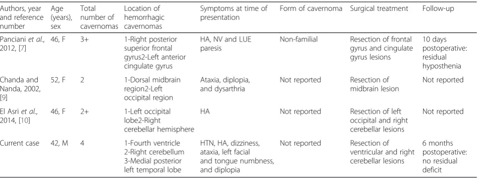
multiple CCMs. Personal communication from Issam Awad suggests that multiple simultaneously hemorrhagic CCM lesions are more common (July 2015). However, to date there have been a total of only three reports on sim-ultaneous hemorrhages of multiple cavernomas (Table 1). Our report is different in that the presented case exhibited simultaneous and sequential rupture patterns in the set-ting of new onset hypertension. What would lead to such a phenomenon remains an enigma, as the exact cause of CCM hemorrhage has yet to be determined. Many studies have focused on the genetic and inflammatory mecha-nisms contributing to CCM rupture, but few have investi-gated the potential role chronic hypertension may play in this complex multifactorial disease process.
Genetic and inflammatory causes have clearly been shown to influence CCM hemorrhage. In mouse model experiments, Cunningham et al. observed that condi-tional inactivation of the CCM2 gene in adult mice produced a cerebral hemorrhage similar to that ob-served in adult human CCMs [1]. Mutation of the
[image:3.595.57.541.544.724.2]CCM3 gene in humans has been linked to a hereditary variant of CCM and demonstrates early-onset cerebral hemorrhage patterns [8]. Shi et al. have reported CCMs to be active inflammation sites infiltrated with B cells and plasma cells [14]. Our patient did not have any familial past medical history of intracerebral hemorrhage or relatives with a diagnosis of CCM. However, he was being treated for sinusitis and it is possible that there was an increased release of inflam-matory cytokines, TNF-αand ILs. These inflammation mediators stimulate angiogenesis and BBB breakdown and are thought to contribute to CCM rupture [5]. It is also possible that rupture of one of the CCMs enhanced recruitment of the inflammatory processes that contributed to the sequential pattern that was observed in our patient. However, inflammation and
Table 1Details of the three published cases of simultaneous multiple cavernoma hemorrhages (and the present case)
Authors, year and reference number Age (years), sex Total number of cavernomas Location of hemorrhagic cavernomas
Symptoms at time of presentation
Form of cavernoma Surgical treatment Follow-up
Pancianiet al., 2012, [7]
46, F 3+ 1-Right posterior
superior frontal gyrus2-Left anterior cingulate gyrus
HA, NV and LUE paresis
Non-familial Resection of frontal gyrus and cingulate gyrus lesions 10 days postoperative: residual hyposthenia Chanda and Nanda, 2002, [9]
52, F 2 1-Dorsal midbrain
region2-Left occipital region
Ataxia, diplopia, and dysarthria
Not reported Resection of midbrain lesion
Not reported
El Asriet al., 2014, [10]
46, F 2+ 1-Left occipital
lobe2-Right
cerebellar hemisphere
HA Not reported Resection of left
occipital and right cerebellar lesions
Not reported
Current case 42, M 4 1-Fourth ventricle 2-Right cerebellum 3-Medial posterior left temporal lobe
HTN, HA, dizziness, ataxia, left facial and tongue numbness, and diplopia
Not reported Resection of ventricular and right cerebellar lesions
6 months postoperative: no residual deficit
genetics may be only part of this complex multifactor-ial disease process and hypertension may play a role.
The studies that have addressed hemodynamic effects on CCMs are narrowly focused and limited. A recent study on the association of cardiovascular risk factors with CCM severity in 185 Hispanic individuals with
CCM1 mutation failed to find any positive correlation between CCM rupture and cardiovascular risks, includ-ing hypertension [15]. However, an experiment by Little
et al. demonstrated that cavernomas are affected by changes in mean arterial blood pressure (MABP) and venous pressure. Direct angioma pressure measurements showed that a mean reduction of 14.7±2.1 mmHg in MABP resulted in a 7.0±0.5 mmHg drop in angioma pressure. Mechanical jugular compression induced real measurable changes in CCM pressure up to 9 mmHg [2]. Although the study did not investigate changes caused by increased MABP, the data clearly demonstrate that systemic blood pressure changes significantly affect CCM pressures.
Few studies to date have directly measured cerebral capillary pressure or determined the direct effects of sys-temic blood pressure on capillary physiology. Classical studies of cerebral blood flow show that the pial arteri-oles autoregulate until systolic blood pressure exceeds approximately 160 mmHg above which smaller pial arte-rioles are differentially affected, become dilated, and lose regulatory control; this results in increases in blood flow [16, 17]. Direct measurement of cerebral capillary pres-sure is problematic but prespres-sure characteristics can be extrapolated from peripheral limb vasculature experi-ments which demonstrate that significantly higher apex pressures are measured in patients with essential hyper-tension when compared to age-matched and sex-matched normotensive controls [18, 19]. Structural abnormalities can also occur in peripheral capillaries as a result of essen-tial hypertension with loss or reduction in the density of vessels per volume of tissue, which is a process known as rarefaction [18, 20]. When considering the histopatho-logical features of CCM architecture, chronic hypertensive changes alter arterial flow and vessel physiology, which could cause meaningful alterations in capillary anatomy as well as hemorrhage propensity and patterns.
Many vascular disease processes are influenced by multi-factorial systemic corporeal changes. An example of this is the development and rupture of cerebral aneurysms. In-flammation, genetics, hypertension, smoking, and age are known risk factors that contribute to the development of aneurysm rupture and hemorrhage [21–25]. Models have been developed to describe the pathophysiology for aneurysm induction and progression and include endothe-lial damage and degeneration of the elastic lamina, inflam-matory cell recruitment and infiltration, and chronic remodeling of vascular wall [26]. Similarly, we argue that
chronic high blood pressure may be a factor in capillary physiology that alters architectural features of abnormal ca-pillary anatomy in CCM which increases the propensity for hemorrhage.
Conclusions
This case report is unusual in that hypertension may have played a role in the simultaneous and sequential hemorrhage pattern noted in this individual. Notwith-standing the many advances made in underNotwith-standing the structure, formation and evolution of CCMs, there is still a lack of understanding concerning the effect of hemodynamic changes on cerebral capillary physiology and cavernomas. Investigative work focusing on the role of hypertension and other hemodynamic factors in CCM rupture is much needed, especially experiments in order to better determine if blood pressure changes affect the incidence of CCM hemorrhage.
Consent
Written informed consent was obtained from the patient for publication of this case report and accompanying images. A copy of the written consent is available for review by the Editor-in-Chief of this journal.
Competing interests
The authors declare that they have no competing interests concerning this case report.
Authors’contributions
NL and RM equally contributed to the creation of this manuscript. Both authors read and approved the final manuscript.
Received: 8 December 2015 Accepted: 24 January 2016
References
1. Cunningham K, Uchida Y, O’Donnell E, Claudio E, Li W, Soneji K, et al. Conditional deletion ofCcm2causes hemorrhage in the adult brain: a mouse model of human cerebral cavernous malformations. Hum Mol Genet. 2011;20:3198–206.
2. Little J, Awad I, Jones S, Ebrahim Z. Vascular pressure and cortical blood flow in cavernous angioma of the brain. J Neurosurg. 1990;73:555–9. 3. Riant F, Bergametti F, Ayrignac X, Boulday G, Tournier-Lasserve E. Recent
insights into cerebral cavernous malformations: the molecular genetics of CCM. FEBS J. 2010;277:1070–5.
4. Robinson JR, Awad IA, Little JR. Natural history of the cavernous angioma. J Neurosurg. 1991;75:709–14.
5. Leblanc GG, Golanov E, Awad IA, Young WL. Biology of vascular malformations of the brain. Stroke. 2009;40:e694–702.
6. Kivelev J, Niemela M, Hernesniemi J. Characteristics of cavernomas of the brain and spine. J Clin Neurosci. 2012;19:643–8.
7. Panciani PP, Agnoletti A, Fornaro R, Fontanella M, Ducati A. Multiple cavernomas of the brain: simultaneous hemorrhage of two lesions in a non-familial form. Turk Neurosurg. 2012;22:671–4.
8. Riant F, Bergametti F, Fournier HD, Chapon F, Michalak-Provost S, Cecillon M.CCM3mutations are associated with early-onset cerebral hemorrhage and multiple meningiomas. Mol Syndromol. 2013;4:165–72.
9. Chanda A, Nanda A. Multiple cavernomas of brain presenting with simultaneous hemorrhage in two lesions: a case report. Surg Neurol. 2002; 57(5):340–4.
11. Steinberg GK, Chang SD, Gewirtz RJ, Lopez JR. Microsurgical resection of brainstem, thalamic, and basal ganglia angiographically occult vascular malformations. Neurosurgery. 2000;46:260–70.
12. Del Curling JO, Kelly Jr DL, Elster AD, Craven TE. An analysis of the natural history of cavernous angiomas. J Neurosurg. 1991;75:702–8.
13. Kondziolka D, Lunsford L, Kestle J. The natural history of cerebral cavernous malformations. J Neurosurg. 1995;83:820–4.
14. Shi C, Shenkar R, Batjer HH, Check IJ, Awad IA. Oligoclonal immune response in cerebral cavernous malformations. J Neurosurg. 2007;107:1023–6.
15. Choquet H, Nelson J, Pawlikowska L, McCulloch C, Akers A, Baca B, et al. Association of cardiovascular risk factors with disease severity in cerebral cavernous malformation type 1 subjects with common Hispanic mutation. Cerebrovasc Dis. 2014;37:57–63.
16. Kontos HA, Wei EP, Navari RM, Levasseur JE, Rosenblum WI, Patterson Jr JL. Responses of cerebral arteries and arterioles to acute hypotension and hypertension. Am J Physiol. 1978;234:H371–83.
17. Mackenzie ET, Strandgaard S, Graham DI, Jones JV, Harper AM, Farrar JK. Effects of acutely induced hypertension in cats on pial arteriolar caliber, local cerebral flow, and the blood-barrier. Circ Res. 1976;39:33–41. 18. Landau J, Davis E. Capillary thinning and high capillary blood-pressure in
hypertension. Lancet. 1957;269:1327–30.
19. Williams SA, Boolell M, MacGregor GA, Smaje LH, Wasserman SM, Tooke JE. Capillary hypertension and abnormal pressure dynamics in patients with essential hypertension. Clin Sci (Lond). 1990;79:5–8.
20. Noon JP, Walker BR, Webb DJ, Shore AC, Holton DW, Edwards HV, et al. Impaired microvascular dilatation and capillary rarefaction in young adults with a predisposition to high blood pressure. J Clin Invest. 1997;99:1873–9. 21. Jamous MA, Nagahiro S, Kitazato KT, Tamura T, Aziz HA, Shono M, et al.
Endothelial injury and inflammatory response induced by hemodynamic changes preceding intracranial aneurysm formation: experimental study in rats. J Neurosurg. 2007;107(2):405–11.
22. UCAS Japan Investigators, Morita A, Kirino T, Hashi K, Aoki N, Fukuhara S, et al. The natural course of unruptured cerebral aneurysms in a Japanese cohort. N Engl J Med. 2012;366:2474–82.
23. Ortiz R, Stefanski M, Rosenwasser R, Veznedaroglu E. Cigarette smoking is a risk factor for recurrence of aneurysms treated by endosaccular occlusion. J Neurosurg. 2008;108:672–5.
24. Rinkel GJ. Natural history, epidemiology and screening of unruptured intracranial aneurysms. J Neuroradiol. 2008;35:99–103.
25. Taylor CL, Yuan Z, Selman WR, Ratcheson RA, Rimm AA. Cerebral arterial aneurysm formation and rupture in 20,767 elderly patients: hypertension and other risk factors. J Neurosurg. 1995;83:812–9.
26. Hosaka K, Hoh BL. Inflammation and cerebral aneurysms. Transl Stroke Res. 2014;2:190–8.
• We accept pre-submission inquiries
• Our selector tool helps you to find the most relevant journal • We provide round the clock customer support
• Convenient online submission • Thorough peer review
• Inclusion in PubMed and all major indexing services • Maximum visibility for your research
Submit your manuscript at www.biomedcentral.com/submit