Hierarchical Contributions of Allorecognition Pathways
in Chronic Lung Rejection
Worakij Chalermskulrat, Isabel P. Neuringer, W. June Brickey, Nathan J. Felix, Scott H. Randell, Jenny P. Ting, and Robert M. Aris
Division of Pulmonary Disease and Critical Care Medicine and the Lung Transplantation Program; and Department of Microbiology and Immunology, Lineberger Comprehensive Cancer Center, University of North Carolina, Chapel Hill, North Carolina
The role of allorecognition in initiating lung graft rejection is not the pathology of human OB (2). Studies have validated the clearly defined. Using the heterotopic tracheal transplantation use of the mouse heterotopic tracheal transplant model and model, we examined the contributions of the indirect and direct yielded important initial observations showing that alloim-allorecognition pathways in chronic airway rejection. Fully mis- mune injury that leads to OB (3–8).
matched, wild-type grafts were transplanted into major histocom- The initial and central event that ultimately leads to graft patibility complex (MHC) II⫺/⫺, class II-like accessory molecule
(H2-rejection is allorecognition. Allospecific T cells may be acti-DM␣)⫺/⫺using MHC I⫺/⫺and wild-type allorecipients as control
vated by two distinct pathways (9–13). The direct pathway subjects. Similarly, MHC I⫺/⫺, MHC II⫺/⫺, or MHC I/II⫺/⫺allografts
is defined by the allorestricted activation of recipient T cells were transplanted into wild-type mice with appropriate control
by donor antigen-presenting cells, whereas the indirect path-subjects. Grafts from nonimmunosuppressed recipients were
evalu-way refers to activation of recipient T cells through the inter-ated at Weeks 2, 4, and 6. Grafts transplanted into MHC II⫺/⫺and
action with preprocessed allopeptides presented by recipient H2-DM␣⫺/⫺allorecipients showed a more intact epithelium and
antigen-presenting cells in a self major histocompatibility reduced lumen obliteration compared with grafts transplanted into
complex (MHC) II–restricted manner. Although the mecha-wild-type or MHC I⫺/⫺ allorecipients (p⬍ 0.05 for each). These
nism for indirect allorecognition is similar to the physiologic grafts exhibited abundant CD4⫹and CD8⫹cell infiltrates similar
mechanism of host defense, the mechanism of direct allorec-to control allografts. MHC I⫺/⫺and MHC I/II⫺/⫺but not MHC II⫺/⫺
ognition pathway is less well characterized. Overall, the rela-allografts placed in wild-type animals demonstrated less severe
re-jection compared with allograft control subjects (p⬍0.05 for each). tive contributions of the indirect and direct allorecognition Although the indirect allorecognition pathway has the strongest pathways to organ graft rejection remain largely unknown influence on rejection, the direct pathway is sufficient to ultimately and have not been well studied in the tracheal transplant cause chronic airway rejection. In addition, these results suggest model.
that MHC class I molecules are the principal alloantigens in the Genetically engineered mice have been generated and mouse heterotopic tracheal model of obliterative bronchiolitis. used to examine the biologic function of components of the antigen processing pathway. The MHC class II molecules Keywords:allorecognition; alloantigen; lung transplant; trachea
trans-are heterodimers consisting of ␣ and  chains, which are plant model; obliterative bronchiolitis
assembled in the endoplasmic reticulum, associated with the invariant chain and transported into the endosomal compart-Lung transplantation has become a successful clinical therapy
ment where the invariant chain is proteolytically degraded for individuals with diverse end-stage pulmonary diseases.
into class II-associated invariant chain peptide (CLIP). The Despite advances in several aspects of pulmonary transplant
class II-like accessory molecule HLA-DM or its murine medicine, chronic lung allograft rejection, manifested as
pro-equivalent, H2-DM, catalyzes the dissociation of CLIP and gressive airway obstruction, namely obliterative bronchiolitis
assists in the loading of the antigenic peptides, thereby play-(OB), remains the leading cause of morbidity and mortality
ing an important role in the orderly trafficking of MHC II-among long-term survivors of lung transplantation (1). The
peptide complexes to the cell surface. Mice deficient in MHC treatment of OB is ineffective, mainly because of our lack
class II expression have been generated by deleting the A of knowledge of the pathogenesis of this disease. In the mouse
gene, resulting in undetectable MHC class II expression (14, heterotopic tracheal transplant model, allografts, but not
iso-15). Mice deficient in MHC class I expression have also been grafts, develop a defined, predictable succession of airway
generated by disruption of the gene encoding the 2-micro-inflammation, epithelial injury and denudation, and lumen
globulin, a polypeptide required for proper assembly and cell fibroproliferation, resulting in airway obliteration replicating
surface expression of MHC class I molecules (16, 17). In addition, mice deficient in H2-DM have been generated by disruption of the H2-DM␣gene, which encodes the␣subunit for H2-DM. In the absence of H2-DM␣, CLIP cannot be
(Received in original form September 25, 2002; accepted in final form November 20, 2002)
dissociated from and exogenous antigens cannot be
com-Supported by a Cystic Fibrosis Foundation research grant (R.M.A. and W.C.), an American plexed with newly synthesized MHC class II molecules (18– Lung Association research grant (I.P.N.), and National Institutes of Health grants
21). H2-DM␣deficiency on the C57BL/6 background results
HL67178 (I.P.N.), HL58345 (S.H.R.), AI29564 ( J.P.T.), and DK38108 ( J.P.T.). W.C. is
in an almost complete blockade of alloantigen processing
the recipient of an American Lung Association Fellowship Award. This work was
re-ported, in part, in the form of an abstract (55, 56) and was presented at the 96th and and presentation by MHC class II molecules (21–23). 98th American Thoracic Society International Conferences (May 8, 2000, in Toronto, Previous studies using MHC II-deficient (skin) allografts
Ontario, Canada, and May 20, 2002, in Atlanta, Georgia). (i.e., absence of donor MHC II-bearing antigen-presenting
Correspondence and requests for reprints should be addressed to Robert M. Aris, M.D., cells) to eliminate the direct allorecognition pathway have 420 Burnett-Womack Building, CB# 7020, Chapel Hill, NC 27599. E-mail: aris@med. concluded that indirect allorecognition is sufficient to cause unc.edu
rejection (24, 25). However, recent studies have indicated Am J Respir Crit Care Med Vol 167. pp 999–1007, 2003
that allograft antigen-presenting cell cells can directly acti-Originally Published in Press as DOI: 10.1164/rccm.200209-1099OC on November 21, 2002
H2dmice were transplanted into fully mismatched MHC I⫺/⫺, MHC the absence of CD4⫹ T cell “help” (26–28), resulting in
II⫺/⫺, H2-DM␣⫺/⫺, and wild-type H2bmice. For isograft control subjects,
rejection (26, 27). Therefore, the use of MHC II-deficient
tracheae from wild-type H2bwere transplanted into wild-type H2bmice.
grafts cannot entirely exclude direct (MHC I-dependent)
al-Second, tracheal grafts were obtained from MHC I⫺/⫺, MHC II⫺/⫺, lorecognition (24, 25). In the experiments described herein,
MHC I/II⫺/⫺, and wild-type H2bor H2dmice and placed into wild-type
we pursued an alternative and comprehensive approach to
H2dmice. All mice were maintained following the National Institutes
evaluate the role and contribution of each allorecognition of Health guidelines for the care and use of laboratory animals under pathway and donor MHC molecules in lung transplant rejec- specific pathogen-free conditions.
tion. First, we transplanted wild-type tracheal allografts into
MHC II⫺/⫺or H2-DM␣⫺/⫺mice, which have different disrup- Mouse Heterotopic Tracheal Transplantation
tions in the class II-dependent antigen presentation process As previously described (4–6), tracheae were transplanted into a subcu-that, in effect, eliminates indirect allorecognition. Second, taneous pocket behind the neck of the recipient after anesthesia was we transplanted MHC I-, II-, and I/II-deficient allografts into achieved by injecting Domitor (5 mg/kg)/Ketamine (100 mg/kg)
(Divi-wild-type recipients to assess the role of MHC I and MHC sion of Laboratory Animal Medicine, UNC) intraperitoneally and then
II-mediated direct allorecognition and MHC molecules as reversed with Antisedan (2.5 mg/kg) (Division of Laboratory Animal Medicine), introduced subcutaneously. No immunosuppressant was
alloantigens.
used. METHODS
Morphometry
Mice and Study Design Grafts were harvested at Weeks 2, 4, and 6. Tissue processing, staining, and image acquisition have been previously described (4–6). Detach-The following female mice between 8 and 12 weeks of age were used:
ment of the differentiated (ciliated) epithelium (epithelial injury) and wild-type BALB/c (H2d) and C57BL/6 (H2b) (Harlan Labs,
Indianapo-lumen obliteration (fibroproliferation) were examined to determine lis, IN), H2bMHC class I deficient (MHC I⫺/⫺) (17), H2bMHC class
airway rejection. The intact differentiated epithelium and total airway II deficient (MHC II⫺/⫺) (14), and H2bMHC class I and II deficient
circumference were measured at the level of the sub-basement mem-(MHC I/II⫺/⫺) (29) (Taconic Labs, Germantown, NY). In addition,
H2-brane. Quantitation of the epithelialization and fibroproliferation was DM␣⫺/⫺ mice were backcrossed onto the H2bbackground for nine
performed using the Metamorph Image analysis program (West Ches-generations and were bred and maintained at the University of North
Carolina at Chapel Hill (UNC) (18). First, allografts from wild-type ter, PA) by three independent, blinded reviewers and was expressed
as the fraction of total airway circumference or of the total lumen cross-sectional area, respectively (4–6).
Immunohistochemistry
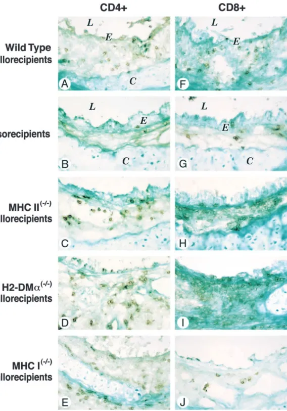
Frozen sections were air dried, fixed in chilled acetone, and blocked with 5% normal goat serum (Jackson ImmunoResearch, West Grove, PA) and avidin-biotin block (Vector Labs, Burlingame, CA). The sec-tions were incubated with monoclonal rat anti-mouse CD4⫹or CD8⫹ antibodies, then biotinylated goat anti-rat antibody, and finally streptav-idin-horseradish-peroxidase (Pharmingen, San Diego, CA). Optimal dilutions and incubation periods were determined empirically. Dupli-cate sections on the same slide were incubated with rat IgG2a (Jackson ImmunoResearch) as a negative isotype control. The diaminobenzidine substrate (Sigma, St. Louis, MO) was used, and sections were counter-stained with Light Green (Fisher Scientific Co., Pittsburgh, PA). Cells were counted within the area outlined by an equidistant line, 10m outward from the sub-basement membrane. In accordance with the previously described profile of immune cellular infiltration into allo-grafts in this model (4), allo-grafts were examined for CD4⫹and CD8⫹T cells at Week 2.
Statistical Analysis
Analyses were performed using SigmaStat software (SPSS Inc., Chi-cago, IL). Parameters were compared at each time point using the student’st-test. A two-tailed␣level of p⬍0.05 was considered signifi-cant. Repeated-measures analyses of variance were used (8) to confirm the results from thet-test analyses.
RESULTS
Allogeneic Transplantation into Fully Mismatched, Wild-type H2bor H2dMice
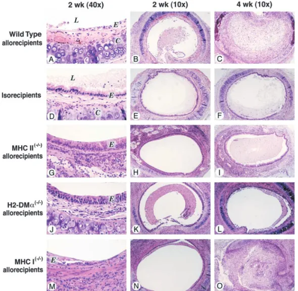
The wild-type H2dand H2btracheal isografts showed a complete, intact, fully differentiated epithelium and patent lumen at 2, 4, and 6 weeks (n⫽4, at each time point for both arms; Figures 1 and 2). Therefore, the initial ischemic insult by itself had no impact on the subsequent repair of the epithelium or the OB in this transplant model.
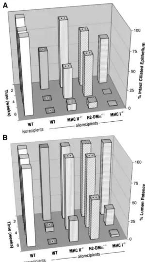
Figure 2. Airway rejection in wild-type (WT) grafts transplanted into
At 2, 4 and 6 weeks, the allografts (wild-type H2d grafts
wild-type, MHC-, or H2-DM␣–deficient recipients is shown as (A) the
transplanted into fully mismatched, wild-type H2brecipients, n⫽
mean percentage of intact ciliated airway epithelium and (B) the mean
3 to 4, at each time point) had less intact, fully differentiated percentage of airway lumen patency. *Comparisons (p⬍0.05) between (ciliated) epithelium compared with the isografts (wild-type H2b
the control allografts and the control isografts at the same time point.
grafts transplanted into wild-type isorecipients, n⫽4, at each **Comparisons (p⬍0.05) between the experimental arms and the
time point) (p⫽0.03, p ⬍0.001, and p⬍0.001, respectively; control allografts placed in wild-type recipients at the same time point.
Figures 1 and 2A). After epithelial denudation, the lumen of Grafts transplanted into MHC II- and H2-DM␣–deficient allorecipients, but not grafts transplanted into MHC I–deficient allorecipients, showed
the allografts displayed markedly decreased patency compared
significantly less epithelial denudation and airway obliteration from 2
with the isografts (n⫽3, p⫽0.002 at 4 weeks, and n⫽4, p⬍
to 6 weeks when compared with grafts transplanted into wild-type
0.001 at 6 weeks; Figures 1 and 2B).
recipients.
Similar results were seen in wild-type H2b allografts trans-planted into wild-type H2d recipients as compared with H2d allografts transplanted into wild-type H2brecipients (n⫽ 3 to
weeks, wild-type grafts transplanted into fully mismatched MHC 5, each arm and time point; p⭐0.005 for ciliated epithelium at II⫺/⫺allorecipients (n⫽3) underwent epithelial denudation and 2, 4, and 6 weeks and p⬍0.005 for lumen patency at 4 or 6 lumen obliteration similar to wild-type grafts transplanted into weeks; Figures 5, 6A, and 6B), which reproduced the results from wild-type allorecipients (p⭓0.2 each; Figures 1, 2A, and 2B). previous studies (3–8). Therefore, we demonstrated a uniform Allogeneic transplantation into fully mismatched, H2-DMa–
profile of airway rejection across two strains of mouse recipients. deficient mice. At 2 weeks, wild-type grafts transplanted into H2-DM␣⫺/⫺allorecipients (n⫽6) had more intact ciliated
epi-Testing the Indirect Pathway of Allorecognition thelium than those transplanted into wild-type allorecipients
Allogeneic transplantation into fully mismatched, MHC II-defi- (p⫽0.007). There was no lumen obliteration (Figures 1, 2A,
cient mice.At 2 weeks, wild-type grafts transplanted into fully and 2B). At 4 weeks, wild-type grafts transplanted H2-DM␣⫺/⫺ mismatched MHC II⫺/⫺allorecipients (n⫽4) had more intact,
allorecipients (n⫽6) showed significantly more intact, ciliated ciliated epithelium than those transplanted into wild-type allore- epithelium (p⫽0.005) and more airway lumen patency (p⫽ cipients (p⫽0.01). There was no lumen obliteration observed 0.001) than grafts transplanted into wild-type allorecipients. At in any of the grafts (Figures 1, 2A, and 2B). At 4 weeks, wild- 6 weeks, wild-type grafts transplanted into H2-DM␣⫺/⫺ allore-type grafts transplanted into fully mismatched MHC II⫺/⫺allore- cipients (n⫽4) showed epithelial loss similar to wild-type trans-cipients (n⫽4) had significantly more intact ciliated epithelium planted into wild-type allorecipients (p⭓0.2; Figure 2A); how-(p⫽ 0.04) and more airway lumen patency (p⫽ 0.005) than ever, they showed significantly less lumen obliteration (p⫽0.05;
Figure 3. Immunohistochemical staining for CD4⫹ (A–E) and CD8⫹(F–J) cells in wild-type tra-cheal grafts transplanted into MHC- or H2-DM␣–deficient allo-recipients at 2 weeks. Magnifica-tion⫻40. E ⫽epithelium, L ⫽ airway lumen, C⫽tracheal carti-lage.
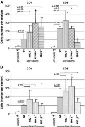
Allogeneic transplantation into fully mismatched, MHC I-defi- as compared with the wild-type grafts transplanted into isoreci-pients (n⫽3) (CD4⫹: 109⫾56.3 versus 23⫾12.1, p⫽0.01;
cient mice.At 2, 4, and 6 weeks (n⫽4 for each), wild-type grafts
transplanted into MHC I⫺/⫺ allorecipients showed a similar CD8⫹: 230.7⫾25.7 versus 16⫾6.1, p⬍0.001; Figures 3 and 4A). Trends toward higher mean CD4⫹cell counts were found amount of intact, ciliated epithelium and lumen obliteration as
wild-type grafts transplanted into wild-type allorecipients (p⬎ in wild-type grafts transplanted into MHC I⫺/⫺(n ⫽5), H2-DM␣⫺/⫺(n⫽4), and MHC II⫺/⫺allorecipients (n⫽6) in com-0.1 for ciliated epithelium and lumen obliteration at each time
point; Figures 1, 2A, and 2B). parison to wild-type grafts transplanted into wild-type allorecipi-ents (p⫽0.07, p⫽0.09, and p⫽0.13, respectively; Figures 3
Graft-infiltrating lymphocytes in wild-type grafts transplanted
into fully mismatched, MHC- and H2-DMa–deficient mice.As and 4A). A similar mean CD8⫹ cell count was observed in wild-type grafts transplanted into MHC II⫺/⫺and H2-DM␣⫺/⫺ expected, the wild-type grafts transplanted into wild-type
tion compared with wild-type allografts (p⫽0.05 and p⫽0.004, respectively). By 6 weeks, MHC I⫺/⫺allografts (n⫽5) developed lumen obliteration similar to wild-type allografts (n ⫽4, p⬎ 0.2 for each; Figures 5, 6A, and 6B).
Transplantation of MHC I/II⫺/⫺tracheal allografts into fully
mismatched, wild-type mice.At 2 weeks, MHC I/II⫺/⫺allografts (n⫽4) had more intact ciliated epithelium than wild-type allo-grafts (p ⫽0.05) and did not develop lumen obliteration. At Week 4, MHC I/II⫺/⫺allografts (n⫽6) had more intact ciliated epithelium and less lumen obliteration compared with wild-type allografts (p ⫽0.04 and p ⫽0.006, respectively). At Week 6, although MHC I/II⫺/⫺allografts (n⫽6) lost their intact ciliated epithelium resembling wild-type allografts (p ⬎ 0.2), they showed significantly less airway lumen obliteration compared with wild-type (p⫽0.04), MHC I⫺/⫺(p⫽0.04), or MHC II⫺/⫺ (p⫽0.05) allografts (Figures 5, 6A, and 6B).
Graft-infiltrating lymphocytes in MHC-deficient allografts.
Wild-type allografts transplanted into wild-type H2dmice (n⫽ 5) showed an abundance of CD4⫹and CD8⫹cells compared with isografts (99⫾10.4 versus 23⫾7.5, p⬍0.001, and 191⫾ 104.5 versus 16⫾5.1, p⫽0.01, respectively; Figure 4B). MHC I⫺/⫺, II⫺/⫺, and I/II⫺/⫺allografts placed in wild-type mice had a trend toward higher CD4⫹(p⫽0.14, p ⫽0.07, and p⬎0.2, respectively) and CD8⫹ (p ⫽ 0.17, p ⫽ 0.06, and p ⫽ 0.17, respectively) cells when compared with wild-type allografts (Figure 4B).
DISCUSSION
In this study, we report on the hierarchical importance of the indirect alloantigen recognition pathway over the direct pathway in the mouse model of chronic airway rejection. First, in the presence of the direct allorecognition pathway, we showed that the disruption of the indirect allorecognition pathway using Figure 4. The mean number (⫾ SD) of graft-infiltrating CD4⫹ and
MHC II⫺/⫺or H2-DM␣⫺/⫺recipient mice, which lack cell surface
CD8⫹ lymphocytes: (A) wild-type grafts transplanted into MHC- or
MHC II molecules or have disrupted loading of peptide antigens,
H2-DM␣–deficient allorecipients. H2-DM␣⫺/⫺ and MHC II⫺/⫺ mice
respectively, led to less chronic airway rejection. Because equal
showed similar numbers of allograft-infiltrating CD4⫹and CD8⫹
lym-or greater numbers of allograft-infiltrating CD4⫹and CD8⫹T
phocytes in comparison to the control allografts (p⬎0.05, each). A
lower mean CD8⫹cell count was observed in wild-type grafts trans- lymphocytes were found in wild-type allografts transplanted into
planted into MHC I⫺/⫺mice (p⬍0.01). (B) MHC-deficient allografts H2-DM␣⫺/⫺, and to our surprise, MHC II⫺/⫺recipients
(com-transplanted into wild-type allorecipients. MHC I⫺/⫺, II⫺/⫺, and I/II⫺/⫺ pared with wild-type control allografts), the attenuation of
rejec-allografts showed trends toward higher numbers of CD4⫹and CD8⫹ tion was not due to the inability of the recipient’s T lymphocytes infiltrating cells when compared with the control allografts (p⬎0.05, to access these grafts. Second, in the presence of the indirect each). Bracketed p values show comparisons between the experimental pathway, MHC II⫺/⫺ allografts (and passenger leukocytes),
arms or the control isografts with the control allografts. *Comparisons which lack the exogenous (MHC II-mediated) direct allorecogni-(p⬍0.05) between the experimental arms and the control isografts. tion pathway, developed chronic rejection in the same manner WT⫽wild type.
as control allografts. Third, because MHC I- and MHC I/II-deficient allografts placed into wild-type animals showed dimin-ished rejection, MHC I peptides are important alloantigens in this model of chronic lung rejection. Endogenous (MHC I-medi-into wild-type allorecipients (p⫽0.08 and p⬎0.2, respectively).
ated) direct allorecognition was not directly tested in this model A lower mean CD8⫹cell count was observed in wild-type grafts
but, if present, is a less important pathway based on the finding transplanted into MHC I⫺/⫺mice (p⬍0.001; Figures 3 and 4A).
that wild-type grafts are rejected more slowly in recipients who Testing the Direct Pathway of Allorecognition and the Role have impaired indirect allorecognition (but intact MHC I direct of Donor MHC Molecules allorecognition). Finally, the fact that MHC I/II⫺/⫺grafts were eventually rejected suggested that non-MHC antigens (e.g.,
mi-Transplantation of MHC II⫺/⫺tracheal allografts into fully
mis-nor histocompatibility antigens) were sufficient to cause chronic
matched, wild-type mice.At each time point (n⫽5 for each),
rejection through the indirect pathway in this model. the MHC II⫺/⫺allografts had a similar amount of ciliated
epithe-The absence of indirect allorecognition pathway did not lead lium and lumen obliteration as the wild-type allografts (p⬎0.2
to indefinite survival of fully MHC-mismatched airway allografts for ciliated epithelium or lumen obliteration at each time point;
as grafts placed into MHC II⫺/⫺and H2-DM␣⫺/⫺mice but dis-Figures 5, 6A, and 6B).
played a 2-week delay in epithelial denudation and lumen
oblit-Transplantation of MHC I⫺/⫺tracheal allografts into fully
mis-eration (Figure 2). This observation maybe explained by the fact
matched, wild-type mice.At 2 weeks, MHC I⫺/⫺allografts (n⫽
that the direct allorecognition pathway, which triggers a less 5) had more ciliated epithelium than wild-type allografts (p⫽
robust alloimmune response, was sufficient to result in complete 0.05) and no lumen obliteration. At 4 weeks, these grafts (n⫽7)
Figure 5. Histopathology of wild-type, MHC-deficient H2b tracheal allografts transplanted into wild-type H2drecipients at Weeks 2 (⫻10 and⫻40) and 4 (⫻10) (hematoxylin and eo-sin). An identical profile of air-way rejection was observed in wild-type H2ballografts trans-planted into wild-type H2d re-cipients (A–C) in comparison with that in wild-type H2d allo-grafts transplanted into wild-type H2b recipients (Figures 1A–1C). MHC I (G–I) and I/II⫺/⫺ (M–O) but not MHC II⫺/⫺(J–L) allografts showed significantly less severe chronic airway rejec-tion. E⫽epithelium, L⫽airway lumen, C⫽tracheal cartilage.
or greater numbers of infiltrating lymphocytes in these grafts allografts in a pattern similar to MHC II⫺/⫺mice confirms the importance of the indirect allorecognition and antigenic peptide compared with wild-type allografts may signify weaker donor
specificities, a characteristic of direct allorecognition (30). Alter- loading processes in mediating airway allograft rejection. Our results are also consistent with human studies, which have re-natively, cross-presentation of antigens (occurring with MHC
II⫺/⫺ and H2-DM␣⫺/⫺ antigen-presenting cells to recipient ported indirect evidence for the importance of the indirect allo-recognition in OB (34–37) and with animal models (38–40) and CD8⫹ T cells via MHC I molecules) may be responsible for
this late rejection. However, this form of indirect allorecognition human studies (41, 42) showing the importance of the indirect pathway in other organ transplants. In addition, our findings help is almost certainly inconsequential in our experiments, as fully
MHC-mismatched grafts were used. Therefore, recipient CD8⫹ explain the previous contradictory results showing that either the direct (43) or indirect (44) pathway may cause chronic airway T cells sensitized by allopeptides in the context of recipient MHC
I molecules will not find such determinants on the donor grafts. rejection in the murine tracheal transplant model by demonstra-ting that each pathway contributes in a different way.
MHC II⫺/⫺mice, due to deficient thymic selection of CD4⫹
T cells during embryonic development, have limited T-cell reper- Previous studies, using MHC-deficient grafts, have demon-strated the importance of MHC I molecules in pancreatic trans-toires and very low numbers of resting and stimulated CD4⫹
lymphocytes (14, 15). In these mice, deficient T-cell populations, plantation (45, 46), MHC II molecules in heart transplantation (47), and both MHC I and II molecules in skin transplantation rather than abnormal antigen processing, might have attenuated
the rejection process that we and others (14, 15) have observed. (24, 29). Although the MHC molecules expressed on the graft may function as alloantigens, they may also participate in the In contrast, H2-DM␣⫺/⫺mice have slightly reduced numbers of
CD4⫹lymphocytes in the lymph nodes and spleens, but express direct allorecognition pathway as antigen-presenting molecules. Therefore, efforts to understand the importance of MHC alloan-a very diverse T cell receptor repertoire, quite similalloan-ar to thalloan-at of
wild-type animals (18–21, 31, 32). In addition, H2-DM␣⫺/⫺CD4⫹ tigens in previous studies using MHC⫺/⫺ grafts only (and not using MHC⫺/⫺hosts) may have been confounded by the variable lymphocytes display an allogeneic proliferative response (18–21,
MHC I⫺/⫺and MHC I/II⫺/⫺tracheal grafts (17, 52, 53). Likewise, although H2-DM␣⫺/⫺mice express predominantly surface self CLIP-bound (and not allopeptide-bound) MHC II molecules, not all cell surface MHC II molecules are bound to CLIP (32, 54). However, the significance of non–CLIP-bound MHC II in graft rejection is not clear (18). In addition, the mechanism of H2-DM–dependent peptide loading is allele specific. The ex-change of CLIP for allopeptides in H2-Abmice that we employed is highly dependent on H2-DM expression in comparison to any other mouse strains (e.g., H2-Ak, H2-Ad) (21–23). Third, because the pretransplant and post-transplant graft-specific microenvi-ronment may account for the different contributions of allorec-ognition pathways among organ transplants and models, the result in this lung transplant model may not be applicable to others and requires further investigation. Finally, the effect of immunosuppressants used in clinical transplant, which may have different influences on direct and indirect alloreacitivity, was not tested here. We are making plans to include immunosuppres-sants in future experiments.
In conclusion, this comprehensive study of alloantigen recog-nition in the mouse heterotopic tracheal transplant model dem-onstrated a hierarchical order of allorecognition pathways for initiating chronic airway rejection. Although the indirect allorec-ognition pathway is more important, the significance of both allorecognition pathways should be taken into consideration when developing strategies to protect recipients from allograft rejection. This is the first study that takes advantage of H2-DM␣⫺/⫺mice to demonstrate the importance of the allopeptide loading process in indirect recognition in lung transplant rejec-tion. In addition, we demonstrated that MHC class I molecules are the principle alloantigens triggering rejection in the mouse model of OB, whereas MHC II and minor antigens are likely to play a less important role. Further studies in other models, as well as in the clinical arena, are essential to confirm these findings. Studies to assess the donor-directed specificities of air-Figure 6. Airway rejection in wild-type (WT), MHC-deficient tracheal way graft-infiltrating lymphocytes are also necessary and are allografts transplanted into wild-type allorecipients, shown as (A) the
under current investigation.
mean percentage of intact ciliated airway epithelium and (B) the mean
percentage of airway lumen patency. *Comparisons (p⬍0.05) between Acknowledgment: The authors thank Steven B. Wagoner for his professional the control allografts and the control isografts at the same time point. graphical assistance and continued support, Tracy L. Eldred and Kim A. Burns for
**Comparisons (p⬍0.05) between the experimental arms and the their exceptional and long-term assistance in histologic studies, and the investiga-tors and staff at the Cystic Fibrosis Research and Treatment Center, University of
control allografts at the same time point. MHC I- and I/II-deficient
North Carolina at Chapel Hill.
allografts, but not MHC II-deficient allografts, showed significantly less epithelial denudation and airway obliteration from 2 to 6 weeks when
compared with wild-type allografts. References
1. Hosenpud JD, Bennett LE, Keck BM, Boucek MM, Novick RJ. The Registry of the International Society for Heart and Lung Transplanta-tion: eighteenth official report-2001.J Heart Lung Transplant2001;20:
both donors and recipients in separate experiments, our study 805–815.
design is unique in its ability to assess the role of MHC molecules 2. Hertz MI, Jessurun J, King MB, Savik SK, Murray JJ. Reproduction of both as antigen-presenting molecules and as alloantigens. This the obliterative bronchiolitis lesion after heterotopic transplantation has allowed us to demonstrate unquestionably that MHC I pep- of mouse airways.Am J Pathol1993;142:1945–1951.
tides are important alloantigens in this model of chronic lung 3. Kelly KE, Hertz MI, Mueller DL. T-cell and major histocompatibility complex requirements for obliterative airway disease in
heterotopi-rejection. This finding is consistent with clinical studies in lung
cally transplanted murine tracheas.Transplantation1998;66:764–771.
transplant recipients that mismatches at HLA class I increase
4. Neuringer IP, Mannon RB, Coffman TM, Parsons M, Burns K,
Yan-the risk and severity of chronic lung rejection (48–51). Taken
kaskas JR, Aris RM. Immune cells in a mouse airway model of
oblitera-together, these observations suggest that matching donor and
tive bronchiolitis.Am J Respir Cell Mol Biol1998;19:379–386.
recipient HLA may reduce chronic lung rejection clinically.
5. Neuringer IP, Walsh SP, Mannon RB, Gabriel S, Aris RM. Enhanced
Some limitations are worthy of note in this study. First,
al-T cell cytokine gene expression in mouse airway obliterative
bronchio-though the heterotopic mouse airway model has been success- litis.Transplantation2000;69:399–405.
fully employed to study the pathogenesis of OB (2–8, 43, 44), 6. Neuringer IP, Aris RM, Burns KA, Bartolotta TL, Chalermskulrat W, it does not perfectly replicate human OB due to the heterotopic Randall SH. Epithelial kinetics in mouse heterotopic tracheal allo-position of the graft and the rapid course of rejection. Second, grafts.Am J Transplant2002;2:410–419.
in MHC I⫺/⫺mice, low levels of MHC I heavy chains may reach 7. Rumbley CA, Silver SJ, Phillips SM. Dependence of murine obstructive
airway disease on CD40 ligand.Transplantation2001;72:1616–1625.
8. Aris RM, Walsh S, Chalermskulrat W, Hathwar V, Neuringer IP. Growth 32. Tourne S, Miyazaki T, Oxenius A, Klein L, Fehr T, Kyewski B, Benoist C, Mathis D. Selection of a broad repertoire of CD4⫹T cells in H-factor upregulation during obliterative bronchiolitis in the mouse
2Ma0/0 mice.Immunity1997;7:187–195. model.Am J Respir Crit Care Med2002;166:417–422.
33. Ardehali A, Fischbein MP, Yun J, Irie Y, Fishbein MC, Laks H. Indirect 9. Rogers N, Lechler R. Allorecognition.Am J Transplant2001;1:97.
alloreactivity and chronic rejection. Transplantation 2002;73:1805– 10. Shoskes DA, Wood KJ. Indirect presentation of MHC antigens in
trans-1807. plantation.Immunol Today1994;15:32–38.
34. SivaSai KS, Smith MA, Poindexter NJ, Sundaresan SR, Trulock EP, 11. Auchincloss H Jr, Sultan H. Antigen processing and presentation in
Lynch JP, Cooper JD, Patterson GA, Mohanakumar T. Indirect recog-transplantation.Curr Opin Immunol1996;8:681–687.
nition of donor HLA class I peptides in lung transplant recipients with 12. Benichou G. Direct and indirect antigen recognition: the pathways to
bronchiolitis obliterans syndrome.Transplantation1999;67:1094–1098. allograft immune rejection.Front Biosci1999;4:D476–D480.
35. Leonard CT, Soccal PM, Singer L, Berry GJ, Theodore J, Holt PG, Doyle 13. Sherman LA, Chattopadhyay S. The molecular basis of allorecognition.
RL, Rosen GD. Dendritic cells and macrophages in lung allografts: a
Annu Rev Immunol1993;11:385–402.
role in chronic rejection?Am J Respir Crit Care Med2000;161:1349– 14. Grusby MJ, Johnson RS, Papaioannou VE, Glimcher LH. Depletion of
1354. CD4⫹T cells in major histocompatibility complex class II-deficient
36. Reznik SI, Jaramillo A, Sivasai KSR, Womer KL, Sayegh MH, Trulock mice.Science1991;253:1417–1420.
EP, Alexander PG, Mohanakumar T. Indirect allorecognition of mis-15. Cosgrove D, Gray D, Dierich A, Kaufman J, Lemeur M, Benoist C,
matched donor HLA class II peptides in lung transplant recipients with Mathis D. Mice lacking MHC class II molecules.Cell1991;66:1051–
bronchiolitis obliterans syndrome.Am J Transplant2001;1:228–235. 1066.
37. Duncan SR, Leonard C, Theodore J, Lega M, Girgis RE, Rosen GD, 16. Koller BH, Smithies O. Inactivating the beta 2-microglobulin locus in
Theofilopoulos AN. Oligoclonal CD4(⫹) T cell expansions in lung mouse embryonic stem cells by homologous recombination.Proc Natl
transplant recipients with obliterative bronchiolitis.Am J Respir Crit
Acad Sci USA1989;86:8932–8935.
Care Med2002;165:1439–1444.
17. Zijlstra M, Bix M, Simister NE, Loring JM, Raulet DH, Jaenisch R. Beta
38. Fangmann J, Dalchau R, Fabre JW. Rejection of skin allografts by indi-2-microglobulin deficient mice lack CD4–8⫹cytolytic T cells.Nature
rect allorecognition of donor class I major histocompatibility complex 1990;344:742–746.
peptides.J Exp Med1992;175:1521–1529. 18. Felix NJ, Brickey WJ, Griffiths R, Zhang J, Van Kaer L, Coffman T,
39. Watschinger B, Gallon L, Carpenter CB, Sayegh MH. Mechanisms of Ting JP. H2-DMalpha(⫺/⫺) mice show the importance of major
histo-allo-recognition: recognition byin vivo–primed T cells of specific major compatibility complex-bound peptide in cardiac allograft rejection.J
histocompatibility complex polymorphisms presented as peptides by
Exp Med2000;192:31–40.
responder antigen-presenting cells.Transplantation1994;57:572–576. 19. Fung-Leung WP, Surh CD, Liljedahl M, Pang J, Leturcq D, Peterson PA,
40. Gallon L, Watschinger B, Murphy B, Akalin E, Sayegh MH, Carpenter Webb SR, Karlsson L. Antigen presentation and T cell development in
CB. The indirect pathway of allorecognition: the occurrence of self-H2-M-deficient mice.Science1996;271:1278–1281.
restricted T cell recognition of allo-MHC peptides early in acute renal 20. Miyazaki T, Wolf P, Tourne S, Waltzinger C, Dierich A, Barois N, Ploegh
allograft rejection and its inhibition by conventional immunosuppres-H, Benoist C, Mathis D. Mice lacking H2-M complexes, enigmatic
sion.Transplantation1995;59:612–616. elements of the MHC class II peptide-loading pathway.Cell1996;84:
41. Vella JP, Spadafora-Ferreira M, Murphy B, Alexander SI, Harmon W, 531–541.
Carpenter CB, Sayegh MH. Indirect allorecognition of major histo-21. Martin WD, Hicks GG, Mendiratta SK, Leva HI, Ruley HE, Van Kaer
compatibility complex allopeptides in human renal transplant recipi-L. H2-M mutant mice are defective in the peptide loading of class II
ents with chronic graft dysfunction.Transplantation1997;64:795–800. molecules, antigen presentation, and T cell repertoire selection.Cell
42. Hornick PI, Mason PD, Yacoub MH, Rose ML, Batchelor R, Lechler 1996;84:543–550.
RI. Assessment of the contribution that direct allorecognition makes 22. Wolf PR, Tourne S, Miyazaki T, Benoist C, Mathis D, Ploegh HL. The
to the progression of chronic cardiac transplant rejection in humans. phenotype of H-2M-deficient mice is dependent on the MHC class II
Circulation1998;97:1257–1263.
molecules expressed.Eur J Immunol1998;28:2605–2618. 43. Szeto WY, Krasinskas AM, Kreisel D, Popma SH, Rosengard BR. Donor 23. Stebbins CC, Loss GE Jr, Elias CG, Chervonsky A, Sant AJ. The require- antigen-presenting cells are important in the development of oblitera-ment for DM in class II-restricted antigen presentation and SDS- tive airway disease.J Thorac Cardiovasc Surg2000;120:1070–1077. stable dimer formation is allele and species dependent.J Exp Med 44. Smith MA, Jaramillo A, SivaSai KS, Naziruddin B, Kaleem Z, Patterson
1995;181:223–234. GA, Mohanakumar T. Indirect recognition and antibody production 24. Auchincloss H Jr, Lee R, Shea S, Markowitz JS, Grusby MJ, Glimcher against a single mismatched HLA-A2-transgenic molecule precede LH. The role of “indirect” recognition in initiating rejection of skin the development of obliterative airway disease in murine heterotopic grafts from major histocompatibility complex class II-deficient mice. tracheal allografts.Transplantation2002;73:186–193.
Proc Natl Acad Sci USA1993;90:3373–3377. 45. Markmann JF, Bassiri H, Desai NM, Odorico JS, Kim JI, Koller BH,
25. Benichou G, Takizawa PA, Olson CA, McMillan M, Sercarz EE. Donor Smithies O, Barker CF. Indefinite survival of MHC class I-deficient major histocompatibility complex (MHC) peptides are presented by murine pancreatic islet allografts.Transplantation1992;54:1085–1089. recipient MHC molecules during graft rejection.J Exp Med1992;175: 46. Osorio RW, Ascher NL, Jaenisch R, Freise CE, Roberts JP, Stock PG. 305–308. Major histocompatibility complex class I deficiency prolongs islet allo-26. Jones ND, Van Maurik A, Hara M, Spriewald BM, Witzke O, Morris graft survival.Diabetes1993;42:1520–1527.
PJ, Wood KJ. CD40–CD40 ligand-independent activation of CD8⫹ 47. Campos L, Naji A, Deli BC, Kern JH, Kim JI, Barker CF, Markmann T cells can trigger allograft rejection.J Immunol2000;165:1111–1118. JF. Survival of MHC-deficient mouse heterotopic cardiac allografts. 27. Kreisel D, Krupnick AS, Gelman AE, Engels FH, Popma SH, Krasinskas Transplantation1995;59:187–191.
AM, Balsara KR, Szeto WY, Turka LA, Rosengard BR. Non-hemato- 48. Kroshus TJ, Kshettry VR, Savik K, John R, Hertz MI, Bolman RM III. poietic allograft cells directly activate CD8⫹T cells and trigger acute Risk factors for the development of bronchiolitis obliterans syndrome rejection: an alternative mechanism of allorecognition.Nat Med2002; after lung transplantation.J Thorac Cardiovasc Surg1997;114:195–202. 8:233–239. 49. Sundaresan S, Mohanakumar T, Smith MA, Trulock EP, Lynch J, Phelan 28. Boisgerault F, Liu Y, Anosova N, Ehrlich E, Dana MR, Benichou G. D, Cooper JD, Patterson GA. HLA-A locus mismatches and develop-Role of cd4(⫹) and cd8(⫹) T cells in allorecognition: lessons from ment of antibodies to HLA after lung transplantation correlate with corneal transplantation.J Immunol2001;167:1891–1899. the development of bronchiolitis obliterans syndrome.Transplantation
29. Grusby MJ, Auchincloss H Jr, Lee R, Johnson RS, Spencer JP, Zijlstra 1998;65:648–653.
M, Jaenisch R, Papaioannou VE, Glimcher LH. Mice lacking major 50. Schulman LL, Weinberg AD, McGregor CC, Suciu-Foca NM, Itescu S. histocompatibility complex class I and class II molecules.Proc Natl Influence of donor and recipient HLA locus mismatching on
develop-Acad Sci USA1993;90:3913–3917. ment of obliterative bronchiolitis after lung transplantation.Am J
30. Benichou G, Valujskikh A, Heeger PS. Contributions of direct and indi- Respir Crit Care Med2001;163:437–442.
rect T cell alloreactivity during allograft rejection in mice.J Immunol 51. Chalermskulrat W, Neuringer IP, Schmitz JL, Catellier DJ, Gurka MJ, 1999;162:352–358. Randell SH, Aris RM. HLA Mismatches Predispose to the Severity 31. Surh CD, Lee DS, Fung-Leung WP, Karlsson L, Sprent J. Thymic selec- of Bronchiolitis Obliterans Syndrome after Lung Transplantation.
tion by a single MHC/peptide ligand produces a semidiverse repertoire Chest(In press)
on the surface of beta 2 microglobulin negative cells. J Exp Med tion of CD4 T cells in mice displaying altered repertoires of MHC class II-bound self-peptides.Immunity1997;7:197–208.
1992;176:829–834.
55. Neuringer IP, Chalermskulrat W, Mannon RB, Bartolotta TL, Burns 53. Lee RS, Grusby MJ, Laufer TM, Colvin R, Glimcher LH, Auchincloss
KA, Randell SH, Aris RM. Suppress donor MHC I expression inhibits H Jr. CD8⫹effector cells responding to residual class I antigens, with
mouse tracheal graft obliteration.Am J Respir Crit Care Med2000; help from CD4⫹cells stimulated indirectly, cause rejection of “major 161:A370.
histocompatibility complex-deficient” skin grafts.Transplantation1997; 56. Chalermskulrat W, Neuringer IP, Ting JP, Felix NJ, Randell SH, Hathwar 63:1123–1133. VK, Aris RM. Alloantigen recognition in the mouse heterotopic model