High-throughput small molecule screen identifies
inhibitors of aberrant chromatin accessibility
Samantha G. Pattendena,b,1, Jeremy M. Simonc,d,e,1, Aminah Walic,d,f,1, Chatura N. Jayakodya,b, Jacob Troutmand,g, Andrew W. McFaddend, Joshua Wootenc,d,f, Cameron C. Wooda,b, Stephen V. Fryea,b, William P. Janzena,b,2, and Ian J. Davisc,d,g,h,3
aCenter for Integrative Chemical Biology and Drug Discovery, University of North Carolina at Chapel Hill, Chapel Hill, NC 27302;bDivision of Chemical Biology and Medicinal Chemistry, Eshelman School of Pharmacy, University of North Carolina at Chapel Hill, Chapel Hill, NC 27302;cDepartment of Genetics, School of Medicine, University of North Carolina at Chapel Hill, Chapel Hill, NC 27599;dLineberger Comprehensive Cancer Center, School of Medicine, University of North Carolina at Chapel Hill, Chapel Hill, NC 27599;eCurriculum in Bioinformatics and Computational Biology, University of North Carolina at Chapel Hill, Chapel Hill, NC 27599;fCurriculum in Genetics and Molecular Biology, University of North Carolina at Chapel Hill, Chapel Hill, NC 27599; gDepartment of Pediatrics, School of Medicine, University of North Carolina at Chapel Hill, Chapel Hill, NC 27599; andhCarolina Center for Genome Sciences, School of Medicine, University of North Carolina at Chapel Hill, Chapel Hill, NC 27599
Edited by Steven Henikoff, Fred Hutchinson Cancer Research Center, Seattle, WA, and approved January 21, 2016 (received for review November 4, 2015)
Mutations in chromatin-modifying proteins and transcription factors are commonly associated with a wide variety of cancers. Through gain- or loss-of-function, these mutations may result in characteristic alterations of accessible chromatin, indicative of shifts in the landscape of regulatory elements genome-wide. The identification of compounds that reverse a specific chromatin signature could lead to chemical probes or potential therapies. To explore whether chromatin accessibility could serve as a platform for small molecule screening, we adapted formaldehyde-assisted isolation of regula-tory elements (FAIRE), a chemical method to enrich for nucleosome-depleted genomic regions, as a high-throughput, automated assay. After demonstrating the validity and robustness of this approach, we applied this method to screen an epigenetically targeted small molecule library by evaluating regions of aberrant nucleosome depletion mediated by EWSR1-FLI1, the chimeric transcription fac-tor critical for the bone and soft tissue tumor Ewing sarcoma. As a class, histone deacetylase inhibitors were greatly overrepresented among active compounds. These compounds resulted in diminished accessibility at targeted sites by disrupting transcription of EWSR1-FLI1. Capitalizing on precise differences in chromatin accessibility for drug discovery efforts offers significant advantages because it does not depend on the a priori selection of a single molecular target and may detect novel biologically relevant pathways.
chromatin
|
Ewing sarcoma|
high throughput screening|
FAIRE|
histone deacetylase inhibitorA
growing range of human cancers have been associated with mutations in genes encoding proteins that regulate chro-matin, the assembly of proteins and DNA that control DNA-templated processes, including transcription and replication (1, 2). Small molecule drugs and chemical probes offer an approach to explore the biological consequences of these mutations and are emerging as a therapeutic strategy to target disease pathways. Drugs targeting histone deacetylase (HDAC) enzymes, the bro-modomain reader BRD4, and DNA methylation have already received regulatory approval or have entered clinical testing, and chemical probes have been developed against a broad range of chromatin regulators, such as the methyltransferases (3) DOT1L (4), EZH2 (5–8), and G9a (9, 10), and the reader proteins L3MBTL3 (11) and BRD4 (12–14). However, transcription fac-tors that lack enzymatic activity or binding pockets with targetable molecular features have been considered “undruggable,” and a reductionist approach based on identification of their molec-ular targets has largely failed.The majority of Ewing sarcomas, highly malignant pediatric bone and soft tissue tumors, harbor a chromosomal translocation that joins the amino-terminal domain ofEWSR1with the DNA binding domain of the ETS transcription factor family member
FLI1to generate the chimeric transcription factor EWSR1-FLI1
(15). Translocations with other ETS genes are detected in most of the remaining tumors, yielding similarly functioning fusion proteins (16). We recently found that, despite conservation of the ETS DNA binding domain, the fusion oncoprotein uniquely localizes to specific microsatellite regions (17, 18). Using form-aldehyde-assisted isolation of regulatory elements (FAIRE), a biochemical strategy to enrich for nucleosome-depleted regions of chromatin, we demonstrated that EWSR1-FLI1 binding was necessary to maintain nucleosome depletion at these sites. The mechanism through which EWSR1-FLI1 modifies chromatin re-mains unknown. Because EWSR1-FLI1 does not possess recog-nizable catalytic activity, other yet-to-be-identified proteins likely mediate its ability to remodel chromatin.
The absence of a biochemical mechanism would typically pose challenges for chemical targeting. However, we hypothesized that reversing a unique chromatin signature could serve as a strategy to discover small molecules with activity toward EWSR1-FLI1. To target this activity, we adapted and validated FAIRE as an automated, high-throughput tool and applied this method to
Significance
Transcriptional regulators lacking enzymatic activity or binding pockets with targetable molecular features have typically been considered“undruggable,”and a reductionist approach based on identification of their molecular targets has largely failed. We have demonstrated that the Ewing sarcoma chimeric tran-scription factor, EWSR1-FLI1, maintains accessible chromatin at disease-specific regions. We adapted formaldehyde-assisted isolation of regulatory elements (FAIRE), an assay for accessible chromatin, to screen an epigenetically targeted small molecule library for compounds that reverse the disease-associated sig-nature. This approach can be applied broadly for discovery of chromatin-based developmental therapeutics and offers sig-nificant advantages because it does not require the selection of a single molecular target. Using this approach, we identified a specific class of compounds with therapeutic potential.
Author contributions: S.G.P., J.M.S., S.V.F., W.P.J., and I.J.D. designed research; S.G.P., J.M.S., A.W., C.N.J., J.T., A.W.M., J.W., and C.C.W. performed research; S.V.F. contributed new reagents/analytic tools; S.G.P., J.M.S., A.W., C.N.J., J.T., A.W.M., J.W., W.P.J., and I.J.D. ana-lyzed data; and S.G.P., J.M.S., A.W., S.V.F., W.P.J., and I.J.D. wrote the paper.
The authors declare no conflict of interest.
This article is a PNAS Direct Submission.
Data deposition: The data reported in this paper have been deposited in the Gene Ex-pression Omnibus (GEO) database,www.ncbi.nlm.nih.gov/geo(accession no.GSE61735). 1S.G.P., J.M.S., and A.W. contributed equally to this work.
2Present address: Epizyme Inc., Cambridge, MA 02139.
3To whom correspondence should be addressed. Email: [email protected].
evaluate a focused set of small molecules designed to interact with proteins that regulate chromatin. Because this approach di-rectly assessed the effects of compounds on a specific, disease-associated aberrant chromatin signature, while remaining agnostic about precise molecular mechanisms, it enabled the discovery of agents that affect the underlying molecular defect without re-quiring an a priori selection of molecular targets.
Results
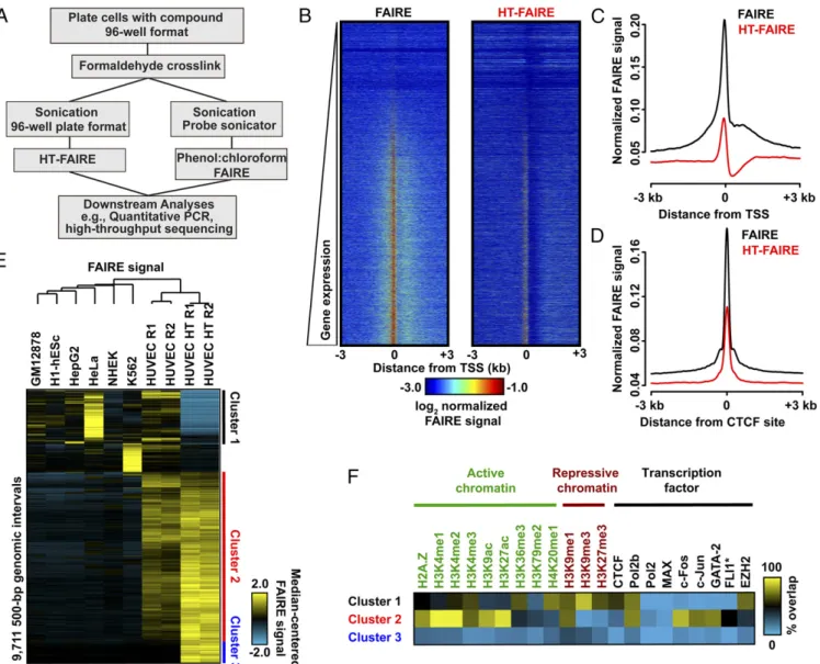
Validation of Column-Based FAIRE.As currently applied, FAIRE, a biochemical assay for the enrichment of nucleosome-depleted regions of the genome, is dependent on organic extraction with a mixture of phenol and chloroform. Because this critical extraction step is not easily automated, we adapted and miniaturized FAIRE by substituting organic extraction with solid-phase selection and robotic automation, hereafter termed high-throughput FAIRE (HT-FAIRE) (Fig. 1A). Because the chromatin fractionation step is central to this technique, we compared the performance of these methods. Quantitative locus-specific testing demonstrated that both approaches offered concordant enrichment at a series of
promoter and enhancer regions (Fig. S1). We then compared the performance of both methods genome-wide. We used primary human umbilical vein endothelial cells (HUVECs) since multiple genomic datasets exploring chromatin features have been gener-ated for these cells permitting subsequent integrative analyses of the FAIRE results [Encyclopedia of DNA Elements (ENCODE) Tier 2] (19). HT-FAIRE demonstrated signal enrichment patterns similar to those observed using standard FAIRE. For example, enrichment at transcriptional start sites (TSSs), including posi-tive correlation with RNA abundance as well as enrichment at CCCTC-binding factor (CTCF) sites demonstrated similar char-acteristics as previously reported FAIRE studies (Fig. 1B–D) (20). Signal enrichment by HT-FAIRE was less robust at TSSs, con-sistent with the quantitative PCR (qPCR) results. Of the top 10,000 nucleosome-depleted regions detected in HUVECs by HT-FAIRE,∼90% overlapped those sites identified by standard FAIRE. In contrast, fewer than 50% of enriched regions over-lapped FAIRE sites from any of six other cell lines (Fig. S2A), demonstrating that HT-FAIRE offers a level of specificity similar to that reported in previous studies (21).
Fig. 1. Comparison of FAIRE methodologies. (A) Flow diagram comparing column-based and standard FAIRE methods. (B) Heatmap representation of normalized FAIRE enrichment (±3 kb from TSS) using standard (Left) or column (Right) FAIRE in HUVEC. (C) Normalized FAIRE signal from both methods±3 kb from TSS. (D) Normalized FAIRE signal from both methods±3 kb around HUVEC CTCF sites (ENCODE). (E) Hierarchical clustering analysis of 500-bp intervals demonstrating differential FAIRE signals across seven cell types, as well as HUVEC HT-FAIRE. Platform-specific (clusters 1 and 3) and cell type-specific (cluster 2) clusters were identified. (F) Fractional overlap annotation of clusters 1–3, with histone modifications and transcription factors (ENCODE).
MEDI
CAL
SCIENC
We then examined those genomic regions that most discrim-inate HT-FAIRE and standard FAIRE and also demonstrate HUVEC cell-type specificity. Hierarchical clustering of these ∼9,700 genomic regions identified three groups (Fig. 1E). Cluster 1 (1,805 regions) consisted of those regions enriched in all cell lines when assayed by standard FAIRE but not by HT-FAIRE. Cluster 2 (6,017 regions), by far the largest, consisted of regions with HUVEC-specific signal enrichment that was detected by both HT-FAIRE and standard FAIRE. Cluster 3 (843 regions) con-sisted of regions selectively identified by HT-FAIRE. Regions in each cluster were then associated with genes [Genome Regions Enrichment of Annotations Tool (GREAT)] (22). As expected, the HUVEC-selective sites detected by both HT-FAIRE and standard FAIRE (cluster 2) were tightly linked with endothelial cell ontologies, including angiogenesis (q=4.4×10⁻¹²) and reg-ulation of cell-substrate adhesion (q=6.5×10⁻⁷) (Fig. S2B). No significant gene ontologies were associated with the regions in the other clusters. To test whether FAIRE-enriched regions were likely to harbor regulatory elements, we annotated the sites from each cluster with active and repressive histone modifications. We also tested for association with targeting of numerous tran-scription factors, including those known to be important in en-dothelial cell biology, as assessed by ChIP-seq (17, 19). (Fig. 1F). Sites in cluster 2 (common to both platforms, HUVEC-specific) were associated primarily with active histone modifications, as well as sites targeted by FLI1, FOS, JUN, GATA2, and RNA Poly-merase II. The prevalence of putative FLI1 and FOS/JUN binding sites was corroborated by the enrichment of ETS and AP1 DNA sequence motifs in these regions (P<1×10−800) (Fig. S2C). ETS (specifically ETV2) and AP1 factors are both known to play prominent roles in endothelial development (23, 24). Regions in cluster 1 were more closely associated with repressive modifica-tions and EZH2 binding (Fig. 1F). Clusters 1 and 3 were distin-guished by enrichment for repetitive regions, with each cluster associated with a specific repetitive element class: satellites (82% of cluster 1 sites) and simple repeats (71% of cluster 3 sites) (Fig. S2D). We also noted a difference in sequence composition be-tween cluster 1 and clusters 2 and 3 (Fig. S2E). The basis of their differential enrichment may reflect chromatin variation at these regions that are distinguished by the biochemical properties spe-cific to organic or solid phase purification. Because we used published standard FAIRE datasets that were generated with a shorter sequencing read length (ENCODE, 36-bp reads), we asked whether the difference in read length could partially ac-count for the variability in mapping and sequence content at clusters 1 and 3 (Fig. S2F). Repeating these analyses after trun-cating the HT-FAIRE sequencing reads to 36 bp did not change the hierarchical clustering or histone modification associations
(Fig. S2GandH). Taken together, these data demonstrate that
HT-FAIRE performs similarly to standard FAIRE and identifies regions that are biologically meaningful.
Application of HT-FAIRE to Small Molecule Screening.We then adapted HT-FAIRE for high-throughput automation and applied it in a targeted screen. The screen was based on two regions selected from a set of Ewing sarcoma-specific sites that we had previously shown were accessible in multiple Ewing sarcoma cell lines with accessi-bility that was dependent on continued expression of EWSR1-FLI1 (17). Regions that consistently demonstrate FAIRE enrichment or lack of enrichment across many cell types were included as controls
(seeTable S3).
We performed the screen using a custom library that consisted of 640 small molecules, including those designed to target his-tone methyltransferases, methyl lysine reader proteins, hishis-tone demethylases and deacetylases, DNA methyltransferases, and acetyl-lysine reader proteins. The compound collection included well-annotated molecular profiles (chemical probes) (25), as well as compounds that were designed around several chemical scaffolds originally directed toward specific targets of interest (methyl-lysine readers and transferases). The latter compounds have only been tested in vitro for activity on the proteins for which they
were designed and likely have effects on other molecular tar-gets. We hypothesized that the inclusion of a large number of uncharacterized compounds would increase the potential to identify previously unanticipated protein targets.
After optimizing conditions for growth and DMSO tolerance
(Fig. S3), Ewing sarcoma patient-derived cells (EWS894) were
exposed to 10μM of each compound (or DMSO control) for 16 h in a 96-well format. The unit automation pipeline separated each of two 384-well compound plates containing the library into four 96-well plates for screening (Fig. S4). After application of HT-FAIRE, chromatin from the 96-well plates was combined into a 384-well plate format for qPCR-based testing of the signature and control regions in two technical replicates. The positive control region demonstrated a significant difference in FAIRE signal over the background control (plate 1,P=7.50e−53; plate 2,P=6.63e−98). Critically, signals from each of the disease-specific regions were highly concordant (Pearsonr=0.8764), which supported combining the values from both sites for subsequent analytics (Fig. 2A).
To quantify the degree to which chromatin accessibility changed after compound treatment, we calculated a “relative chromatin inhibition”(RCI) score that compared the relative enrichment at the oncogene-targeted regions with that at the positive control re-gion. An RCI score of 1.0 was considered “no change.” Com-pounds that similarly affected both the oncogene-targeted and control regions were considered nonspecific. DMSO (vehicle) and UNC0638 (10) (a chemical probe that we had previously de-termined not to affect aberrantly open chromatin in Ewing sarcoma cells) demonstrated an RCI of 1.0 (Fig. 2BandFig. S5A). Because this primary screen was based on a single measurement, we per-mitted greater intersample variability, defining compounds with an RCI value greater than two SDs from the mean RCI score for
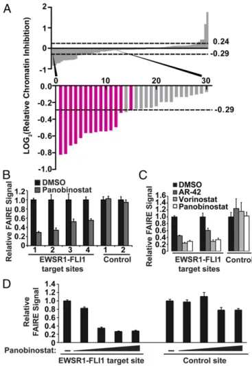
DMSO-treated controls as“hits”(Fig. 2BandFig. S5A) (26). Fifty-eight compounds met criteria for reduced chromatin accessibility (Fig. 2CandD). Forty-three of these compounds were unchar-acterized, 14 of which were analogs within a single chemical scaf-fold, and 15 were characterized chemical probes. Treatment with 20 compounds resulted in increased FAIRE signals, all of which were uncharacterized (Table S1). Because the goal of this study was to inhibit EWSR1-FLI1–mediated aberrant chromatin accessibility, we focused on those compounds that decreased the FAIRE signal. We prepared a secondary screen plate that contained all hit compounds and 16 compounds that failed to show a change in chromatin accessibility in the initial screen (RCI of 1.0, no change). This secondary screen was performed on three in-dependent replicates of EWS894 cells, permitting a more strin-gent threshold of 3 SDs from the mean RCI score to define hits (26, 27). Fifteen compounds (26%) met this enhanced threshold (Fig. 3AandFig. S5B).
Remarkably, of the 21 HDAC inhibitors that were included in our library, 14 of these compounds scored as hits in both the primary and secondary screens (Fig. 3A). We compared the specificity of the active and inactive HDAC inhibitors (Table S2). Although the majority of the inhibitors in our set have known activity against multiple HDAC proteins, making it dif-ficult to identify the specific HDAC target, inhibitors selective for HDAC6 (Tubastatin) (28) and HDAC8 (PCI-34051) (29) did not score in our screen, making it unlikely that either of these HDAC proteins is involved in maintaining aberrant chromatin accessibility in Ewing sarcoma. The overall reproducibility of the screen confirms the robustness of HT-FAIRE as an approach for discovering biologically relevant compounds using a small, focused compound library. A priori selection of inhibitors of HDAC proteins as lead candidates for screening would have been unlikely because histone hyperacetylation is commonly associated with destabilized nucleosomes and open chromatin.
To replicate the results of the screen, we focused on two hit compounds, Vorinostat and Panobinostat because these hydrox-amate derivatives inhibit multiple classes of HDACs and have received FDA approval for oncological indications (30). Treat-ment of EWS894 cells with either of these compounds followed by HT-FAIRE resulted in a dose-dependent decrease in chro-matin accessibility (Fig. S6A). This decrease was not observed with an HDAC inhibitor that did not score as a hit in our screen (Tubastatin), or a compound with an unrelated structure and protein target.
We then validated the compounds that emerged from our screen using standard FAIRE. FAIRE signals from cells treated with Panobinostat decreased at the test regions to a similar extent (approximately twofold) to that observed in the screen and also demonstrated a similar effect on two additional EWSR1-FLI1– tar-geted sites (Fig. 3B). Critically, positive control regions remained unaffected, demonstrating that the decreased RCI was not the result of increased signals at the control regions. We then validated the effect of additional HDAC inhibitors by standard FAIRE. Pan-obinostat, Vorinostat, and AR-42 demonstrated a similar magnitude effect on chromatin (Fig. 3C). Panobinostat also led to a dose-dependent decrease in FAIRE signal at an EWSR1-FLI1–targeted site (Fig. 3D). As further validation, we performed FAIRE-qPCR in two additional patient-derived EWSR1-FLI1–expressing Ewing sarcoma cell lines, EWS502 (Fig. S6B) and RD-ES (Fig. S6C). Panobinostat treatment significantly decreased FAIRE signals at EWSR1-FLI1 binding sites in both of these cell lines. Together, these data confirm that HDAC inhibitors are associated with reduced chromatin accessibility at EWSR1-FLI1–targeted regions as mea-sured by both HT-FAIRE and standard FAIRE.
To explore the mechanism underlying the chromatin signature reversal, we asked whether EWSR1-FLI1 levels were affected by HDAC inhibitor treatment. Panobinostat and Vorinostat treatment resulted in a dose-dependent decrease in EWSR1-FLI1 mRNA levels whereas Tubastatin had no effect (Fig. 4A). Consistent with the decrease in mRNA, EWSR1-FLI1 protein levels were also decreased after treatment with Panobinostat and Vorinostat
(Fig. 4B). Tubastatin did not affect EWSR1-FLI1 protein levels although a slight overall decrease was noted at 10μM (Fig. 4B). These data suggest that Panobinostat and Vorinostat, both hit compounds identified in the HT-FAIRE screen, decrease levels of EWSR1-FLI1 by altering transcription.
We then asked whether ectopically expressed EWSR1-FLI1 would be similarly affected by HDAC inhibition. We generated a Ewing sarcoma cell line in which the endogenous EWSR1-FLI1 was silenced with concurrent stable expression of lentivirally transduced EWSR1-FLI1. Panobinostat or Vorinostat treatment of these cells did not affectEWSR1-FLI1mRNA levels (Fig. 4C). Correspondingly, treatment with multiple HDAC inhibitors had virtually no effect on exogenous EWSR1-FLI1 protein levels (Fig. 4D), in contrast to the endogenous protein (Fig. S7A). These data demonstrate that HDAC inhibition acts through transcription of theEWSR1-FLI1without affecting protein stability.
Fig. 3. HDAC inhibitors affect EWSR1-FLI1 chromatin accessibility in a dose-dependent manner. (A) Secondary screen that included all hit compounds from plate 1 and plate 2 and a selection of compounds that did not score. LOG2ratio of the RCI values as described for Fig. 2CandD. The dashed lines indicate the significance cutoff of RCI values≥3 SDs or≤3 SDs from the average RCI for vehicle-treated controls. The bars representing HDAC inhibi-tors are highlighted in magenta. (B) Standard FAIRE-qPCR was performed on EWS894 cells treated with Panobinostat for EWSR1-FLI1 target sites (1, P1; 2, P7; 3, target 3; 4, target 4) (Table S3) and control sites (1, AURKAIP1; 2, control 2) (Table S3). (C) Standard FAIRE-qPCR (target sites P1, P7; control site AURKAIP1) was performed on EWS894 cells treated with Vorinostat, AR-42, or Panobino-stat. (D) FAIRE-qPCR was performed on an EWSR1-FLI1 binding site (P1) and a control site (AURKAIP1) after treatment of EWS894 cells with DMSO or 10-fold dilutions of Panobinostat (10μM to 0.01μM). All treatments were 10μM for 16 h unless otherwise noted. FAIRE is plotted relative to DMSO control, and error bars represent the SD of three replicates.
MEDI
CAL
SCIENC
We then tested chromatin accessibility in the EWSR1-FLI1– transduced Ewing sarcoma cells. Treatment with multiple HDAC inhibitors failed to affect FAIRE signals at oncogene-targeted regions (Fig. 4E). These results were replicated in a second Ewing sarcoma cell line (EWS502) that similarly ectopically expresses EWSR1-FLI1 (Fig. S7B). Taken together, these data suggest that the loss of chromatin accessibility at EWSR1-FLI1 binding sites results from HDAC inhibition-mediated alteration in EWSR1-FLI1 transcription, thereby suppressing oncoprotein levels, rather than HDAC activity directly on chromatin at target sites.
Discussion
In contrast to previous efforts to inhibit EWSR1-FLI1 activity that have capitalized on individual target gene expression or physical interactions (31–36), the application of HT-FAIRE
offered a strategy to identify therapeutics based on variation in chromatin accessibility, a universal genomic feature deter-mined by the combined effects of transcriptional regulators and chromatin regulatory proteins (37). Indeed, FAIRE has been shown to detect specific chromatin changes resulting from treatment with anthracyclines that directly interact with DNA (38, 39), thereby demonstrating the utility of this assay to explore the function of small molecule compounds. Unlike chromatin immunoprecipitation (ChIP), FAIRE does not de-pend on the prior selection of a specific target. Without the need for an enzymatic processing step, FAIRE does not require the optimization (and consequent variability) associated with DNase, MNase, or Assay for Transposase-Accessible Chromatin (ATAC) (40), other techniques that explore chromatin acces-sibility. A specific chromatin signature associated with multiple molecular mechanisms can form the basis for an HT-FAIRE functional screen.
The identification of HDAC inhibitors was unanticipated be-cause the screen sought to identify compounds associated with a selective decrease in chromatin accessibility, a feature associated with histone hypoacetylation. The unexpected outcome high-lights an advantage of functionally screening a broad compound library to identify molecules that act directly and indirectly. We found that HDAC inhibitors decreased EWSR1-FLI1 levels, which explained the decreased accessibility of chromatin at tar-geted microsatellite repeat regions. The connection between HDAC activity and Ewing sarcoma has been noted previously. Romedepsin had been shown to decreaseEWSR1-FLI1mRNA levels (41), and Romedepsin, Vorinostat, and Entinostat exhibited anti-proliferative activity on Ewing cells (41, 42). In addition, Vorinostat treatment reversed the EWSR1-FLI1–mediated tran-scriptional repressive signature in Ewing sarcoma cells (43). Each of the HDAC inhibitors previously associated with effects on Ewing sarcoma were identified in our screen.
The exact role of HDAC inhibitors on EWSR1-FLI1 tran-scription is not understood. HDAC activity in trantran-scription is complex because HDAC proteins can have both histone and nonhistone protein targets, can function as the catalytic subunits of multiple corepressor complexes including Sin3A, NuRD, NCoR/SMRT, and CoREST (44, 45), and are known to function in transcriptional elongation, which may selectively affect highly expressed genes in cancers (46, 47). Because HDAC inhibitors often target multiple HDAC proteins, each with variable po-tency, they can result in a pleiotropic effect on cellular pathways, making specific effects challenging to interpret. The selective HDAC inhibitors in our library targeted HDAC6 (Tubastatin) (28) or HDAC8 (PCI-34051) (29) and did not alter the Ewing chromatin signature in our screen (Table S2). For the pan-HDAC inhibitors that failed to decrease Ewing FAIRE signals, it is possible that the concentrations used were insufficient to in-hibit the relevant HDAC protein(s).
In conclusion, we developed an approach to screen com-pounds based on changes in chromatin. HT-FAIRE is applicable to any cellular model associated with a specific chromatin ac-cessibility signature and offers a general strategy to disrupt the function of proteins lacking structure amenable to small mole-cule targeting or the absence of complete characterization of the biochemical pathways and partners. The chemical probes iden-tified by this method can offer mechanistic insights into chro-matin dysregulation in disease, can lead to the identification of valid molecular targets, and can serve as starting points for drug discovery efforts.
Methods
Cell Culture and Western Analysis.EWS894 and EWS502 cells (48) were cul-tured in RPMI-1640 supplemented with 15% (vol/vol) FBS and maintained at standard growth conditions of 37 °C and 5% CO2. Proteins were extracted using 2×Laemmli buffer, were separated by SDS/PAGE, and were then transferred onto nitrocellulose and probed for EWSR1-FLI1 (ab15289; Abcam) or tubulin (T9026; Sigma) and fluorescent secondary antibodies and quantified (LiCor).
Standard FAIRE, HT-FAIRE, and RNA Extraction.FAIRE for sequencing was as follows: Replicate samples of chromatin from 2×10⁷cells were divided. Equal portions were used for standard FAIRE, as described (20), or HT-FAIRE, using a ChIP DNA clean and concentrate column (11-379; Zymo Research) as per the manufacturer’s instructions. FAIRE DNA from both replicates was prepared as per the manufacturer’s recommendations (TruSeq DNA Sample Prep Kit; Illumina), and 50-bp reads were sequenced (HiSeq 2000; Illumina) at the University of North Carolina High-Throughput Sequencing Facility. A detailed version of the automated, high-throughput FAIRE screen is outlined inSI Methods. Cells in a 96-well format were incubated for 16 h with compound or vehicle at a final concentration of 10μM. After formaldehyde cross-linking, cells were sonicated in Lysis Buffer A (20). Input was collected from untreated cells in the first column of each plate, and FAIRE was performed on remaining samples using columns (D5207; Zymo Research). Relative chromatin inhibition was de-termined by performing qPCR on FAIRE and input samples. Primer sequences are listed inTable S3. RNA was extracted (74134; Qiagen) followed by cDNA synthesis (Superscript III; Invitrogen). Ten microliters of qPCR reactions (SYBR Green
Mastermix; Bio-Rad or Biotool) were performed according to the manufac-turers’instructions. qPCR for FAIRE and cDNA was performed (ViiA7 Real-Time PCR system; Applied Biosystems) and analyzed using theΔΔCt method (49).
Analysis of Standard FAIRE and HT-FAIRE.Sequencing data were analyzed as described in ref. 37. A detailed description of the analyses is available in SI Methods.
ACKNOWLEDGMENTS.We thank J. Lieb, D. Kireev, L. James, and members of the I.J.D. laboratory for meaningful discussions, and J. Jin for the UNC0638 compound. These studies were supported by the Wide Open Charitable Foundation, the V Foundation for Cancer Research, National Institutes of Health (NIH) Grants R01CA166447 (to I.J.D.) and RC1GM090732 and R01GM100919 (to S.V.F.), the Carolina Partnership, and the University Cancer Research Fund, University of North Carolina at Chapel Hill. A.W. was supported in part by NIH Grants T32CA071341 and T32GM007092 and J.T. was supported in part by Reelin’for Research.
1. Plass C, et al. (2013) Mutations in regulators of the epigenome and their connections to global chromatin patterns in cancer.Nat Rev Genet14(11):765–780.
2. Waldmann T, Schneider R (2013) Targeting histone modifications: Epigenetics in cancer.Curr Opin Cell Biol25(2):184–189.
3. Kaniskan HÜ, Konze KD, Jin J (2015) Selective inhibitors of protein methyl-transferases.J Med Chem58(4):1596–1629.
4. Daigle SR, et al. (2013) Potent inhibition of DOT1L as treatment of MLL-fusion leu-kemia.Blood122(6):1017–1025.
5. Knutson SK, et al. (2012) A selective inhibitor of EZH2 blocks H3K27 methylation and kills mutant lymphoma cells.Nat Chem Biol8(11):890–896.
6. Konze KD, et al. (2013) An orally bioavailable chemical probe of the lysine methyl-transferases EZH2 and EZH1.ACS Chem Biol8(6):1324–1334.
7. McCabe MT, et al. (2012) EZH2 inhibition as a therapeutic strategy for lymphoma with EZH2-activating mutations.Nature492(7427):108–112.
8. Qi W, et al. (2012) Selective inhibition of Ezh2 by a small molecule inhibitor blocks tumor cells proliferation.Proc Natl Acad Sci USA109(52):21360–21365.
9. Liu F, et al. (2013) Discovery of an in vivo chemical probe of the lysine methyl-transferases G9a and GLP.J Med Chem56(21):8931–8942.
10. Vedadi M, et al. (2011) A chemical probe selectively inhibits G9a and GLP methyl-transferase activity in cells.Nat Chem Biol7(8):566–574.
11. James LI, et al. (2013) Discovery of a chemical probe for the L3MBTL3 methyllysine reader domain.Nat Chem Biol9(3):184–191.
12. Filippakopoulos P, et al. (2010) Selective inhibition of BET bromodomains.Nature
468(7327):1067–1073.
13. Dawson MA, et al. (2011) Inhibition of BET recruitment to chromatin as an effective treatment for MLL-fusion leukaemia.Nature478(7370):529–533.
14. Nicodeme E, et al. (2010) Suppression of inflammation by a synthetic histone mimic.
Nature468(7327):1119–1123.
15. Delattre O, et al. (1992) Gene fusion with an ETS DNA-binding domain caused by chromosome translocation in human tumours.Nature359(6391):162–165. 16. Delattre O, et al. (1994) The Ewing family of tumors: A subgroup of small-round-cell
tumors defined by specific chimeric transcripts.N Engl J Med331(5):294–299. 17. Patel M, et al. (2012) Tumor-specific retargeting of an oncogenic transcription factor
chimera results in dysregulation of chromatin and transcription.Genome Res22(2): 259–270.
18. Gangwal K, et al. (2008) Microsatellites as EWS/FLI response elements in Ewing’s sarcoma.Proc Natl Acad Sci USA105(29):10149–10154.
19. Consortium EP, et al.; ENCODE Project Consortium (2012) An integrated encyclopedia of DNA elements in the human genome.Nature489(7414):57–74.
20. Simon JM, Giresi PG, Davis IJ, Lieb JD (2012) Using formaldehyde-assisted isolation of regulatory elements (FAIRE) to isolate active regulatory DNA.Nat Protoc 7(2): 256–267.
21. Song L, et al. (2011) Open chromatin defined by DNaseI and FAIRE identifies regu-latory elements that shape cell-type identity.Genome Res21(10):1757–1767. 22. McLean CY, et al. (2010) GREAT improves functional interpretation of cis-regulatory
regions.Nat Biotechnol28(5):495–501.
23. De Val S, Black BL (2009) Transcriptional control of endothelial cell development.Dev Cell16(2):180–195.
24. Meadows SM, Myers CT, Krieg PA (2011) Regulation of endothelial cell development by ETS transcription factors.Semin Cell Dev Biol22(9):976–984.
25. Frye SV (2010) The art of the chemical probe.Nat Chem Biol6(3):159–161. 26. Malo N, Hanley JA, Cerquozzi S, Pelletier J, Nadon R (2006) Statistical practice in
high-throughput screening data analysis.Nat Biotechnol24(2):167–175.
27. Caraus I, Alsuwailem AA, Nadon R, Makarenkov V (2015) Detecting and overcoming systematic bias in high-throughput screening technologies: A comprehensive review of practical issues and methodological solutions.Brief Bioinform16(6):974–986. 28. Butler KV, et al. (2010) Rational design and simple chemistry yield a superior,
neu-roprotective HDAC6 inhibitor, tubastatin A.J Am Chem Soc132(31):10842–10846. 29. Balasubramanian S, et al. (2008) A novel histone deacetylase 8 (HDAC8)-specific
in-hibitor PCI-34051 induces apoptosis in T-cell lymphomas.Leukemia22(5):1026–1034.
30. Falkenberg KJ, Johnstone RW (2014) Histone deacetylases and their inhibitors in cancer, neurological diseases and immune disorders.Nat Rev Drug Discov13(9): 673–691.
31. Stegmaier K, et al. (2007) Signature-based small molecule screening identifies cyto-sine arabinoside as an EWS/FLI modulator in Ewing sarcoma.PLoS Med4(4):e122. 32. Erkizan HV, et al. (2009) A small molecule blocking oncogenic protein EWS-FLI1
in-teraction with RNA helicase A inhibits growth of Ewing’s sarcoma.Nat Med15(7): 750–756.
33. Sankar S, et al. (2014) Reversible LSD1 inhibition interferes with global EWS/ETS transcriptional activity and impedes Ewing sarcoma tumor growth.Clin Cancer Res
20(17):4584–4597.
34. Owen LA, Kowalewski AA, Lessnick SL (2008) EWS/FLI mediates transcriptional re-pression via NKX2.2 during oncogenic transformation in Ewing’s sarcoma.PLoS One
3(4):e1965.
35. Boro A, et al. (2012) Small-molecule screen identifies modulators of EWS/FLI1 target gene expression and cell survival in Ewing’s sarcoma.Int J Cancer131(9):2153–2164. 36. Kovar H (2014) Blocking the road, stopping the engine or killing the driver? Advances in targeting EWS/FLI-1 fusion in Ewing sarcoma as novel therapy.Expert Opin Ther Targets18(11):1315–1328.
37. Simon JM, et al. (2014) Variation in chromatin accessibility in human kidney cancer links H3K36 methyltransferase loss with widespread RNA processing defects.Genome Res24(2):241–250.
38. Pang B, de Jong J, Qiao X, Wessels LF, Neefjes J (2015) Chemical profiling of the genome with anti-cancer drugs defines target specificities.Nat Chem Biol11(7): 472–480.
39. Pang B, et al. (2013) Drug-induced histone eviction from open chromatin contributes to the chemotherapeutic effects of doxorubicin.Nat Commun4:1908.
40. Tsompana M, Buck MJ (2014) Chromatin accessibility: A window into the genome.
Epigenetics Chromatin7(1):33.
41. Sakimura R, et al. (2005) Antitumor effects of histone deacetylase inhibitor on Ewing’s family tumors.Int J Cancer116(5):784–792.
42. Sonnemann J, et al. (2007) Histone deacetylase inhibitors induce cell death and en-hance the apoptosis-inducing activity of TRAIL in Ewing’s sarcoma cells.J Cancer Res Clin Oncol133(11):847–858.
43. Sankar S, et al. (2013) Mechanism and relevance of EWS/FLI-mediated transcriptional repression in Ewing sarcoma.Oncogene32(42):5089–5100.
44. Singh BN, et al. (2010) Nonhistone protein acetylation as cancer therapy targets.
Expert Rev Anticancer Ther10(6):935–954.
45. Delcuve GP, Khan DH, Davie JR (2013) Targeting class I histone deacetylases in cancer therapy.Expert Opin Ther Targets17(1):29–41.
46. Kim YJ, et al. (2013) HDAC inhibitors induce transcriptional repression of high copy number genes in breast cancer through elongation blockade.Oncogene32(23): 2828–2835.
47. Yokoyama S, et al. (2008) Pharmacologic suppression of MITF expression via HDAC inhibitors in the melanocyte lineage.Pigment Cell Melanoma Res21(4):457–463. 48. Patel M, et al. (2014) PTEN deficiency mediates a reciprocal response to IGFI and
mTOR inhibition.Mol Cancer Res12(11):1610–1620.
49. Livak KJ, Schmittgen TD (2001) Analysis of relative gene expression data using real-time quantitative PCR and the 2(-Delta Delta C(T)) Method.Methods25(4):402–408. 50. Thurman RE, et al. (2012) The accessible chromatin landscape of the human genome.
Nature489(7414):75–82.
51. Zhang Y, et al. (2008) Model-based analysis of ChIP-Seq (MACS).Genome Biol
9(9):R137.
52. Rozowsky J, et al. (2009) PeakSeq enables systematic scoring of ChIP-seq experiments relative to controls.Nat Biotechnol27(1):66–75.
53. Heinz S, et al. (2010) Simple combinations of lineage-determining transcription fac-tors prime cis-regulatory elements required for macrophage and B cell identities.
Mol Cell38(4):576–589.
MEDI
CAL
SCIENC