Introduction
After any kind of surgical manipulation with the nerve, including the operations of entrapment syndromes, a post-operative pain syndrome can develop. Patients complain of severe pain in the area of surgery incision, especially on touch. When moving limbs in adjacent joints they describe irradiations of paraesthesia into the periphery of the given nerve. Sometimes dysaesthesia prevails.
The causes of these post-operative complaints may be as follows:
(1) Neuroma developed due to the interruption of the subcutaneous sensitive nerve passing the incision area
(2) Incomplete discision of the structures compressing the nerve
(3) Cicatricial fixation of the nerve to adjacent structu-res with structu-resulting damage to the nerve gliding or new com-pression caused by hypertrophic scar from the vicinity
(4) Nerve devascularization
With these complaints it is necessary to consider re-operation and reexploration of the given nerve. In most cases external neurolysis with epineurotomy is performed, often even with internal neurolysis. Majority of patients im-prove. Nevertheless, sometimes after reoperation more sig-nificant scar formation with the development of segmental devascularization occurs. These factors then create circulus vitiosus,when movement in the joint causes nerve traction,
due to which there occurs a devascularization and further scar formation with the development of secondary interfas-cicular fibrosis. We most often operate because of carpal tunnel syndrome, and because of entrapment syndromes of ulnar nerve in the cubital area when the nerve has been em-bedded into surrounding tissues (see below).
It is not obvious from the available literature which sur-roundings is the most convenient for the nerve during primary surgery and during re-operation. After the peri-pheral nerve isthmus syndromes surgeries a significant fibrous reaction of the adjoining tissues can occur perineu-rally. This reaction can significantly influence the result of the surgical treatment. Fibrous reaction probably depends, to a certain extent at least, on the degree of soft tissue damage during nerve preparation. In case of more extensive dissection more significant formation of perineural scars can be expected (1,3,5,8).
Less invasive approaches thus mean smaller damage to adjacent structures and, consequently, a smaller possibility of perineural scar formation. Classic operation ways, on the other hand, perfectly disengage the nerve but a wide dis-section brings the danger of perineural fibrosis. The ques-tion is, which anatomical structure produces the most intensive fibrous reaction that impairs nerve gliding and its microvascularization.
The aim of this experimentwas to find the changes in the nerve microstructure and in fibrous reaction of
peri-ORIGINAL ARTICLE
THE CHANGES OF PERIPHERAL NERVE MICROSTRUCTURE
AFTER SURGICAL MANIPULATION – EXPERIMENT ON RAT MODEL
Martin Kanta1, Stanislav Němeček1, Jaroslav Cerman1, Edvard Ehler2
Charles University in Prague, Faculty of Medicine in Hradec Králové and University Hospital Hradec Králové, Czech Republic: Department of Neurosurgery and Department of Medical Biochemistry1; Hospital in Pardubice, Department of Neurology2
Summary: The aim of our study was to describe histopathology of the peripheral nerve after its circular release followed by embedding in different environs. We operated on 18 male rats divided into 3 groups. In the first group right femoral nerve was surgically released. In the second group the nerve was enveloped by the subcutaneous fat flap. In the third one the nerve was wrapped up by the skeletal muscle. Six weeks later the animals were killed by exsanguination.
The femoral nerve, in the first group, did not show any pathological changes. In the second group 3 animals appeared nor-mal or nearly nornor-mal, nevertheless in 3 of them perineural fibrosis and axonal degeneration were observed. Histological reaction in the third group disclosed dispersed axonal injury.
Our experiments using rat model imitate situation in humans. The results obtained will help us in making meaningful de-cision when performing peripheral nerve injury.
neural tissues in the nerve embedded in different environs after its circular freeing.
Material and Methods
The surgical part of the experiment
The surgical part of the experiment was carried out in the animal laboratory at the Military Medical Academy under the leadership of MVDr Stanislav Špelda. The Professional Commission for Animal Protection Against Ill Treatment at the Faculty of Medicine in Hradec Kralove (Charles University in Prague) and the Czech Republic Ministry of Education approved this experiment (Refe-rence No. 19658/2002–30).
In the experiment we used male rats of Wistar strain, and we operated on eighteen animals. We divided them into three groups. The primary operation was carried out under the total anesthesia with Ketamine and Xylazine in i.m. ap-plication into the left thigh.
In supine position with abducted lower extremities in the hip joints we longitudinally dissected the nerve-vascu-lar bundle in the inguina and separated n. femoralis from the artery and the vein. N. femoralis constitutes from se-veral bundles. We released these bundles and the main stem in the whole circumference from adjacent tissues, starting from the inguinal ligament to its distal branching. Further procedure was different in individual monitored groups:
1) In the first group of six rats we did nothing else besides the procedure already described. We only performed su-ture of the subcutaneous tissue and the skin.
2) In the second group of six rats we excised the lobe of subcutaneous of the rectangular shape. We wrapped a portion of the nerve 1 cm distally from the inguinal ligament with this lobe (nerve wrapping with subcuta-neous lobe). We fixed the position of the lobe with two stitches (sutures 8/0).
3) In the third group of six rats we excised the lobe of the rectangular shape from the muscular adductor group and wrapped the nerve circularly with this lobe. The lobe position was fixed with 2 stitches (sutures 8/0).
The first situation imitated a technique of simple de-compression. The second situation imitated a technique of the nerve anteposition with embedding into the subcutane-ous tissue. The third situation imitated submuscular (or in-tramuscular) nerve embedding. The experiment also imitates the extent of dissection in individual procedures, and thus corresponds with the extent of operation injury. Unlike in human medicine, we did not use any coagulation. The whole surgeries were carried out under microscopic 16-fold magnification using microinstruments and the same suture material as in our human operations.
The rats were placed separately into the cages after the operation, the food and fluid intake was ad libitum. No re-striction of the lower extremities mobility was employed, so the nerve gliding was possible.
Histological part of the experiment
Six weeks later thoracotomy was performed under the total ether anesthesia. Cannula was inserted into the left heart ventricle into which 10 ml of saline solution and then a 10% formalin solution were applied. At the same time, we opened the right heart auricle. Rats were killed by exsan-guination.
Then we cut out a block of tissues with the nerve vas-cular bundle and adjacent tissues distally from the inguinal ligament. At the same time we cut out a part of the tissue from the left extremity, which served as a control sample. The blocks were fixed with formalin and embedded into pa-raffin. We have prepared numbered sets of sections both of common thickness (6µm) and 30µm thickness to enhance the intensity of immunohistochemical staining. Moreover, during serial block cutting smaller amount of sections was needed.
Results
Group I:In the group of rats where simple nerve release was performed and where the nerve was left in situwe did not find any pathological changes in comparison with the control sample. Delicate nerve structures were well re-cognizable, axons, fascicles and nerve perineurium and epi-neurium could be easily differentiated. No signs of acute inflammation were found in the nerve surroundings. There appeared only fine fibrous tissue there. Normal nerve glid-ing could be expected in the similar unrestricted way as in the extremity without surgical intervention.
Nerve microvascularization seemed to be without any pathological finding.
Group II: In the group with nerve wrapping by fat lobe the histological findings were heterogenous. Most of the preparations had normal findings or minimum changes in nerve microstructure. In preparation No. 424 a large group of nerves with possible collagenization in one of them could be observed. In preparation No. 432, we found severe changes in the nerve microstructure. Subcutaneous gra-nulation tissue was visible in the nerve surroundings. We observed collagenization of more extent sections of the endoneurium. In some of the preparations axons were prac-tically missing and in other ones axon degeneration with varicosities occurred. In preparation No. 79 giant cell gra-nulomatous reaction was recognizable around the gut ma-terial.
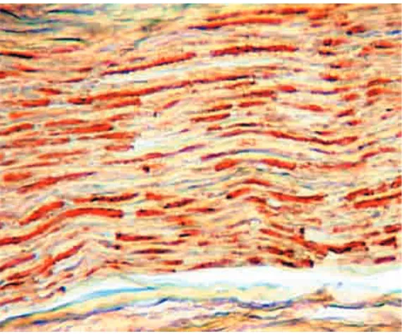
Fig. 1: Normal peripheral nerve bundle. Picture taken in the Group I.
Thin section stained with HE (larger magnification) demonstrates the peripheral nerve fascicle, located in the dermis co-rium. The nerve fascicle is surrounded by the perineurium, a sleeve formed by layers of flattened epithelium –like cells (ar-rows). Elongated nuclei stained in blue with hematoxyline belong mostly to Schwann cells or rarely to fibroblasts. The nerve fibers are only lightly highlighted with eosine while centrally placed axons on cross sections are shown as red spots in the light ring. This ring is after the routine alcohol extraction deprived of myelin so it seems to be “empty” (encircled on the picture).
Fig. 3 A,B:
A: Neurofilaments, normal nerve bundles and cross section of two bundles at medium magnification (thick sections). Specific (IHC) axon demonstration, adjacent fibrous-fat structures remain negative. The picture was taken in the Group I, unoperated control extremity.
B: In the Group I as maximum changes we found a slight defect of architectural arrangement represented by focal irregu-lar varicosities of axons as well as by interstitial widening. The presented picture could eventually correspond with a weak mechanical contusion.
Fig. 4: Diffuse changes seen on the transversal plane of a few nerve bundles (IHC detection of neurofilaments using thick sections).
Fig. 5A,B:
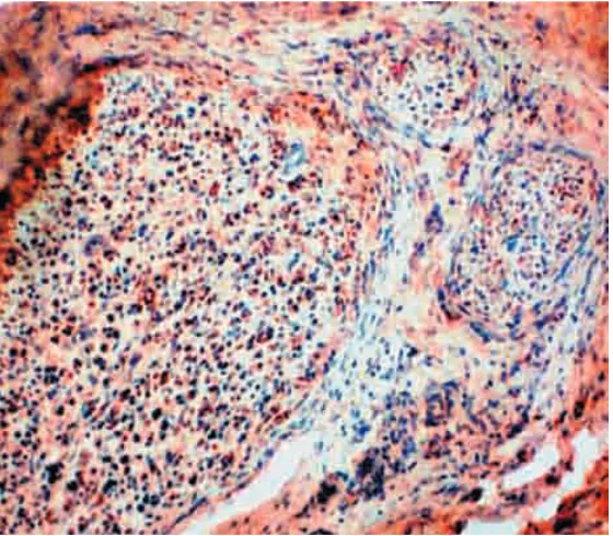
A,B: The same situation as in Fig. 4 using the thick section stained with HE. There is a significant increase of Schwann cell nuclei and probably even of fibroblasts.
A: Nonspecific granulomatous reaction of a subchronical stage spreads around the bundle: at the bottom there are resi-dues of mature fat tissue.
B: The same place with larger magnification. Group II.
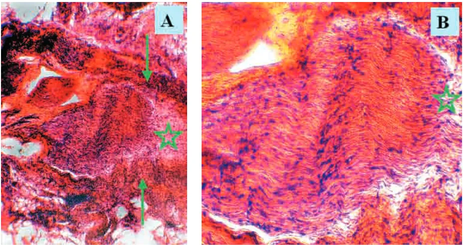
Fig. 6 A,B: Severe distortion and cellular changes. Sharp boundary at the transition into the nerve scar (asterisks). Thick section stained HE. Group II.
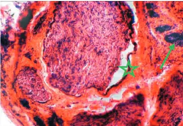
Fig. 7: Focal regression inside heavily affected nerve bundle, which might be of ischemic origin leading to possible oblite-ration of nutritive vessels at the site of adjacent plastic reaction (a thick section, HE staining, medium magnification). Group III.
Commentary on the results: During histological ob-servation of the nerve microstructure and adjacent tissue changes in the group with circular nerve freeing from the nerve-vascular bundle we observed normal findings in most of the preparations, which were quite comparable with the preparations of the control group. In the Group II we found changes in most of the preparations which varied from mi-nor insignificant changes in the nerve microstructure to se-vere pathologies. In the Group III the most sese-vere changes were described by histologist in most of the preparations when compared with previous groups.
The findings are summarized in the following table:
Tab. l
In the table the numbers of animals in which the above-mentioned changes occurred are given.
Discussion
Surgical treatment of peripheral nerve entrapment syn-drome is a topic which is being widely discussed and often mentioned in the medical literature. In spite of the pub-lished excellent or very good outcomes in a high percentage of patients, cases with small or no relief after the operations still occur. In some of the patients the complaints reappear after a short transient improvement. Part of these problems arises from scars in the nerve surroundings affecting micro-vascularization of the nerve and its gliding.
In cubital tunnel syndrome we leave the nerve after its circular release in situ or we transpose it in front of the ulnar epicondyle and fix it in subcutaneous lobe or place it intramuscularly or submuscularly. According to available literature the opinions concerning the nerve placement are far from being in agreement (2,4,7,9,12).
In carpal tunnel syndrome there is a following problem: into which bed the nerve should be placed only in case of reoperation (6,10,11).
In the experimental part of this study we tried to imita-te the same situation as in human medicine.
Differences in our study when compared with human situation:
1) Rat nerves were not injured by previous operation nor was entrapment syndrome formed. This means that we monitored reactions of a primarily healthy undamaged
nerve and its surroundings. In case of entrapment model creation the degree of damage cannot be made sure to be always identical. The results of reoperations with wrapp-ing or simple decompression would be thus very difficult to evaluate. Nevertheless, even in the clinical practice the ulnar nerves are not always seriously damaged by isthmus syndrome. In rats we are neither able to imitate the osteofibrous tunnel with its possible deformities. 2) In transposition techniques of the ulnar nerve, release of
an extent section of the nerve is being demanded, which might cause damage to external blood supply. This can-not be performed in rats.
3) During the human operations the epineurotomy or in-terfascicular neurolysis of the nerve are often performed. Due to very delicate nerve structures in rats these inter-ventions would lead to severe nerve damage. That is why they were not carried out, so we could study a real inter-action of the nerve and surrounding tissues.
4) In human medicine, surgery is very often quite bloody procedure and the use of coagulation is needed. In the animal model, postoperative bleeding is mostly minimal or none. If it occurs, we can manage it using a short ap-plication of gauze swab.
The above-mentioned points show the complexity of problems in humans. In spite of the fact that our study fo-cuses only on part of the problem, it contributes positively to this issue because of the very reason that it does not try to imitate entrapment syndrome in its full length.
From this simplified viewpoint, nerve releasewith leav-ing it in situ seems to be an optimal treatment solution. When histologically monitored the changes in the nerve microstructure were not found. Applying this finding into the human medicine it could be said that simple decom-pression seems to be appropriate, especially in conditions with smooth ulnar sulcus when changes in the nerve at the site of isthmus are small.
Nerve embedding into subcutaneous lobe seems to be less suitable. While histologically monitoring we found structural changes from minimum to significant in majority of preparations. According to published medical sources the low preference of ulnar nerve anteposition with place-ment in subcutaneous tunnel also corresponds with this conclusion. We have given up this approach in our depart-ment during the recent few years, too.
The most significant changes we described in the cases of nerve placement in the muscular lobe. These changes concerned both the muscular tissue and the femoral nerve, which was often embedded in the firmer scar tissue. In case of this approach, nerve gliding is seriously affected. Our ex-periment imitated muscular wrapping in humans. In ani-mals we were not able to imitate submuscular embedding which is currently preferred in entrapment syndromes of the ulnar nerve during reoperations.
During reoperationsit is possible to wrap up the nerve with the fascia, fat or the muscle from the surrounding tis-sues (2,5,6,10,11). Blood supply of the lobe is possible
Group Degree 1 Degree 2 Degree 3
I 6
II 3 2 1
III 1 2 3
through the pedicle. Free transplantations can also be per-formed. We assume that our findings could be also used for planning the approaches in reoperations.
In literature concerning reoperations a possibility of nerve wrapping with autologous vein has also been men-tioned (12,13,14). In the experiment on rats also the ischia-dic nerve wrapping by silicone tube, longitudinally cut through, was used (3). The damaged nerve wrapping by omentum taken from the abdominal cavity was also de-scribed (1). Omentum was in this study compared with the use of the muscle. The extent of perineural fibrosis was five times higher in case when muscle was used.
Preliminary studies also appreciate the ADCON-T/N preparation (a bioabsorbable gel) which was used in the study of reoperations of peripheral nerve entrapment syn-dromes (8).
There is obviously a bright future for biodegradable ma-terials. Different preparations with slow release from the tissue seem promising.
Nevertheless, all the above-mentioned methods of re-operations are laden with uncertain prognosis. It is there-fore important to pay maximum attention to the first opera-tion already. Our study was designed for this purpose.
Conclusion
The peripheral nerve microstructure was investigated both after nerve manipulation and after its embedding into the new environs. We have proven minimum regressive changes after simple deliberation; more significant changes were noticed after its wrapping into the fat lobe, and the most severe changes occurred during intramuscular embedding.
Description of microphotograph
(Fig. 1–8, pages 167–170)
Abbreviations: IHC – immunohistochemistry, immunohistochemically HE – hematoxylin-eosin staining
Histological sections: thin – 6µm thick – 30µm
Magnification – categories: small magnification medium magnification large magnification
Acknowledgement
This study was supported by MSM 0021620820 from Ministry of Education, by IGA MZ NR 8404-3/2005 and by research projects MZO 00179906 from Ministry of Health, Czech Republic
Literature
1. Brunelli GA, Brunelli F, Di_Rosa F. Neurolyzed nerve padding in actinic lesions: omentum versus muscle use. An experimental study. Microsurgery 1988;9: 177–80.
2. Caputo AE, Watson HK. Subcutaneous anterior transposition of the ulnar nerve for failed decompression of cubital tunnel syndrome. J Hand Surg (Am) 2000; 25:544–51.
3. Finsterbush A, Porat S, Rousso M. Prevention of peripheral nerve entrapment following extensive soft tissue injury, using silicone cuffing: an experimental stu-dy. Clin Orthop:276–81.
4. Inserra S, Spinner M. An anatomic factor significant in transposition of the ulnar nerve. J Hand Surg (Am) 1986;11:80–2.
5. Jones NF, Shaw WW, Katz RG. Circumferential wrapping of a flap around a scarred peripheral nerve for salvage of end-stage traction neuritis. J Hand Surg (Am) 1997; 22:527–35.
6. Koncilia H, Kuzbari R, Worseg A. The lumbrical muscle flap: anatomic study and clinical application. J Hand Surg (Am) 1998;23:111–9.
7. Lowe JB, Novak CB, Mackinnon SE. Current approach to cubital tunnel syn-drome. Neurosurg Clin N Am, 2001;12:267–84.
8. McCall TD, Grant GA, Britz GW. Treatment of recurrent peripheral nerve en-trapment problems: role of scar formation and its possible treatment. Neurosurg Clin N Am 2001;12:329–39.
9. Steiner HH, von Haken MS., Steiner Milz HG. Entrapment neuropathy at the cu-bital tunnel: simple decompression is the method of choice. Acta Neurochir (Wien) 1996; 138:308–13.
10. Strickland JW, Idler RS, Lourie GM. The hypothenar fat pad flap for manage-ment of recalcitrant carpal tunnel syndrome. J Hand Surg (Am) 1996; 21: 840–8.
11. Tham SK, Ireland DC, Riccio M. Reverse radial artery fascial flap: a treatment for the chronically scarred median nerve in recurrent carpal tunnel syndrome. J Hand Surg (Am) 1996;21:849–54.
12. Vardakas DG, Varitimidis SE, Sotereanos DG. Findings of exploration of a vein-wrapped ulnar nerve: report of a case. J Hand Surg (Am) 2001;26:60–3. 13. Varitimidis SE, Riano F, Sotereanos DG. Recalcitrant post-surgical neuropathy of
the ulnar nerve at the elbow: treatment with autogenous saphenous vein wrap-ping. J Reconstr Microsurg 2000;16:273–7.
14. Xu J, Varitimidis SE, Fisher KJ. The effect of wrapping scarred nerves with auto-genous vein graft to treat recurrent chronic nerve compression. J Hand Surg (Am) 2000;25:93–103.
Submitted September 2005. Accepted October 2005.
MUDr. Martin Kanta, University Hospital Hradec Králové, Department of Neurosurgery, 500 05 Hradec Králové, Czech Republic.