R E S E A R C H
Open Access
Heterogeneous ribonuclear protein E2
(hnRNP E2) is associated with
TDP-43-immunoreactive neurites in Semantic
Dementia but not with other TDP-43
pathological subtypes of Frontotemporal
Lobar Degeneration
Yvonne S. Davidson
1, Andrew C. Robinson
1, Louis Flood
1, Sara Rollinson
2, Bridget C. Benson
3, Yasmine T. Asi
3,
Anna Richardson
4, Matthew Jones
4, Julie S. Snowden
1,4, Stuart Pickering-Brown
2, Tammaryn Lashley
3and David M. A. Mann
1*Abstract:Frontotemporal Lobar Degeneration (FTLD) encompasses certain related neurodegenerative disorders which alter personality and cognition. Heterogeneous ribonuclear proteins (hnRNPs) maintain RNA metabolism and changes in their function may underpin the pathogenesis of FTLD. Immunostaining for hnRNP E2 was performed on sections of frontal and temporal cortex with hippocampus from 80 patients with FTLD, stratified by pathology into FTLD-tau and FTLD-TDP type A, B and C subtypes, and by genetics into patients withC9orf72expansions,
MAPTorGRNmutations, or those with no known mutation, and on 10 healthy controls. Semi-quantitative analysis assessed hnRNP staining in frontal and temporal cortex, and in dentate gyrus (DG) of hippocampus, in the different pathology and genetic groups. We find that hnRNP E2 immunostaining detects the TDP-43 positive dystrophic neurites (DN) within frontal and temporal cortex, and the neuronal cytoplasmic inclusions (NCI) seen in DG granule cells, characteristic of patients with Semantic Dementia (SD) and type C TDP-43 pathology, but did not detect TDP-43 or tau inclusions in any of the other pathological or genetic variants of FTLD. Double immunofluorescence for hnRNP E2 and TDP-43 showed most TDP-43 immunopositive DN to contain hnRNP E2. Present findings indicate an
association between TDP-43 and hnRNP E2 which might underlie the pathogenetic mechanism of this form of FTLD.
Introduction
Frontotemporal Lobar Degeneration (FTLD) is a clinically, pathologically and genetically heterogeneous disorder affecting principally the frontal and temporal lobes of the brain. Three major clinical syndromes are recognised [34]. One syndrome, behavioural variant frontotemporal de-mentia (bvFTD), is characterised by changes in behaviour and personality and accounts for around 75% of all cases of FTLD, whereas the other two syndromes are disorders
of language [34]. Semantic dementia (SD) (also known as semantic variant of primary progressive aphasia (svPPA)) is a disorder characterized by loss of conceptual know-ledge of the meaning of words and objects [14, 34], whereas Progressive Non-Fluent Aphasia (PNFA) (also known as nfvPPA) is represented by an inability to con-struct language such that speech becomes hesitant and stuttering, becoming grammatically and contextually incorrect [14, 34]. The amyotrophic lateral sclerosis (ALS) form of motor neurone disease (MND) is seen in about 15% of patients with bvFTD, but is only rarely combined with either SD or PNFA [32].
Three different pathologies, characterised by abnormal neuronal, and sometimes glial, accumulations of aggregated * Correspondence:[email protected]
1Division of Neuroscience and Experimental Psychology, School of Biological
Sciences, Faculty of Biology, Medicine and Health, University of Manchester, Salford Royal Hospital, M6 8HD, Salford, UK
Full list of author information is available at the end of the article
proteins, are seen. Neuronal intracytoplasmic inclu-sions (NCI), composed of the microtubule associated protein tau, occur in about 45% cases as neurofibrillary tangle-like structures, or more rounded inclusions known as Pick bodies [33] and termed FTLD-tau [19]. The RNA and DNA binding protein, TDP-43, is present within NCI, neuritic processes (dystrophic neurites, DN) or neuronal intranuclear inclusions (NII) in about 50% of cases [2, 7, 26]. The relative proportions of NCI, DN and NII provide a neuropathological classification of FTLD-TDP subtypes [19]. FTLD-TDP subtype A is applied when NCI and short DN are both commonly present, mostly in outer cortical laminae, type B when NCI present throughout all cortical layers numerically predominate over DN, type C when long thick DN are present throughout all cortical layers and predominate over NCI and type D when NII are most common type of pathological change [19]. Most of the remaining 5% cases show NCI composed of the protein, Fused in Sarcoma (FUS), and are known as FTLD-FUS [19].
TDP-43 and FUS are heterogeneous nuclear riboproteins (hnRNP) [5, 30] and serve as RNA-splicing and transcrip-tion regulators, shuttling between nucleus and cytoplasm, thereby controlling cellular levels of protein synthesis. In the nucleus, TDP-43 binding encourages RNA stability, whereas in the cytoplasm it associates with stress granules and non-coding RNAs for post-transcriptional metabolism of RNA and transport. In FTLD-TDP there is a‘clearing’of normal physiological TDP-43 from the nucleus with its ac-cumulation within the cytoplasm as NCI, DN or NII. How-ever, the precise mechanism(s) directing this pathological change remain unclear.
Previous studies have pointed to specific interactions between particular hnRNPs and the pathological inclu-sions of FTLD. For example, we [9] and others [3, 23, 24] have shown that hnRNP A3 is present in the aggregates of dipeptide repeat proteins (DPR) in FTLD patients bearing expansions in C9orf72 gene. Elsewhere, Gami-Patel and colleagues reported the presence of various hnRNPs, but especially hnRNP A1, within NCI in patients with the Neuronal Intermediate Filament Inclusion Body Disease form of FTLD-FUS [12].
TDP-43 is a stress responsive protein, and the TDP-43 aggregates in FTLD-TDP are thought to arise from stress granules [6, 18, 37]. Stress granules are transient cytoplasmic structures composed of mixed protein-RNA complexes, formed in response to cellular stress and believed to act as a sorting station, triaging mRNAs and sequestering transcripts not needed for coping with the stress [10]. Their composition and morphology varies according to the cell and stress type, but are generated by a reversible aggregation of prion-like core compo-nents such as the T-cell intracellular antigen-1 (TIA-1) and the poly A binding protein 1 (PABP1) [13] in order
to regulate mRNA metabolism and protein translation [1, 18]. Biochemical studies have shown that TDP-43 associates with stress granules by interacting with TIA-1 [18]. Furthermore, using a yeast two-hybrid screen against TIA-1, hnRNP E2 was reported to be present in stress granules and processing bodies in HeLa cells, and an interaction between hnRNP E2 and TIA-1 was dem-onstrated by double immunofluorescence [11]. Finally, it has been reported that TDP-43 pathological inclusions in patients with bvFTD and FTLD-TDP pathology, and in others with ALS, co-localise with multiple markers of stress granules including TIA-1 [18]. Hence, it seems likely that both TDP-43 and hnRNP-E2 can be incorpo-rated into stress granules under stress conditions.
With this in mind, we sought possible relationships between hnRNP E2 and the TDP-43 pathological inclu-sions of FTLD since, because of their shared links with stress granules, it is plausible that hnRNP E2 might be a component of the TDP-43 pathological inclusions that form within neuronal cell bodies (NCI) and processes (DN). We find that hnRNP E2 immunostaining co-local-ises with TDP-43 pathological changes, but only in patients with SD and type C TDP-43 histology. Collectively, present and previous [3, 9, 23, 24] data suggest that although pathological accumulations of TDP-43 or FUS are hall-mark characteristics of FTLD, the fundamental biological mechanisms leading to these molecular end-points may differ between pathological subtypes.
Materials and methods Patients
The study consisted of 2 groups comprising 90 subjects in total. One group had been recruited through Manchester Brain Bank (MBB), 54 with a clinical diagnosis of FTLD (30 males, 24 females; cases #1-54), and 10 healthy control subjects (3 males, 7 females; cases #55-64) (Table 1). The brains of these patients had been consecutively acquired by MBB over the years 1986 to present. All patients were from the North West of England and North Wales, and tissues were obtained through appropriate consenting procedures for the collection and use of the human brain tissues. All patients fulfilled relevant clinical diagnostic cri-teria [14, 25, 31], having been investigated longitudinally within a specialist dementia clinic using the Manchester Neuropsychological Profile (Man-NP) [35, 36] to deter-mine and characterise the nature of their dementia.
Table 1Selected clinical, neuropathological and genetic details on patients studied
case ID# MRC ID# clinical TDP subtype mutation gender PMI (h) Onset (y) Death (y) Duration (y)
1* BBN_5681 bvFTD FTLD-TDP A C9ORF72 M 74 49 58 9
2* BBN_5706 bvFTD FTLD-TDP A C9ORF72 M 28 60 68 8
3* BBN_5719 bvFTD FTLD-TDP A C9ORF72 F 74 59 64 5
4* BBN_5752 bvFTD FTLD-TDP A C9ORF72 M na 64 72 8
5 BBN_14793 bvFTD FTLD-TDP A C9ORF72 M 81 54 65 11
6 BBN_5756 FbvTD FTLD-TDP B C9ORF72 F 48 52 70 18
7* BBN_5771 FTD + MND FTLD-TDP B C9ORF72 F 50 63 65 2
8* BBN_5772 FTD + MND FTLD-TDP B C9ORF72 F 50 68 73 5
9 BBN_5691 FTD + MND FTLD-TDP B C9ORF72 M 72 60 62 2
10* BBN_5663 FTD + MND FTLD-TDP B C9ORF72 M 36 57 59 2
11* BBN_5742 PNFA FTLD-TDP A GRN V452WfsX38 M 57 66 71 5
12 BBN_10260 PNFA FTLD-TDP A GRN V452WfsX38 M 25 62 72 10
13* BBN_5660 bvFTD FTLD-TDP A GRN V452WfsX38 F 13 53 71 18
14 BBN_5773 bvFTD FTLD-TDP A GRN Q130SfsX124 M 74 66 73 7
15 BBN_5715 PNFA FTLD-TDP A GRN Q130SfsX124 F 24 63 71 8
16* BBN_5727 PNFA FTLD-TDP A GRN C31LfsX34 M 104 66 73 7
17 BBN_5718 bvFTD FTLD-TDP A GRN R493X M 35 59 66 7
18 BBN_5675 bvFTD FTLD-TDP A GRNR493X F 12 51 61 10
19 BBN_5686 bvFTD FTLD-TDP A GRN Q468X F 48 60 66 6
20 BBN_5734 bvFTD FTLD-TDP A none M 48 69 75 6
21* BBN_5685 PNFA FTLD-TDP A none M 19 68 78 10
22 BBN_5757 PNFA FTLD-TDP A none F 72 66 77 11
23 BBN_5753 bvFTD FTLD-TDP A none F 46 66 72 6
24 BBN005.28193 bvFTD FTLD-TDP A none F 144 67 72 5
25 BBN005.29059 bvFTD FTLD-TDP A none M 52 67 71 4
26 BBN_5661 FTD + MND FTLD-TDP B none M 21 43 45 2
27* BBN_5676 FTD + MND FTLD-TDP B none M na 60 68 8
28* BBN_5701 FTD + MND FTLD-TDP B none M 30 45 51 6
29* BBN_5721 FTD + MND FTLD-TDP B none M 87 58 69 11
30 BBN_5732 FTD + MND FTLD-TDP B none F 164 50 52 3
31* BBN_5764 FTD + MND FTLD-TDP B none M 110 61 65 4
32 BBN_24314 FTD + MND FTLD-TDP B none F 133 61 63 2
33 BBN005.28645 FTD + MND FTLD-TDP B none M 114 64 69 5
34 BBN_5678 SD FTLD-TDP C none M 48 54 68 14
35 BBN_5708 SD FTLD-TDP C none F 28 55 66 11
36* BBN_5720 SD FTLD-TDP C none M 59 60 75 15
37 BBN_5726 SD FTLD-TDP C none F 104 56 67 11
38 BBN_5736 SD FTLD-TDP C none F 168 70 72 2
39* BBN_5731 SD FTLD-TDP C none M 44 71 77 6
40 BBN_16418 SD FTLD-TDP C none M 29 56 68 12
41 BBN_19623 SD FTLD-TDP C none F 86 68 82 14
42 BBN005.26066 SD FTLD-TDP C none M 59 52 66 14
43 BBN005.28698 SD FTLD-TDP C none M 35 57 77 20
Table 1Selected clinical, neuropathological and genetic details on patients studied(Continued)
45 BBN_5696 bvFTD FTLD-tau MAPT exon 10 + 16 F 52 46 58 12
46 BBN_5699 bvFTD FTLD-tau MAPT exon 10 + 16 M 30 43 55 12
47 BBN_5717 bvFTD FTLD-tau MAPT exon 10 + 16 F 23 52 65 13
48 BBN_5744 bvFTD FTLD-tau MAPT exon 10 + 16 F 33 50 60 10
49* BBN_5733 bvFTD FTLD-tau MAPT exon 10 + 16 M 24 46 53 7
50 BBN_5760 bvFTD FTLD-tau MAPT exon 10 + 16 F 96 48 63 15
51 BBN_5763 bvFTD FTLD-tau MAPT exon 10 + 16 F 48 52 58 6
52 BBN_6081 bvFTD FTLD-tau MAPT exon 10 + 16 F 26 53 63 10
53 BBN005.29180 bvFTD FTLD-tau MAPT exon 10 + 16 F 83 60 69 9
54 BBN_5710 bvFTD FTLD-tau MAPT exon 10 + 13 M 43 65 70 5
55 BBN_3109 Control none none F 72 na 76 na
56 BBN_3124 Control none none F 44 na 82 na
57 BBN_3126 Control none none M 52 na 80 na
58* BBN_3378 Control none none F 48 na 77 na
59 BBN_3447 Control none none F 41 na 80 na
60 BBN_20608 Control none none F 130 na 76 na
61 BBN_3337 Control none none F 12 na 87 na
62* BBN_3430 Control none none M 49 na 84 na
63 BBN_25922 Control none none F 103 na 100 na
64 BBN_25974 Control none none M 93 na 91 na
65 BBN_8556 bvFTD FTLD-TDP A C9orf72 M 77 53 63 10
66 BBN_8109 PNFA FLTD-TDP A C9orf72 F 85 56 67 11
67 BBN_20082 PNFA FTLD-TDP A C9orf72 F 63 57 62 5
68 BBN007.29537 bvFTD FTDL-TDP A C9orf72 M 52 66 71 5
69 BBN007.26830 bvFTD FTLD-TDP A C9orf72 F 107 58 66 8
70 BBN_11628 bvFTD FLTD-TDP A TBK1 M 97 62 72 10
71 BBN_11698 bvFTD FTLD-TDP A none F 85 57 63 6
72 BBN_11574 bvFTD FTDL-TDP A none F 8 70 83 13
73 BBN_12507 bvFTD FTLD-TDP A none M 93 57 62 5
74 BBN007.29538 bvFTD FLTD-TDP A GRN C31LfsX34 M 29 49 55 6
75 BBN_12169 bvFTD FTLD-TDP B C9orf72 F 94 64 66 2
76 BBN_12136 MND FTLD-TDP B none M 70 67 69 2
77 BBN_11773 MND FTLD-TDP B none F 30 75 77 2
78 BBN_12451 FTD/MND FTLD-TDP B none F 46 63 67 4
79 BBN_12331 bvFTD FTLD-TDP B none F 45 63 83 20
80 BBN_12480 SD FTLD-TDP C none F 38 58 73 15
81 BBN_12524 SD FTLD-TDP C none F 84 59 73 14
82 BBN_11594 SD FTLD-TDP C none M 27 64 78 14
83 BBN_11883 SD FTLD-TDP C none M 19 64 74 10
84 BBN_8614 SD FTLD-TDP C none M 52 50 65 15
85 BBN_12300 SD FTLD-TDP C none M 71 61 66 5
86 BBN007.26814 SD FTLD-TDP C none M 76 44 67 23
human brain tissues. Again, all patients fulfilled relevant clinical diagnostic criteria [14, 25, 31] having been referred to the Dementia Research Unit, Queen Square, London.
Of the combined 80 FTLD patients, 36 had been clinically diagnosed with bvFTD (17 males, 19 females; cases #1-6,13,14,17-20,23-25,44-54,65,68-75,79), 13 with bvFTD + MND or MND alone (9 males, 4 females; cases #7-10,26-33,76-78), 9 with PNFA (4 males, 5 females; cases #11,12,15,16,21,22,66,67,87) and 20 with SD (12 males, 8 females; cases #34-43, 80-86, 88-90) (Table 1). Pathologically, the FTLD group comprised 30 patients with FTLD-TDP type A (cases #1-5,11-25, 65-74), 18 with FTLD-TDP type B (cases #6-10,26-33,75-79), 21 with FTLD-TDP type C (cases #34-43,80-90), 11 with FTLD-tau (cases #44-54) (Table 1). Furthermore, within the FTLD group there were 16 patients with expansions in C9orf72 (cases #1-10,65-69,75), 10 with GRN muta-tions (cases #11-19,74), 1 patient with TBK1 mutation (case #70), 11 patients with intronic mutations in
MAPT (cases #44-54) and 42 without known mutation
(cases #20-43,71-73,76-90) (Table 1).
Histological methods
Standard blocks of frontal (BA 8/9) and temporal (BA21/22) lobe, the latter to include the posterior hippo-campus at the level of the geniculate bodies, were cut from the formalin fixed brains. Where possible, and in order to preserve maximum antigenicity, the original blocks taken for diagnosis were employed since these had been cut from brains which had been fixed in for-malin for no more than 3-6 months from the time of ac-quisition. However, it was not possible to do this in all instances, especially in respect of some of the more long-standing cases (ie those acquired before 2007) where this block was no longer available and new blocks had to be cut from 20 cases for the purpose of this study (see Table 1 for those cases where new blocks were cut).
Paraffin sections were cut from these blocks at a thick-ness of 6 μm. Preliminary titration experiments were performed at dilutions 1:100 to 1:3000 on 5 randomly chosen cases, one from each pathological subgroup, in order to determine optimal specific nuclear and DN/ NCI immunostaining for each antibody. When dilutions for hnRNP E2 staining less than what was optimal for nuclear and inclusion body staining were employed, im-munostaining for nuclei and DN/NCI was still observed,
but less strongly so, as would be expected given the lower antibody concentrations. Subsequently, all antibodies were employed at these optimised dilutions (as specified below) in a standard IHC protocol, as described previously [7–9, 22]. The following antibodies were employed: hnRNP E2 (also known as PCBP2) (mouse monoclonal, Santa Cruz, 23G: sc101136, 1:100 and Novus, mouse monoclonal H00005094-M07, 5F12 clone, 1:500), non-phosphorylated TDP-43 (rabbit polyclonal, 10,782-2-AP antibody, Protein-tech, Manchester, UK, 1:3000) and phosphorylated TDP (pS409/410-2 antibody, Cosmo Biotech Ltd., Tokyo, Japan, 1:2000) and tau (mouse monoclonal, AT8, Innogenetics, Antwerp, Belgium, 1:750) proteins. Both the Santa Cruz and Novus hnRNP E2 antibodies are raised against recom-binant hnRNP E2 of human origin, and on western blot-ting detect a protein with molecular mass of around 40 kDa. For all antibodies employed, antigen unmasking was performed by pressure cooking in citrate buffer (pH 6.0, 10 mM) over a 30-min period to include warming and cooling times, reaching 123 degrees Celsius for 30 s, and >15 psi pressure.
Double immunohistochemical staining was performed using phosphorylated TDP-43 antibody (pS409/410-2 antibody, Cosmo Biotech Ltd., Tokyo, Japan, 1:2000) and hnRNP E2 (Novus, 5F12, 1:500) antibody to investigate the co-localisation of the two proteins. Sections were cut, pre-treated and incubated in primary antibody as described above. Two Alexa Fluor secondary antibodies (Alexa Fluor 488 and Alexa Fluor 568; Molecular Probes, 1:300, with incubation for 1 hour at room temperature) were used to visualise sites of protein de-position. 4′-6-diamidino-2-phenylindol (DAPI) was used for nuclear counterstaining.
Pathological assessment
Immunostained sections were examined microscopically for the appearance of intracellular distribution of stain-ing within neurones of the temporal cortex, dentate gyrus and CA4 region of the hippocampus. These re-gions were chosen since it was known from previous work [7, 33] that the temporal cortex and dentate gyrus of the hippocampus are involved in all forms of FTLD-TDP or tau pathology in those patients withMAPT mu-tation [29]. Moreover, the CA4 region of the hippocam-pus was included because this is one of the principal regions affected by DPR pathology in patients with Table 1Selected clinical, neuropathological and genetic details on patients studied(Continued)
88 BBN007.26876 SD FTLD-TDP C none F 31 58 72 14
89 BBN007.26890 SD FTLD-TDP C none M 40 67 76 9
90 BBN007.29539 SD FTLD-TDP C none F 25 52 65 13
expansions in C9orf72 [8, 22, 23]. The degree of neur-onal nuclear and/or cytoplasmic hnRNP E2 immuno-staining in each region was scored semi-quantitatively [9] at an objective magnification of ×25 (overall micro-scope magnification of ×250) employing the following rating scale:
0 = No staining present.
0.5 = rare (ie 1-5) cells per section showing weak nuclear and/or cytoplasmic staining.
1 = few (1-5) cells showing weak nuclear and/or cytoplasmic staining per ×250 microscope field. 2 = moderate number (5-10) cells showing moderate nuclear and/or cytoplasmic staining per ×250 microscope field.
3 = more than 10 cells showing strong nuclear and/or cytoplasmic staining per ×250 microscope field.
The severity of hnRNP E2-immunoreactive, and TDP-43-immunoreactive, inclusion body immunostaining (ie NCI and DN) in frontal and temporal cortex, and in granule cells of the dentate gyrus of the hippocampus, was separately graded at an objective magnification of ×25 (overall microscope magnification of ×250), employ-ing the followemploy-ing ratemploy-ing scale:
0 = no inclusions present.
0.5 = rare (ie 1-5 inclusions per section).
1 = few (ie 1-5 inclusions per ×250 microscope field). 2 = moderate (ie 5-10 inclusions per ×250 microscope field).
3 = many (ie 10-50 inclusions per ×250 microscope field). 4 = very many (ie more than 50 inclusions per ×250 microscope field).
Scoring of staining was performed by a single observer (DMAM) blinded to clinical, histopathological and genetic status. Previous use of this particular scoring system has shown robust agreement in assessments when employed by both highly and lesser experienced observers [9].
Double immunolabelled sections were viewed with a Leica TCS4D confocal microscope using a 3-channel scan head and argon/krypton laser.
Statistical analysis
Rating data was entered into an excel spreadsheet and analyzed using Statistical Package for Social Sciences (SPSS) software (version 17.0). The 80 FTLD patients were stratified according to genetic and pathological subtype for statistical analysis of the effect of each muta-tion and underlying pathology on the degree and pattern of hnRNP E2 staining. Comparisons of semi-quantitative scores for the intensity of neuronal hnRNP E2 immuno-staining in nucleus and cytoplasm of neurones of the
frontal and temporal cortex, and dentate gyrus of the hippocampus, with respect to pathological type or gen-etic mutation, were performed using Kruskal-Wallis test with post-hoc Mann-Whitney test where Kruskal-Wallis yielded a significant difference between antibody staining scores. Comparisons of semi-quantitative scores for the severity of hnRNP E2- and TDP-43-immunoreactive inclusions in frontal and temporal cortex, and dentate gyrus of the hippocampus, with respect to pathological subtype, were also performed using Kruskal-Wallis test. Comparison of scores for the severity of hnRNP E2- and TDP-43-immunoreactive inclusions in the same cases was made using Wilcoxon matched pairs test. Group comparisons of age at onset, age at death, post mortem delay interval and duration of illness were made using ANOVA with post hoc Tukey test. In all instances, sig-nificance levels were set atp< 0.05.
Results
Demographic comparisons
Comparison of the 4 FTLD pathology patient groups showed significant differences in mean age at onset of disease (F3,76 = 4.9, p = 0.004), mean age at death (F3,76= 7.0, p< 0.001) and duration of illness did differ (F3,76= 8.3, p < 0.001). Patients with FTLD-tau had an earlier age at onset than those with FTLD-TDP type A (p = 0.002), type B (p= 0.016) and type C (p = 0.012) pathology (Table 2) and an earlier age at death than
those with FTLD-TDP type A (p = 0.014) and type C
(p < 0.001) pathology, but not those with FTLD-TDP type B (p= 0.449) pathology. None of the 3 FTLD-TDP groups differed from each other in terms of age at onset, though patients with FTLD-TDP type B pathology died at an earlier age than those with FTLD-TDP type C pathology (p = 0.015). Patients with FTLD-TDP type B had a shorter disease duration than those with
FTLD-TDP type C (p = 0.001) and those with FTLD-tau
(p= 0.040); those with FTLD-TDP type A pathology also had a shorter disease duration than those with FTLD-TDP type C (p= 0.006), but there were no other differ-ences between the other sub-types (Table 2). The healthy control group was also significantly older at death (p< 0.001) than each of the FTLD subgroups (Table 2).
significantly earlier in MAPT than the no mutation group (p = 0.002), but otherwise there were no signifi-cant differences between all other groups (Table 2).
There were no significant differences between mean post mortem intervals for each pathological (F4,83 = 1.0,
p= 0.401) or genetic (F4,73 = 1.6, p= 0.181) group (see Table 2), nor were there any significant correlations between each pathological measure and post mortem interval (p= 0.351-0.993).
TDP-43 immunostaining
TDP-43- and tau-immunostaining was employed to clas-sify the 80 FTLD patients into their respective patho-logical subgroups (FTLD-TDP subtypes A, B or C and FTLD-tau) according to the form and distribution of the TDP-43 or tau-immunoreactive inclusions (NCI, DN) present (see [19] for criteria). Median scores (with inter-quartile range) from TDP-43 immunostaining of patho-logical inclusions and neuronal nuclei and cytoplasm, derived from semi-quantitative scoring are shown in Table 3. As expected, given the inclusion of FTLD-tau group, the degree of TDP-43 inclusion body immuno-staining (irrespective of whether these were in the form of NCI or DN, or both) differed between the 4 FTLD pathological groups in both the frontal (χ2 = 34.1,
p < 0.001) and temporal (χ2 = 36.8, p < 0.001) cortex, and in the dentate gyrus (χ2= 35.3,p< 0.001) with all 3 TDP subtypes differing significantly from FTLD-tau group (p< 0.001 in every instance). However, there were no significant differences in the overall degree of TDP-43 immunostaining between FTLD-TDP type A, type B or type C subgroups.
hnRNP E2 immunostaining
When using Santa Cruz hnRNP E2 antibody, 58/80 FTLD patients, and 8/10 controls, showed a variable level of cellular staining within nerve cells of the frontal and temporal cortex, and in granule cells of the dentate
Table 2Mean +/−SD age at onset, death and duration of illness, and gender composition, for each neuropathological and genetic subgroup of patients. Mean +/−SD and median (in parentheses) post mortem delay interval (PMI) is also presented for each group
Group M/F Onset (y) Death (y) Duration (y) PMI (h)
FTLD-TDP type A (n= 30) 17/13 60.7 ± 6.2 68.7 ± 6.1 8.0 ± 3.0 59.5 ± 33.3 (57)
FTLD-TDP type B (n= 18) 9/9 59.7 ± 8.0 65.2 ± 9.2 5.6 ± 5.5 70.6 ± 40.6 (50)
FTLD-TDP type C (n= 21) 12/9 59.7 ± 7.9 71.8 ± 5.3 12.1 ± 5.1 54.7 ± 35.1 (44)
FTLD-tau (n= 11) 4/7 51.4 ± 6.4 61.4 ± 5.4 10.0 ± 3.1 45.8 ± 24.0 (44)
FTLDC9orf72expansion (n= 16) 8/8 58.8 ± 5.3 65.7 ± 4.4 6.9 ± 4.6 66.1 ± 21.9 (72)
FTLDGRNmutation (n= 10) 6/4 59.5 ± 6.4 67.9 ± 5.9 8.4 ± 3.7 42.1 ± 29.2 (32)
FTLD No mutation (n= 42) 23/19 60.7 ± 7.9 70.0 ± 8.1 9.3 ± 5.5 62.6 ± 40.2 (48)
FTLDMAPTmutation (n= 11) 4/7 51.4 ± 6.4 61.4 ± 5.4 10.0 ± 3.1 45.8 ± 24.0 (44)
FTLD TBK1 mutation (n= 1) 1/0 62 72 10 97
Healthy Controls (n= 10) 3/7 na 83.3 ± 7.6 na 64.4 ± 35.0 (50.5)
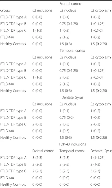
Table 3Median values with interquartile range for hnRNP E2 immunostaining of pathological inclusions, neuronal nuclei and cytoplasm in frontal and temporal cortex, and dentate gyrus of hippocampus, for each pathological subgroup
Frontal cortex
Group E2 inclusions E2 nucleus E2 cytoplasm
FTLD-TDP type A 0 (0-0) 1 (0-1) 1 (0-2)
FTLD-TDP type B 0 (0-0) 0.75 (0-1.25) 1 (0-1.25)
FTLD-TDP type C 1 (1-2) 1 (0-3) 1 (0.5-2)
FTLD-tau 0 (0-0) 2 (1-2) 1 (0-2)
Healthy Controls 0 (0-0) 1.5 (0-3) 1.5 (0-2.25)
Temporal cortex
E2 inclusions E2 nucleus E2 cytoplasm
FTLD-TDP type A 0 (0-0) 1 (0-1) 1 (0-2)
FTLD-TDP type B 0 (0-0) 0.75 (0-1.25) 1 (0-1.25)
FTLD-TDP type C 1 (1-3) 2 (0-3) 2 (0.5-3)
FTLD-tau 0 (0-0) 2 (1-2) 1 (0-2)
Healthy Controls 0 (0-0) 1.5 (0-3) 1.5 (0-2.25)
Dentate Gyrus
E2 inclusions E2 nucleus E2 cytoplasm
FTLD-TDP type A 0 (0-0) 1 (0-1) 1 (0-2)
FTLD-TDP type B 0 (0-0) 0.75 (0-2) 1 (0-2)
FTLD-TDP type C 2 (0-3) 2 (0-3) 2 (0-3)
FTLD-tau 0 (0-0) 1 (0-3) 1 (0-2)
Healthy Controls 0 (0-0) 1.5 (0-3) 1.5 (0-2.25)
TDP-43 inclusions
Frontal cortex Temporal cortex Dentate Gyrus
FTLD-TDP type A 3 (2-3) 3 (2-3) 1 (1-1.25)
FTLD-TDP type B 2 (2-3) 2 (2-3) 2 (1-3)
FTLD-TDP type C 2 (2-3) 3 (2-3) 3 (2-3)
FTLD-tau 0 (0-0) 0 (0-0) 0 (0-0)
gyrus of hippocampus. When present, this was usually observed in both nucleus and cytoplasm, and ranged in intensity from very weak to strong. Conversely, 22 FTLD patients and 2 controls showed no nuclear or cytoplas-mic staining at all in any region examined. Twenty of these cases had been stored for long periods (in excess of 10 years) in formalin fixation before blocks had been taken for this study, whereas the brains of those cases based on diagnostic blocks had been stored for a shorter time in formalin fixation before blocking (3-6 months), and all these, except 4 cases, showed variable degrees of nuclear and cytoplasmic staining. There did not appear to be any pathological or genetic group preference for presence/absence of this kind of staining. Hence, some degree of positive (nuclear or cytoplasmic, or both) staining was seen in 22/30 (73%) patients with TDP type A histology, 11/18 (61%) patients with FTLD-TDP type B, 16/21 (76%) patients with FTLD-FTLD-TDP type C, 9/11 (81%) patients with FTLD-tau and 8/10 (80%) controls. According to FTLD genetics, some positive degree of positive staining was seen in 9/16 (56%) patients with C9orf72 expansion, 7/10 (70%) patients with GRN mutation, 32/42 (76%) patients with no mutation and 9/11
(80%) patients with MAPT mutation. There were no
significant differences between the proportions of FTLD patients showing some degree of hnRNP E2 immunostain-ing when stratified either by pathological (χ2 = 2.09,
p= 0.718) or genetic (χ2= 1.98,p= 0.577) groupings. Median scores (with interquartile range) from hnRNP E2 immunostaining of pathological inclusions and neur-onal nuclei and cytoplasm, derived from semi-quantitative scoring, for each of the pathological groups are shown in Table 3. Statistical analysis showed no significant differ-ences in scores for nuclear and cytoplasmic hnRNP E2
staining in frontal cortex (χ2= 5.8,p= 0.120 andχ2= 2.5,
p = 0.479, respectively), temporal cortex (χ2 = 6.7,
p= 0.080 andχ2= 3.6,p= 0.312, respectively) or dentate gyrus (χ2= 5.7,p= 0.128 andχ2= 2.7,p= 0.444, respect-ively). Similarly, no significant differences in scores for nu-clear and cytoplasmic hnRNP E2 staining in the 4 genetic groups were seen in frontal cortex (χ2= 4.0,p= 0.400 and
χ2
= 1.1, p = 0.891, respectively), temporal cortex (χ2= 3.6,p= 0.463 andχ2= 1.1,p= 0.889, respectively) or dentate gyrus (χ2 = 4.5, p = 0.338 and χ2 = 1.2,
p= 0.882, respectively).
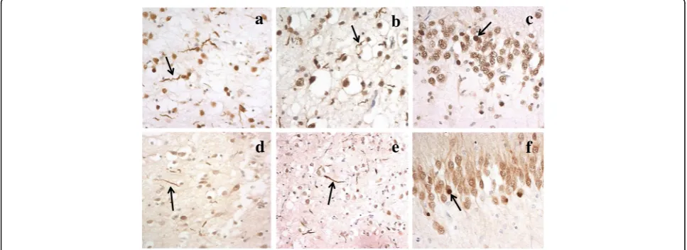
DN with morphology akin to that seen on TDP-43 munostaining (Fig. 1a and b) were seen on hnRNP E2 im-munostaining (Fig. 1d and e) in the frontal (Fig. 1a and c) and temporal (Fig. 1b and e) cortex of 15/21 FTLD-TDP type C cases. In the same cases similar rounded, solid-appearing NCI seen on TDP-43 immunostaining in den-tate gyrus granule cells (Fig. 1c) were also seen on hnRNP E2 immunostaining (Fig. 1f). Only FTLD-TDP type C cases had pathological inclusions labelled by hnRNP E2.There was no immunolabelling by hnRNP E2 of TDP-43 or tau inclusions in FTLD-TDP type A or type B cases or those with FTLD-tau. Interestingly, there was no appar-ent loss of physiological immunostaining of the nucleus for hnRNP E2 in those cells of the dentate gyrus bearing NCI (Fig. 1f), in contrast to that seen with TDP-43 where there was loss of normal nuclear staining in those cells containing TDP-43 immunoreactive NCI (Fig. 1c).
There were no clinical or neuropathological differ-ences between the 15 hnRNP E2 positive cases (cases #37,40-43,81-90) and the 6 hnRNP E2 negative cases (cases #34-36,38,39,80). Moreover, there were no differ-ences in age at onset (p= 0.542), age at death (p= 0.966), duration of illness (p = 0.405) or post mortem interval
a
b
c
d
e
f
(p= 0.285) between the 15 positive and 6 negative cases. The only difference between the two groups was that the negative FTLD-TDP type C cases employed tissue blocks that had been newly cut for this study from long-standing (before 2007) archived fixed tissues as the original diag-nostic blocks were no longer available for study. All other cases employed the original diagnostic blocks cut after 3-6 months fixation. Furthermore, within those FTLD cases bearing expansions in C9orf72 gene, there was no immunostaining of the TDP-43-negative, p62-positive DPR inclusions within neurones of frontal and temporal cortex, or dentate gyrus granule cells and CA4 neurones of the hippocampus.
Consequently, semi-quantitative analysis within 5 pathological groups showed a significant difference in scores for hnRNP E2 positive inclusions in the frontal cortex (χ2= 46.6,p< 0.001), temporal cortex (χ2= 50.6,
p< 0.001) and dentate gyrus (χ2= 50.7,p< 0.001), with the number of inclusions in both areas being signifi-cantly different in FTLD-TDP type C cases than all other pathological subtypes (p< 0.001 in every instance). This result would be expected given the observations that only in FTLD-TDP type C cases were there pathological inclusions immunostained by hnRNP E2.
The results of immunostaining with the Novus hnRNP E2 antibody appeared broadly similar to those obtained with Santa Cruz antibody on all cases of FTLD-TDP type C, and selected cases of the other pathological subtypes, where both antibodies were employed, both with respect to the intensity of neuronal staining and severity of inclusion body staining. Comparison of semi-quantitative scores for the intensity of nuclear and cytoplasmic staining, and inclu-sion body staining, on all 21 cases of FTLD-TDP type C showed that Novus antibody immunostained nuclei less in-tensely than Santa Cruz antibody (frontal cortexp= 0.008, temporal cortex p = 0.005; dentate gyrus p = 0.005), but there were no significant differences between the antibodies for level of cytoplasmic staining (p= 0.617,p= 0.120 and
p = 0.104, respectively). There were no significant differ-ences between scores for inclusion body staining with both antibodies for frontal cortex (p= 0.054) and temporal cor-tex (p= 0.655) with only a trend for scores in dentate gyrus to be greater (p= 0.023) for Novus antibody.
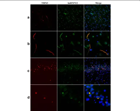
In general, the number of DN or NCI visualised on hnRNP E2 immunostaining appeared fewer than those seen in TDP-43 immunostaining (compare Fig. 1a with d, and b with e and c with f ). Indeed, comparison of scores for TDP-43- and hnRNP E2- immunostaining of DN in FTLD-TDP type C cases showed that the number of DN immunostained for hnRNP E2 was significantly less than that immunostained for TDP-43 in frontal cortex (p= 0.004), temporal cortexp= 0.002) and dentate gyrus (p= 0.010). Consequently, double immunofluorescence la-belling for TDP-43 and hnRNP E2 (using either the Santa
Cruz or the Novus antibody) was performed on selected cases of FTLD-TDP type C and showed a good degree of colocalisation between the two proteins (Fig. 2).
Discussion
In the present study, we have investigated the pattern of hnRNP E2 immunostaining across a range of clinical, pathological and genetic forms of FTLD. We found no sig-nificant changes in the amount, or intraneuronal distribu-tion pattern, of neuronal hnRNP E2 immunostaining in any of the clinical, pathological or genetic FTLD sub-groups. On the other hand, a strong immunostaining of DN and NCI, resembling that seen on TDP-43 immuno-staining, was seen in 15/21 cases of SD with FTLD-TDP type C histology. No immunostaining of NCI, DN or NII for hnRNP E2 was seen in any of the other histological forms of FTLD-TDP, or in cases of FTLD-tau. However, it is not clear why only 15/21 cases of SD showed hnRNP E2-immunoreactive DN and NCI, despite there being plentiful TDP-43-immunoreactive DN and NCI present in the 6 other negatively staining cases. Long term storage of tissues in formalin rather than variations in clinical or pathological features, or differences in post mortem inter-val, is most likely responsible for the lack of hnRNP E2 immunostaining of DN and NCI, since it was necessary to prepare new samples from these 6 cases from archived fixed tissues as original freshly cut tissue blocks were no longer available for study. The complete lack of neuronal nuclear or cytoplasmic hnRNP E2 immunostaining in these same 6 cases supports this interpretation.
Interestingly, while incorporation of TDP-43 into NCI and DN was associated with loss of normal physiological TDP-43 immunostaining, there was no apparent loss of nuclear hnRNP E2 immunostaining in those cells of the dentate gyrus of the hippocampus containing hnRNP E2 immunoreactive NCI. Although, by analogy with TDP-43, this loss might have been anticipated to have occurred, this cannot be necessarily assumed to be the case, since while some hnRNP E2 protein will be binding to TDP inclusions during the formation/evolution of NCI (and DN), at the same time the remainder will still be available to participate in its normal physiological role (and generating nuclear staining) up until the time when the cell dies. It therefore cannot be assumed that all hnRNP E2 protein will be mis-localised into the cytoplasm, as is the case with TDP-43.
recruitment of hnRNP E2 protein, along with other hnRNPs, into the aggregating protein conglomerate. It is not clear why co-localization was not complete, but this again could be due to technical reasons such as antigen preservation or accessibility. Alternatively, it might indi-cate that hnRNP E2 protein is incorporated into pre-existing TDP-43 pathological inclusions at later points in time, with those inclusions that are hnRNP E2 negative not having had sufficient time to accrue enough hnRNP E2 protein to be detectable by immunohistochemistry.
The results of this study imply an important role for hnRNP E2 in the pathogenesis of SD. hnRNP E1 and E2 are the most highly expressed and well characterised iso-forms of hnRNP E proteins in human tissues with 89% amino acid homology. They belong to the hnRNP K pro-tein family and have a triple hnRNP K (KH) domain, des-ignated KH1, KH2 and KH3, through which they can bind both poly(rC) regions and low rC mRNAs [20, 27, 28].
They can shuttle between nucleus and cytoplasm and par-ticipate in the regulation of mRNA stability and translation [20, 27]. It is believed that hnRNP E1 is encoded by an intronless gene that is a product of a retrotransposition event of a fully processed minor isoform of hnRNP E2 [21]. Each of the KH domains is able to interact independ-ently with a target RNA sequence which gives this protein a potentially high number of complex specific RNA inter-actions. Woolaway and colleagues demonstrated that de-pletion of either hnRNP E1 or hnRNP E2 lead to increased production of HIV-1 structural proteins, whereas overpression of hnRNP E1, but not hnRNP E2, inhibited ex-pression of Rev.-dependent RNAs encoding gp120 and p24 [38]. Other work has demonstrated a higher affinity of hnRNP E1 for hnRNP D than hnRNP E2 [17], and hnRNP E1 and hnRNP E2 have differential responses to hypoxic stress [39]. Both hnRNP E1 and hnRNP E2 can regulate BC200 RNA-mediated translation inhibition but not
through the same control mechanism [16]. Therefore, despite the high degree of sequence similarities between hnRNP E1 and hnRNP E2 isoforms, they each have dis-tinct non-redundant cellular functions.
Little is known concerning any specific role for hnRNP E2 within the nervous system in health or neurodegener-ative disease, beyond that of control of mRNA stability and mRNA translation. Broderick and coworkers reported that hnRNP E2 can bind to exon 10 ofMAPTand acti-vate/regulate alternative splicing [4]. Mis-splicing of exon 10 is a cause of that form of FTLD-tau, known as Frontotemporal Dementia with parkinsonism linked to chromosome 17 (FTDP-17) in which there is increased use of this splice site leading to an imbalance in the ra-tio of 3-repeat and 4-repeat tau isoforms, in favour of 4-repeat tau, with aggregation of the excess 4-repeat tau into neurofibrillary tangle-like structures [15, 29]. In the present study we investigated 11 cases of FTDP-17 associated with intronic mutations affecting exon 10, but did not find any changes in neuronal hnRNP E2 immunoreactivity or binding of hnRNP E2 to aggre-gated tau, implying that the functional disturbances leading to increased splicing of exon 10 in this form of FTLD are not mediated by changes in hnRNP E2. Such a conclusion would be supported by the lack of tauopathy in patients with SD where hnRNP E2 pathological changes are associated with TDP-43 proteinopathy instead.
To our knowledge, there have been no previous re-ports documenting any direct functional or pathological association between hnRNP E2 protein and TDP-43, and the mechanism leading to binding of hnRNP E2 to DN and NCI in patients with SD remains unclear. It is plaus-ible that this scenario could underpin the presence of hnRNP E2 and TDP-43 in DN in patients with SD. However, it is not clear why, in the present report, hnRNP E2 was not also seen in the TDP-43 pathological inclusions of other forms of FTLD-TDP (ie FTLD-TDP types A and B) when these have also been shown to con-tain markers of stress granules such as TIA-1 [18].The absence of hnRNP E2 in the TDP-43 pathological inclu-sions of other forms of FTLD-TDP points to a disease mechanism which is specific to patients with SD and FTLD-TDP type C pathology, and one not shared by other forms of FTLD-TDP despite all pathological forms being linked by the same pathological‘end product’.
Conclusions
In the present study we have shown that a high propor-tion of TDP-43-positive DN in patients with SD contain hnRNP E2 protein; no other histological forms of TDP showed this association, nor were NCI in FTLD-tau hnRNP E2-immunoreactive despite evidence that hnRNP E2 may function as a modulator of alternative splicing of MAPT. The association between hnRNP E2
and TDP-43 in DN in SD, when taken in conjunction with previous findings showing specific interactions between hnRNP A1 and FUS-positive NCI [12] and hnRNP A3 and DPR inC9orf72expansion carriers [3, 9, 23, 24], suggests that specific changes in different hnRNPs might underlie each pathological form of FTLD. The exact nature of how these proteins (hnRNP E2, TDP) might interact is outside the scope of the present study and requires further work, employing expression studies, western blotting or pull-down methodologies, for example, to support the present argument that an increase in hnRNP E2 protein in NCI is specific to FTLD-TDP type C.
Acknowledgements
We acknowledge the support of the Manchester Brain Bank by Alzheimer’s Research UK and Alzheimer’s Society through their funding of the Brains for Dementia Research (BDR) Programme. Manchester Brain Bank also receives Service Support costs from Medical Research Council. This work was supported by Medical Research Council of UK, grant number G0701441. TL is funded by an Alzheimer’s Research UK senior fellowship. BCB and YTA are supported by the Leonard Wolfson Centre for Experimental Neurology. The Queen Square Brain Bank is supported by the Reta Lila Weston Institute for Neurological Studies and the Progressive Supranuclear Palsy (Europe) Association.
Authors’contributions
YD and LF performed all immunohistochemical staining, AR provided technical support and carried out all statistical analyses. SR and SPB provided genetic analyses. BCB, YTA and TL provided technical help with the double immunolabeling work, advice concerning the London cases and assisted with writing of the paper. AR, MJ and JSS helped with patient
characterization and review of the manuscript. DMAM conceived the study, performed the semiquantitative assays, prepared the figures and wrote and revised the manuscript. All authors read and approved the final manuscript.
Competing interests
The authors declare that they have no competing interests.
Publisher’s Note
Springer Nature remains neutral with regard to jurisdictional claims in published maps and institutional affiliations.
Author details
1
Division of Neuroscience and Experimental Psychology, School of Biological Sciences, Faculty of Biology, Medicine and Health, University of Manchester, Salford Royal Hospital, M6 8HD, Salford, UK.2Division of Neuroscience and Experimental Psychology, School of Biological Sciences, Faculty of Biology, Medicine and Health, University of Manchester, A V Hill Building, Manchester M13 9PT, UK.3Institute of Neurology, Queen Square Brain Bank for
Neurological Disorders, Department of Molecular Neuroscience, University College London, 1 Wakefield St, London WC1N 1PJ, UK.4Cerebral Function
Unit, Greater Manchester Neurosciences Centre, Salford Royal Hospital, Stott Lane, M6 8HD, Salford, UK.
Received: 14 June 2017 Accepted: 14 June 2017
References
1. Anderson P, Kedersha N (2008) Stress granules: the Tao of RNA triage. Trends Biochem Sci 33:141–150
2. Arai T, Hasegawa M, Akiyama H, Ikeda K, Nonaka T, Mori H, Mann D, Tsuchiya K, Yoshida M, Hashizume Y, Oda T (2006) TDP-43 is a component of ubiquitin-positive tau-negative inclusions in frontotemporal lobar degeneration and amyotrophic lateral sclerosis. Biochem Biophys Res Commun 351:602–611
Schymkowitz J, Cruts M, Van Broeckhoven C, Van Damme P, Gitler AD, Robberecht W, Van Den Bosch L (2016) Drosophila screen connects nuclear transport genes to DPR pathology in c9ALS/FTD. Sci Rep 6:20877
4. Broderick J, Wang J, Andreadis A (2004) Heterogeneous nuclear ribonucleoprotein E2 binds to tau exon 10 and moderately activates its splicing. Gene 331:107–114 5. Chaudhury A, Chander P, Howe PH (2010) Heterogeneous nuclear
ribonucleoproteins (hnRNPs) in cellular processes: Focus on hnRNP E1's multifunctional regulatory roles. RNA 16:1449–1462
6. Colombrita C, Zennaro E, Fallini C, Weber M, Sommacal A, Buratti E, Silani V, Ratti A (2009) TDP-43 is recruited to stress granules in conditions of oxidative insult. J Neurochem 111:1051–1061
7. Davidson Y, Kelley T, Mackenzie IR, Pickering-Brown SM, Du Plessis D, Neary D, Snowden JS, Mann DMA (2007) Ubiquitinated pathological lesions in frontotemporal lobar degeneration contain the TAR DNA-binding protein, TDP-43. Acta Neuropathol 113:521–533
8. Davidson Y, Barker H, Robinson AC, Troakes C, Smith B, Al-Sarraj S, Shaw C, Rollinson S, Masuda-Suzukake M, Hasegawa M, Pickering-Brown S, Snowden JS, Mann DMA (2014) Brain distribution of dipeptide repeat proteins in Frontotemporal Lobar Degeneration and Motor Neurone Disease associated with expansions inC9ORF72. Acta Neuropathol Comm 2:70
9. Davidson YS, Flood L, Robinson AC, Nihei Y, Mori K, Rollinson S, Richardson A, Benson BC, Jones M, Snowden JS, Pickering-Brown S, Haass C, Lashley T, Mann DMA (2017) Heterogeneous ribonuclear protein A3 (hnRNP A3) is present in dipeptide repeat protein containing inclusions in Frontotemporal Lobar Degeneration and Motor Neurone disease associated with expansions inC9orf72gene. Acta Neuropathol Commun 5:31 10. Dewey CM, Ceni B, Sephton CF, Johnson BA, Herz J, Yu G (2012) TDP-43
aggregation in neurodegeneration: are stress granules the key? Brain Res 1462:16–25
11. Fujimura K, Kano F, Murata M (2008) Identification of PCBP2, a facilitator of IRES-mediated translation, as a novel constituent of stress granules and processing bodies. RNA 14:425–431
12. Gami-Patel P, Bandopadhyay R, Brelstaff J, Revesz T, Lashley T (2016) The presence of heterogeneous nuclear ribonucleoproteins in Frontotemporal Lobar degeneration with FUS-positive inclusions. Neurobiol Aging 46:192–203 13. Gilks N, Kedersha N, Ayodele M, Shen L, Stoecklin G (2014) Stress
granule assembly is mediated by prion-like aggregation of TIA-1. Mol Biol Cell 15:5383–5398
14. Gorno-Tempini ML, Hillis AE, Weintraub S, Kertesz A, Mendez M, Cappa SF, Ogar JM, Rohrer JD, Black S, Boeve BF, Manes F, Dronkers NF,
Vandenberghe R, Rascovsky K, Patterson K, Miller BL, Knopman DS, Hodges JR, Mesulam MM, Grossman M (2011) Classification of primary progressive aphasia and its variants. Neurology 76:1006–1014
15. Hutton M, Lendon CL, Rizzu P, Baker M, Froelich S, Houlden M, Pickering-Brown SM, Chakraverty S, Isaacs A, Grover A, Hackett J, Adamson J, Lincoln S, Dickson D, Davies P, Petersen RC, Stevens M, de Graaf E, Wauters E, van Baren J, Hillebrand M, Joosse M, Kwon JM, Nowotny P, Che LK, Norton J, Morris JC, Reed LA, Trojanowski JQ, Basun H, Lannfelt L, Neystat M, Fahn S, Dark F, Tannenberg T, Dodd P, Hayward N, JBJ K, Schofield PR, Andreadis A, Snowden J, Craufurd D, Neary D, Owen F, Oostra BA, Hardy J, Goate A, van Swieten J, Mann DM, Lynch T, Heutink P (1998) Association of missense and 5′–splice-site mutation in tau with inherited dementia FTDP-17. Nature 393:702–705 16. Jang S, Shin H, Lee J, Kim Y, Bak G, Lee Y (2017) Regulation of BC200
RNA-mediated translation inhibition by hnRNP E1 and E2. FEBS Lett 591:393–405
17. Kiledjian M, Wang X, Liebhaber SA (1995) Identification of two KH domain proteins in the alpha-globin mRNP stability complex. EMBO J 14:4357–4364 18. Liu-Yesucevitz L, Bilgutay A, Zhang YJ, Vanderweyde T, Citro A, Mehta T,
Zaarur N, McKee A, Bowser R, Sherman M, Petrucelli L, Wolozin B (2010) Tar DNA binding protein-43 (TDP-43) associates with stress granules: analysis of cultured cells and pathological brain tissue. PLoS One 5:e13250
19. Mackenzie IRA, Neumann M, Baborie A, Sampathu DM, Du Plessis D, Jaros E, Perry RH, Trojanowski JQ, Mann DMA, Lee VM-Y (2011) A harmonized classification system for FTLD-TDP pathology. Acta Neuropathol 122:111–113
20. Makeyev AV, Liebhaber SA (2002) The poly(C)-binding proteins: a multiplicity of functions and a search for mechanisms. RNA 8:265–278 21. Makeyev AV, Chkheidze AN, Liebhaber SA (1999) A set of highly conserved
RNA-binding proteins, alpha CP-1 and alpha CP2, implicated inmRNA stabilization, are co-expressed from an intronless gene and its intron-containing paralog. J Biol Chem 274:24849–24857
22. Mann DMA, Rollinson S, Robinson A, Callister J, Snowden JS, Gendron T, Petrucelli L, Masuda-Suzukake M, Hasegawa M, Davidson YS, Pickering-Brown S (2013) Dipeptide repeat proteins are present in the p62 positive inclusions in patients with Frontotemporal Lobar Degeneration and Motor Neurone Disease associated with expansions in C9ORF72. Acta Neuropathol Comm 1:68 23. Mori K, Lammich S, Mackenzie IRA, Forne I, Zilow S, Kretzschmar H, Edbauer D,
Jannssens J, Kleinberger G, Cruts M, Herms J, Neumann M, Van Broekhoven C, Arzberger T, Haass C (2013) hnRNP A3 binds to GGGGCC repeats and is a constituent of p62-positive/TDP-43 negative inclusions in the hippocampus of patients with C9orf72 mutations. Acta Neuropathol 125:413–423
24. Mori K, Nihei Y, Arzberger T, Zhou Q, Mackenzie IR, Hermann A, Hanisch F, German Consortium for Frontotemporal Lobar Degeneration, Bavarian Brain Banking Alliance, Kamp F, Nuscher B, Orozco D, Edbauer D, Haass C (2016) Reduced hnRNPA3 increases C9orf72 repeat RNA levels and dipeptide-repeat protein deposition. EMBO Rep 17:1314–1325
25. Neary D, Snowden JS, Gustafson L, Passant U, Stuss D, Black S, Freedman M, Kertesz A, Robert PH, Albert M, Boone K, Miller BL, Cummings J, Benson DF (1998) Frontotemporal lobar degeneration: a consensus on clinical diagnostic criteria. Neurology 51:1546–1554
26. Neumann M, Sampathu DM, Kwong LK, Truax AC, Micsenyi MC, Bruce J, Schuck T, Grossman M, Clark CM, McCluskey LF, Miller BL, Masliah E, Mackenzie IR, Feldman H, Feiden W, Kretzschmar HA, Trojanowski JQ, Lee VM (2006) Ubiquitinated TDP-43 in frontotemporal lobar degeneration and amyotrophic lateral sclerosis. Science 314:130–133
27. Ostareck-Lederer A, Ostareck DH (2004) Control of mRNA translation and stability in haematopoietic cells: the function of hnRNPs K and E1/E2. Biol Cell 96:407–411
28. Ostareck-Lederer A, Ostareck DH, Hentze MW (1998) Cytoplasmic regulatory functions of the KH domain proteins hnRNPs K and E1/E2. TIBS 23:409–411 29. Pickering-Brown SM, Richardson AMT, Snowden JS, McDonagh AM, Burns A,
Braude W, Baker M, Liu W-K, Yen S-H, Hardy J, Hutton M, Davies Y, Allsop D, Craufurd D, Neary D, Mann DMA (2002) Inherited frontotemporal dementia in 9 British families associated with intronic mutations in the tau gene. Brain 125:732–751
30. Piñol-Roma S, Choi YD, Matunis MJ, Dreyfuss G (1988) Immunopurification of heterogeneous nuclear ribonucleoprotein particles reveals an assortment of RNA-binding proteins. Genes Dev 2:215–227
31. Rascovsky K, Hodges JR, Knopman D, Mendez MF, Kramer JH, Neuhaus J, van Swieten JC, Seelaar H, Dopper EG, Onyike CU, Hillis AE, Josephs KA, Boeve BF, Kertesz A, Seeley WW, Rankin KP, Johnson JK, Gorno-Tempini ML, Rosen H, Prioleau-Latham CE, Lee A, Kipps CM, Lillo P, Piguet O, Rohrer JD, Rossor MN, Warren JD, Fox NC, Galasko D, Salmon DP, Black SE, Mesulam M, Weintraub S, Dickerson BC, Diehl-Schmid J, Pasquier F, Deramecourt V, Lebert F, Pijnenburg Y, Chow TW, Manes F, Grafman J, Cappa SF, Freedman M, Grossman M, Miller BL (2011) Sensitivity of revised diagnostic criteria for the behavioural variant of frontotemporal dementia. Brain 134:2456–2471 32. Saxon JA, Harris JM, Thompson JC, Jones M, Richardson AMT, Langheinrich
T, Neary D, DMA M, Snowden JS (2017) Semantic dementia, progressive non-fluent aphasia and their association with amyotrophic lateral sclerosis. J Neurol Neurosurg Psychiatry (in press)
33. Shi J, Shaw CL, Richardson AMT, Bailey K, Tian J, Varma AR, Neary D, Snowden JS, Mann DMA (2005) Histopathological changes underlying frontotemporal lobar degeneration with clinicopathological correlation. Acta Neuropathol 110:501–512
34. Snowden JS, Neary D, Mann DMA (2005) Frontotemporal dementia. Lancet Neurol 4:771–780
35. Snowden JS, Thompson JC, Stopford CL, Richardson AMT, Gerhard A, Neary D, Mann DMA (2011) The clinical diagnosis of early-onset dementias: diagnostic accuracy and clinicopathological relationships. Brain 135:693–708 36. Thompson JC, Stopford CL, Snowden JS, Neary D (2005) Qualitative
neuropsychological performance characteristics in frontotemporal dementia and Alzheimer’s disease. J Neurol Neurosurg Psychiatry 76:920–927 37. Wang IF, Wu LS, Chang HY, Shen CK (2008) TDP-43, the signature protein of
FTLD-U, is a neuronal activity-responsive factor. J Neurochem 105:797–806 38. Woolaway K, Asai K, Emili A, Cochrane A (2007) hnRNP E1 and E2 have
distinct roles in modulating HIV-1 gene expression. Retrovirology 4:28 39. Zhu Y, Sun Y, Mao XO, Jin KL, Greenberg DA (2002) Expression of